© 2018 Turkish Journal of Plastic Surgery | Published by Wolters Kluwer - Medknow
34
Abstract
I
ntroductIonLymphedema can occur following axillary dissection performed as part of surgical treatment in breast cancer cases. The systemic condition in which angiosarcoma and lymphedema arising from chronic lymphedema occur concomitantly has been defined as the Stewart–Treves syndrome (STS). Open wounds and vascular lesions can be seen in lymphangioma caused by chronic lymphedema.
In this article, we presented the case of a patient with angiosarcoma arising from lymphedema and was diagnosed with STS. In our patient, the lesion in the arm was excised, and histopathologic examination reported angiosarcoma. The patient was diagnosed with STS in the light of morphologic findings, immune profile, and clinical picture. In this article, together with the case of our patient, we further describe the clinical diagnosis, stages, and prognosis of the syndrome.
c
aser
ePortA 79-year-old female patient presented to our clinic with a bleeding mass on her left arm that had been rapidly growing for the past 2 months. Six years earlier, the patient had undergone modified radical mastectomy and axillary lymph node dissection for breast cancer in an external center. The patient had insulin-dependent diabetes for 20 years and hypertension for 25 years. After her breast
surgery, the patient had taken Femara® (letrozole, Novartis Pharmaceuticals) as antineoplastic and immunomodulator for 1 month, and it was in this period that lymphedema developed. Her physical examination showed an ulcerated lesion of 3 cm ×4 cm on the posterior surface of the distal third left arm [Figure 1].
The 3 cm ×4 cm ulcerated mass was excised with 5-mm surgical margins. No complications were experienced during the operation and the skin was primarily closed without any problems. No problems were observed in the follow-up period. Sutures were removed on day 14.
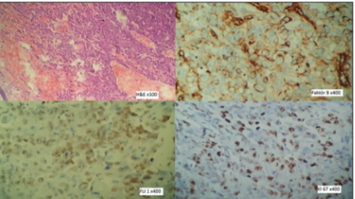
Histologic sections of the excised tissue showed malignant neoplastic developments with ulceration and hyperkeratosis infiltrating the dermis, as well as cavernous vascular structures containing solid groups of distinct nucleoli, vesicular nuclei, and atypical cells with distinct cytoplasm. Multiple (about 40) mitosis were observed under ×10 objective lens. Immunohistochemically, the tumor stained positive for Fli-1 and Factor VIII [Figure 2]. Ki-67 proliferation index was found 30%. The lesion, which was
Stewart–Treves syndrome (STS) is defined as angiosarcoma arising in the setting of chronic lymphedema. It is typically presented in breast cancer patients who underwent axillary dissection. A 79-year-old woman presented to our clinic with a wound that developed secondary to lymphedema. Pathologic examinations diagnosed the lesion as angiosarcoma. STS is a rare and deadly entity. It is hard to diagnose and has 10% mean survival rate for 5 years. We emphasize the importance of early diagnosis that can be a lifesaver.
Keywords: Angiosarcoma, breast cancer, cancer diagnosis, Stewart–Treves syndrome
Access this article online
Quick Response Code:
Website:
http://www.turkjplastsurg.org
DOI:
10.4103/tjps.tjps_18_18
Address for correspondence: Dr. Atilla Adnan Eyuboglu,
Department of Plastic, Reconstructive and Aesthetic Surgery, School of Medicine, Baskent University, Ankara, Turkey. E‑mail: [email protected]
This is an open access article distributed under the terms of the Creative Commons Attribution-NonCommercial-ShareAlike 3.0 License, which allows others to remix, tweak, and build upon the work non-commercially, as long as the author is credited and the new creations are licensed under the identical terms.
For reprints contact: [email protected]
How to cite this article: Eyüboǧlu AA, Cologlu H, Bal N, Albayati A,
Ertas NM. Stewart–Treves syndrome: A case report and review of literature. Turk J Plast Surg 2018;26:34-6.
Stewart–Treves Syndrome: A Case Report and Review of
Literature
Atilla Adnan Eyüboğlu, Harun Cologlu, Nebil Bal1, Abbas Albayati, Nilgun Markal Ertas
Departments of Plastic, Reconstructive and Aesthetic Surgery and 1Pathology, School of Medicine, Baskent University, Adana, Turkey
Case Report
Eyuboglu, et al.: Stewart–Treves syndrome
Turkish Journal of Plastic Surgery ¦ Volume 26 ¦ Issue 1 ¦ January-March 2018 35 excised with intact surgical margins, both in depth and width,
was reported as angiosarcoma. The patient was diagnosed with STS based on morphologic findings, immune profile, and clinical picture.
Increased fluoro-2-deoxy-d-glucose involvement was identified under the skin to the lateral of the surgical site in positron emission tomography/computed tomography examinations that were performed after diagnosis for systemic screening. The radiation oncology clinic was consulted when a nodular lesion possibly consistent with a lymph node was seen posterior to the surgical site. Decision was made to re-excise the lesion and apply radiotherapy.
The surgical site was re-excised with 2-cm margins. The skin defect was primarily closed. Two-dimensional conformal plan, curative radiotherapy with 200 cGy/Fr (total 60 Gy) was applied after sutures were removed. Patient’s general condition worsened in the 3rd month after re-excision. Magnetic resonance (MR) imaging found metastatic developments in regional lymph nodes. One-and-a-half year after the diagnosis, the patient’s overall condition worsened due to systemic hypertension, diabetic coma, and vascular complications caused by diabetes and she passed away. The patient wore a compression garment during the treatment to reduce arm lymphedema. Extremity elevation was also advised. There was no clinically visible improvement even though the patient adhered to the therapy.
d
IscussIonThe STS was first described in 1948 by Stewart and Treves.[1] There are about 400 cases reported in the literature.[2] STS is a highly rare and fatally progressing condition, and as reported in the first article, commonly presents with angiosarcoma arising from lymphedema that develops in 5–11 years after radical mastectomy.[1] Lymphedema develops in 14% of the patients undergoing total axillary lymph node dissection.[3] The rate of STS development within 5 years after mastectomy is reported between 0.07% and 0.45%.[4] STS can be seen not only after a mastectomy but also after burns, traumas, and radiotherapy,
or can be congenital.[1] Reports in the literature indicate about 5–15 years after radical mastectomy until sarcoma presents itself in breast cancer patients. The mean time from radical mastectomy to lymphangioma development is 11 years.[4] Our patient had undergone mastectomy 6 years earlier, after which angiosarcoma had developed following lymphedema. Our patient passed away 1.5 years after the STS diagnosis. Hemorrhagic, red-purple-colored spots in the lymphedema site can be confused with cellulitis in the early period. Lesions can grow over time and lead to bleeding. Lesions that are clinically similar to infection can resist to antibiotherapy. Ultrasonography or MR imaging can be performed for diagnosis.[2] These imaging techniques are also used in follow-up.
Histopathological and immunohistochemical examinations following incisional or excisional biopsy are important for definitive diagnosis. Growth of lymph vessels secondary to lymphatic obstruction is seen in sections. Irregularly proliferating vascular structures invade dermal collagen tissue. Hyperchromatic and macronucleoli cells undergo mitosis.[5] The epidermis over the lymphatic lesion appears acanthotic, hyperkeratotic, or atrophic. Immunohistochemically, endothelial cell markers laminin, Type IV collagen, CD31, CD34, FLI-1, von Willebrand factor and Factor VIII express positive, and increased proliferation of Ki-67 is observed.[6,7] Antibodies targeting lymphatic tissues have been developed with the advancement of molecular biology. Podoplanin (D2-40), lymphatic vascular endothelial hyaluronan receptor-1, prospero homeobox protein 1, and vascular endothelial growth factor receptor 3 help for a more definitive diagnosis of sarcoma.[8] Examination of structures such as pinocysts and intercellular junctions under electron microscope supports malignancy diagnosis.
Medical and surgical approaches are used in the treatment of the STS, but the treatment is nevertheless a difficult and complex process. The best surgical approach is excision of the lesion with wide margins or early amputation the limb.[3,4] Radical ablative surgery should be performed in the early period after diagnosis to increase the chance of survival.[3-5] Surgical margins should be 2–3 cm.[5] Local
Figure 1: Macroscopic view of lesion, hemorrhagic, black‑purple‑colored tumoral lesion on the skin. Distinct edema on the arm
Figure 2: Microscopic view of lesion. Staining of tumoral cells with immune markers
Eyuboglu, et al.: Stewart–Treves syndrome
Turkish Journal of Plastic Surgery ¦ Volume 26 ¦ Issue 1 ¦ January-March 2018
36
recurrence has been reported to be less in cases which amputation is performed than those treated with wide local excision.[2] Mean survival after diagnosis is 2.5 years and patients are expected to die within 2 years after metastasis.[3,8,9] Grobmyer et al. report to have found no significant differences in terms of survival among patients treated with chemotherapy and radiotherapy.[10] Chemotherapeutic agents used in the treatment of STS are bleomycin, 5-fluorouracil, methotrexate, vincristine, actinomycin, doxorubicin, cyclophosphamide, or dacarbazine. Local recurrence and systemic metastasis are frequent despite these treatments.[11]
c
onclusIonSTS is a rare and deadly disease. With delayed diagnosis and poor prognosis, 5-year mean survival rate is about 10%.[4] History of mastectomy should be questioned in cases presenting with chronic lymphedema, and the patient should be informed about the prognosis of the lesions and the disease itself. Excision of early-stage angiosarcoma using wide surgical margins can be a life-saver.
Patient consent
The patient was informed about the usage of her photographs and her verbal consent was obtained. Written consent was not deemed necessary.
Financial support and sponsorship Nil.
Conflicts of interest
There are no conflicts of interest.
r
eferences1. Stewart FW, Treves N. Lymphangiosarcoma in postmastectomy lymphedema; a report of six cases in elephantiasis chirurgica. Cancer 1948;1:64-81.
2. Berebichez-Fridman R, Deutsch YE, Joyal TM, Olvera PM, Benedetto PW, Rosenberg AE, et al. Stewart-Treves syndrome: A case report and review of the literature. Case Rep Oncol 2016;9:205-11. 3. Tabareau F, de Muret A, Miquelestorena E, Decouvelaere AV, de
Pinieux G. Cutaneous epitheloid clear cells angiosarcoma in a young woman with congenital lymphedema. Case Rep Pathol 2013;1:1-6. 4. Sharma A, Schwartz RA. Stewart-Treves syndrome: Pathogenesis and
management. J Am Acad Dermatol 2012;67:1342-8.
5. Lee R, Saardi KM, Schwartz RA. Lymphedema-related angiogenic tumors and other malignancies. Clin Dermatol 2014;32:616-20. 6. Harrison WD, Chandrasekar CR. Stewart-Treves syndrome following
idiopathic leg lymphoedema: Remember sarcoma. J Wound Care 2015;24:S5-7.
7. Wysocki WM, Komorowski A. Stewart-Treves syndrome. J Am Coll Surg 2007;205:194-5.
8. Young RJ, Brown NJ, Reed MW, Hudges D, Woll PJ. Angiosarcoma. Lancet Oncol 2010;11:983-91.
9. McHaffe DR, Kozak KR, Warner TF, Cho CS, Heiner JP, Attia S. Stewart-Treves syndrome on the lower extremity. J Clin Oncol 2010;28:351-2.
10. Grobmyer SR, Daly JM, Glotzbach RE, Grobmyer AJ 3rd. Role of
surgery in the management of postmastectomy extremity angiosarcoma (Stewart-Treves syndrome). J Surg Oncol 2000;73:182-8.
11. Shon W, Ida CM, Boland-Froemming JM, Rose PS, Folpe A. Cutaneous angiosarcoma arising in massive localized lymphedema of the morbidly obese: A report of five cases and review of the literature. J Cutan Pathol 2011;38:560-4.