REVIEW
Evaluation of hydrocephalus patients with 3D‑SPACE technique
using variant FA mode at 3T
Oktay Algin1,2
Received: 18 July 2017 / Accepted: 18 September 2017 / Published online: 26 September 2017 © Belgian Neurological Society 2017
Introduction
Progressive developments in magnetic-resonance imaging (MRI) technology allow us to assess the cerebrospinal-fluid (CSF) circulation better [1]. Three-dimensional sampling perfection with application optimized contrasts using dif-ferent flip-angle evolution (3D-SPACE) sequence (Siemens, Erlangen, Germany) is a recently developed technique [2]. This technique is being used more and more in daily prac-tice and related papers are being increasing in the literature. 3D-SPACE technique (GE for CUBE, Philips for VISTA) can be helpful in the diagnosis and follow-up of the patients with hydrocephalus without need for an additional MR examination (such as phase-contrast cine, heavily T2W imaging or cisternography) [1, 3].
I have been working particularly on the CSF applica-tion of 3D-SPACE techniques for more than 9 years. We would like to share our experience related to the value of this novel MRI technique for the radiological evaluation of hydrocephalus. To the best of our knowledge; there is no previous comprehensive review or pictorial essay that explains the role of the 3D-SPACE technique for the evalu-ation of hydrocephalus.
In this pictorial essay, I discussed the inclusive role and the advantages of 3D-SPACE (especially T2W variant flip-angle type) technique for the evaluation of patients with hydrocephalus in the light of the data regarding to the recent literature.
Details of 3D‑SPACE technique
3D-SPACE technique leads to the formation of a specific subclass, under the classification of current sequences (Fig. 1). This technique is basically a variant of turbo Abstract The major advantages of three-dimensional
sam-pling perfection with application optimized contrasts using dif-ferent flip-angle evolution (3D-SPACE) technique are its high resistance to artifacts that occurs as a result of radiofrequency or static field, the ability of providing images with sub-millimeter voxel size which allows obtaining reformatted images in any plane due to isotropic three-dimensional data with lower spe-cific absorption rate values. That is crucial during examination of cerebrospinal-fluid containing complex structures, and the acquisition time, which is approximately 5 min for scanning of entire cranium. Recent data revealed that T2-weighted (T2W) 3D-SPACE with variant flip-angle mode (VFAM) imaging allows fast and accurate evaluation of the hydrocephalus patients during both pre- and post-operative period for monitoring the treatment. For a better assessment of these patients; radiologists and neurosurgeons should be aware of the details and implica-tions regarding to the 3D-SPACE technique, and they should follow the updates in this field. There could be a misconception about the difference between T2W-VFAM and routine heavily T2W 3D-SPACE images. T2W 3D-SPACE with VFAM imag-ing is only a subtype of 3D-SPACE technique. In this review, we described the details of T2W 3D-SPACE with VFAM imaging and comprehensively reviewed its recent applications.
Keywords Hydrocephalus · CSF · 3D-SPACE · Third ventriculostomy · Ventriculo-peritoneal shunt · Variant flip-angle · MR · 3T
* Oktay Algin
1 Atatürk Training and Research Hospital, Bilkent, Ankara, Turkey
2 National MR Research Center (UMRAM), Bilkent University, Ankara, Turkey
spin-echo technique with extremely larger turbo factors (> 100) and relatively longer time of relaxation (TR) [2]. The major advantages of this technique are the lower specific absorption rate (SAR) value which makes it safe for using in 3-tesla (3T) scanners and the reasonable acquisition time which takes approximately 5 min for scanning the entire cranium. It provides isotropic three-dimensional (3D) data which is resistant to the most of the artifacts, particularly the susceptibility artifacts [2–5].
Flip angle mode (FAM), echo train duration/design, TR and time of echo (TE) values are modified for changing con-trast behavior in T2W images in three different ways [2]. For instance, if TE is selected as 580 ms and FAM is selected as variant; variant FAM (VFAM) images are acquired in which mobile structures such as CSF and blood is evaluated with a low signal. In this sequence, when FAM is switched
to a constant FAM without changing any other parameters, heavily T2W images are obtained. In the other hand, when TE is fixed to 140 ms and the FAM is kept constant; images, equivalent to conventional T2W images, are obtained [2]. This flexibility of the 3D-SPACE technique should be well known to use the right nomenclature and assess accurately.
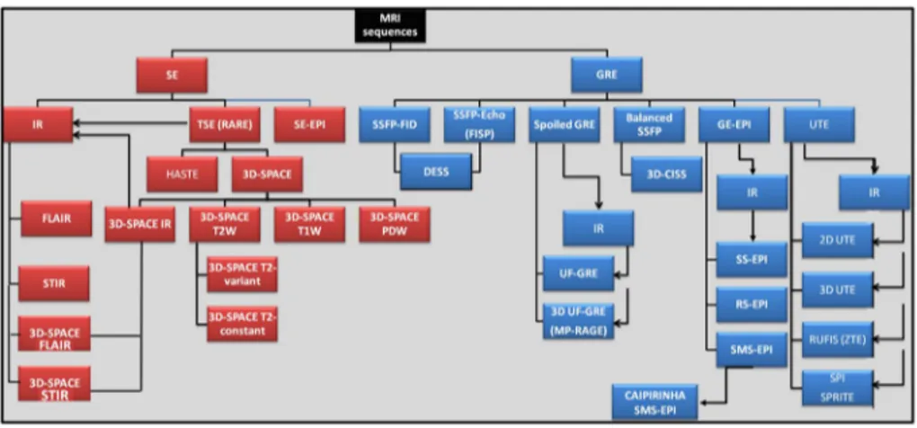
The mobile or flowing CSF is seen hypointense on T2W 3D-SPACE VFAM images because of the motion sensitivity is due to the low flip-angle values in the series of variable flip-angles [2] (Fig. 2). This feature is useful for differentia-tion of flowing CSF from stagnant or trapped CSF. Thus, it can provide fast, effective and problem solving data about CSF circulation [1, 3]. Isotropic data covering the entire cra-nium allows obtaining accurate reformatted images, which is helpful in standardizing the protocol, reduces the need for additional scanning and, therefore, decreases total imaging Fig. 1 Taxonomy of MRI sequences, and place of the 3D-SPACE
technique. SE spin-echo, GRE gradient-echo, IR inversion-recovery, TSE turbo spin-echo, EPI echo-planar imaging, HASTE half-Fourier acquisition single-shot TSE, FLAIR fluid attenuated IR (long tau IR), STIR short tau IR, PDW proton-density weighted, SSFP steady-state free precession, DESS dual-echo steady-state, 3D-CISS 3D construc-tive interference in steady state, UTE ultra-short TE, MPRAGE (3D
UF-GRE) three dimensional T1-weighted magnetization-prepared rapid acquisition of GRE (3D ultrafast GRE), SS-EPI single-shot EPI, RS-EPI readout-segmented EPI, SMS-EPI simultaneous multi-slice EPI, CAIPIRINHA controlled aliasing in parallel imaging results in higher acceleration, SPI single point imaging, SPRITE single point ramped imaging with T1 enhancement, RUFIS rotating ultra-fast imaging sequences
Fig. 2 A figure of normal 3D-SPACE VFAM appearances for comparison for later figures. There is prominent hypointense CSF flow in the aqueduct (arrow, a), foramen Monroe (arrows, b and c), and fourth ventricle (dashed arrows, a and b)
time and increases patient compliance [5–9]. As a result, T2W 3D-SPACE VFAM acquisition at 3T is a fast, flexible and efficacious technique with substantial SAR reduction to evaluate almost all of the patients with hydrocephalus accu-rately [1, 3]. Therefore; in the last few years, in our institu-tion, T2W 3D-SPACE VFAM images have been obtained for the evaluation of the patients with diagnosis or suspicion of hydrocephalus, as a crucial part of the protocol which is described in detail in Table 1.
As described above; image contrast of T2W 3D-SPACE VFAM is far different from routine conventional T2W images. Therefore, there is a significant learning curve for fast and accurate interpretation of the images. As we showed previously, T2W 3D-SPACE VFAM imaging is a reliable tool with good to perfect intra-observer and inter-observer agreement values [3, 9].
In daily routine clinical practice; 3D-heavily T2W images are also required for an optimal evaluation of some patients with obstructive hydrocephalus [8–11]. So far 3D constructive-interference in steady-state (3D-CISS) has been the mostly used sequence for this purpose [11]. Although this sequence is useful to demonstrate the anatomic details, evaluation of luminal patency and distinction of fluid from tissue thanks to its high SNR, it has some limitations includ-ing poor soft-tissue resolution, limited slab thickness and long acquisition times [6–9]. Other disadvantages of this technique are dark banding, slab boundary, and suscepti-bility artifacts due to its gradient-echo schema [12, 13].
3D-heavily T2W images, which are obtained as mentioned above on the basis of 3D-SPACE technique with constant FA mode and a relatively higher effective TE, can overcome those limitations and may be used instead of gradient-echo based 3D-heavily T2W sequences [1]. T2W 3D-SPACE VFAM is a problem solving technique in most of the cases but heavily T2W 3D-SPACE images may be complementary with equivocal results in complex cases [3].
It has been shown that there is a good correlation between the T2W 3D-SPACE VFAM and PC-MRI based evaluations [3, 9]. Therefore, T2W 3D-SPACE VFAM imaging reduced the need for PC-MRI images attributed to its own advan-tages. In some cases T2W 3D-SPACE VFAM imaging may avoid misinterpretation due to false positive results from PC-MRI [7–9]. In Table 2, advantages and disadvantages T2W 3D-SPACE VFAM imaging were given when com-pared with PC-MRI and gradient-echo based 3D-heavily T2W images.
Hydrocephalus
Hydrocephalus is called ventriculomegaly (Evans index > 0.3) and it affects 1–2% of the population [14]. Establishment of the diagnosis and management of hydro-cephalus are still challenging issues mainly due to the insufficient data regarding to the physiology and patho-physiology of the process [15]. Therefore, classification, Table 1 3 Tesla MRI
protocol used for patients with hydrocephalus
FOV can be adjusted according to patient size
We suggest 3D-MPRAGE should be obtained for an optimum morphologic analysis and tissue characteri-zation. Also, heavily T2W 3D-SPACE should be obtained if necessary (e.g. for patients with suspicious findings or complex pathologies)
TI time of inversion, 3D-SPACE three-dimensional sampling perfection with application-optimized con-trasts using different flip angle evolutions, 3D-MPRAGE 3D T1W magnetization prepared rapid acquisition gradient-echo, NEX number of excitations, FOV field of view, PAT parallel acquisition technique, GRAPPA generalized auto calibrating partially parallel acquisitions
Sequences/parameters 3D-MPRAGE 3D-SPACE (with VFAM) Heavily T2W 3D-SPACE
TR/TE (ms) 2130/3.45 3000/579 3000/423
TI (ms) 1100 – –
Slice thickness 0.8 mm 0.6 mm 0.8 mm
FOV (mm) 230 × 230 240 × 240 210 × 210
Acquisition time 5 min 5 min 5 min
Velocity encoding – – –
NEX 1 2 1
Number of slices 240 240 192
Flip angle 8° 100° 100°
Imaging plane Sagittal Sagittal Sagittal
Distance factor 50% – –
PAT factor 2 2 2
PAT mode GRAPPA GRAPPA GRAPPA
Voxel size (mm) 0.8 × 0.8 × 0.8 0.6 × 0.6 × 0.6 0.8 × 0.8 × 0.8
diagnostic features and treatment plans vary in a wide range between centers [16]. Hydrocephalus is basically classified as obstructive type and communicating type. Nevertheless, there are complex cases in which both types of hydrocepha-lus play role in the ethiopathogenesis of the disease [16]. Among the treatment alternatives; surgery has come to fore and in daily practice ventriculo-peritoneal shunt (VPS) and endoscopic third ventriculostomy (ETV) are the most com-monly used procedures [15].
T2W-VFAM 3D-SPACE imaging demonstrates the mor-phologic features of hydrocephalus (e.g. ventriculomegaly, normal or narrowed cortical sulci, enlargement of the third ventricular recesses and lateral ventricular horns, perive-ntricular interstitial edema, thinning and elevation of the corpus callosum) by a fast and accurate way [3]. In addition, T2W-VFAM 3D-SPACE findings are important for planning the management as well as follow-up of the patients [1].
Obstructive hydrocephalus
Tumor, cyst, adhesion, web/membrane, infection, inflam-mation or vascular anomalies may lead to direct or indi-rect obstruction at any level but particularly at the level of foramina [17, 18]. T2W 3D-SPACE VFAM imaging helps to determine the etiology and location of blockage in obstruc-tive hydrocephalus [1, 19] (Fig. 3). Although aqueduct is the place where obstruction is mostly detected, disorders may lead to complete or partial obstruction at the level of all the other foramina (e.g. foramen of Monro, foramen magnum, fourth ventricle’s outlet) as well [8, 17]. T2W 3D-SPACE VFAM may play a pivot role as the problem solving method during diagnosis, since it allows multiplanar evaluation of vascular structures with a complex morphology and CSF distribution-volume analysis [6, 18].
Table 2 Advantages and disadvantages of T2W 3D-SPACE imaging were given when compared with PC-MRI and gradient-echo based 3D-heavily T2W images
FAM flip-angle mode, ECG electrocardiography
Sequences 3D-SPACE (variant FAM) 3D-SPACE (constant FAM) PC-MRI 3D-heavily T2W (e.g. 3D-CISS)
Physiological data Yes No Yes No
Morphological data Yes Yes No Yes
Whole brain imaging Yes Yes No No
Experience requirement Yes No Yes No
Mean acquisition time (min) 4–5 4–5 6–10 4–5
Artifact susceptibility No No Yes Yes
Necessity of ECG or pulse triggering No No Yes No
Two or three dimensional acquisition 3D 3D 2D (single slice) 3D
Fig. 3 Sagittal 3D-SPACE VFAM images of three different patients with obstructive hydrocephalus. A 42-year-old man with complete aqueduct stenosis due to distal web and tectal glioma (black arrow, a). The patient who undergone ETV, has patent stoma (white arrow, a). In the second case complete stenosis in the distal aqueduct is seen
due to a tectal glioma (white arrow) and venous vascular variation (black arrow) (b). In the third case fourth ventricle outlet and foramen magnum stenosis are demonstrated secondary to Chiari malformation (arrow in c)
Evaluation of endoscopic or spontaneous third ventriculostomy (ETV or STV)
Endoscopic third ventriculostomy has become the primary CSF diversion method used in the treatment of obstructive hydrocephalus [20]. Occasionally the diversion is sponta-neously formed by tearing of one of the walls of the third ventricle, which is mostly the inferior wall. This entity is called STV. If STV leads to a regression in patient’s symp-toms, an additional intervention is usually unnecessary [9]. Spontaneous third ventriculostomy can be detected with high accuracy using T2W 3D-SPACE VFAM imaging in preoperative period. Thus, unnecessary surgical interven-tions can be avoided in patients who would undergo CSF diversion procedures [9].
T2W 3D-SPACE VFAM imaging is useful in determin-ing the CSF diversion methods that should be used in case
of obstructive hydrocephaly. For instance, in patients with existing membranes in the prepontine cistern or in the cervi-cal subarachoid space, which will prevent CSF circulation or in patients with narrow foramen magnum, ETV should not be considered [6]. Endoscopic aqueductoplasty can be preferred as the initial restorative method in patients with occlusion within the aqueduct secondary to the webs [8]. Interpedincular, suprasellar and prepontine cisterns are very narrow and can be obstructed easily in patients with subarachnoid hemorrhage or meningeal infection [21]. In these cases, failure rate is high in ETV procedure. Factors affecting the failure risk of ETV can be summarized as fol-lows; complex third ventricular anatomy, thickened/twisted Liliequist membrane or third ventricular floor, thickened prepontine or cervical cistern membranes, adhesions in the subarachnoid space, non-visible aqueduct obstruction, nor-mal or mininor-mally dilated third ventricular recesses, and/or Fig. 4 3D-SPACE VFAM
images of two different patients with aqueduct stenosis and ETV. On sagittal image of the first patient, there is a complete aqueduct stenosis due to tectal glioma (black arrow, a). ETV was patent or functional on sag-ittal and coronal images (white arrows in a and b). On sagittal image of a 5-year-old girl with progressive headache, there is a partial (nearly complete) aqueduct stenosis (black arrow) and nonfunctional/closed stoma (white arrow) (c). After redo ETV, open and functional stoma was observed on coronal refor-matted image (arrow in d)
small stoma [6, 20, 21]. All of these findings can be dem-onstrated using only T2W 3D-SPACE VFAM technique in a fast, effective and a more convenient way for the patients [6, 7].
Qualitative and quantitative analyses of PC-MRI acquisi-tions may not always give accurate data about the success of ETV due to various reasons (such as artifacts due to complex CSF flow, inaccurate slice position, inappropriate velocity encoding value, or heart arrhythmia) [22]. In addition, there is not a definite cut-off value that evaluates the patency of ETV. Due to these limitations, 3D-heavily T2W images which can increase the acquisition time are frequently added in the MRI protocol, when PC-MRI is necessary [6, 7]. It is also possible to evaluate the presence of STV and/or the con-dition of ETV with MR ventriculography or cisternography as previously suggested [17, 22]. However, these techniques are highly invasive, require experience and carry some risks due to the injection of contrast agent into the subarachnoid space [17, 22]. T2W 3D-SPACE VFAM technique over-comes all of those limitations of PC-MRI, gradient-echo
based 3D-heavily T2W, and MR ventriculography/cisternog-raphy [6, 7] (Fig. 4).
Liliequist membrane patency and its morphology
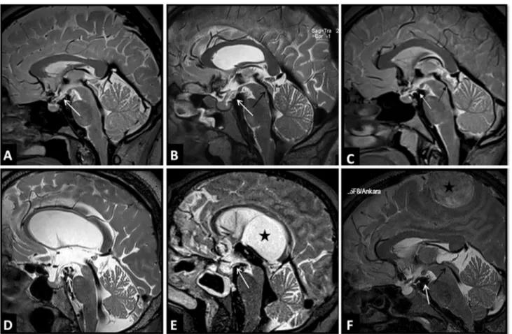
Liliequist membrane (LM) forms a complete division in the basal subarachnoid space [21]. Thus; LM fenestration has crucial importance for success of ETV [23]. LM is thicker and more resistant in patients with long-standing hydroceph-alus when compared with LM in patients who have newly developed hydrocephaly. LM may not be always visualized during ETV procedure [23]. Unless LM is fenestrated dur-ing ETV procedure, accurate CSF flow from third ventricle into the prepontine cistern or foramen magnum cannot be established [20]. Although the importance of LM is well understood; morphology, attachment sites and variation regarding to this structure is not fully known and there have been several conflicting reports published [20–23]. This may be attributed to the fact that LM is a very thin membrane and shows variations, thus it is challenging to establish Fig. 5 On figures a–f, LM in different patients is shown (white
arrows). As seen in these figures, LM may show several variations. Besides it may be patent in some patients (a), whereas it may be
fenestrated in others (e and f). Asterisk in e non-communicating intra-ventricular arachnoid cyst, asterisk in f menengioma, black arrows aqueductal flow voids
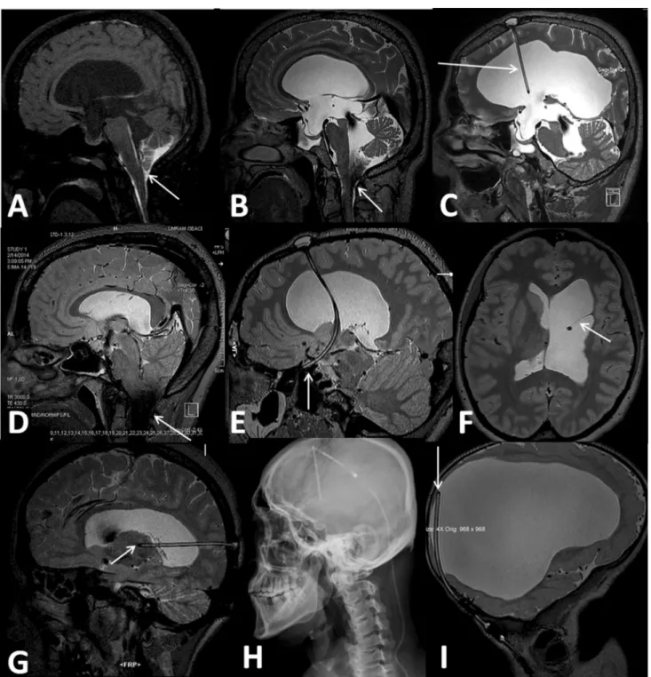
Fig. 6 Four different patients with VPS. Postcontrast MR cister-nography image of a 34 year old male who presented with rhinor-rhea, fourth ventricular outlet obstruction is seen (arrow, a). Sag-ittal 3D-SPACE VFAM images of the same patient demonstrate the obstruction (arrow, b) and functional shunt catheter (arrow, c). After the shunt operation rhinorrhea is completely regressed. Sec-ond patient is a 20-year old female with Chiari malformation. She has progressive headache and previously implanted VPS (d–f). On 3D-SPACE VFAM images of the patient, decreased fourth ven-tricular CSF flow, excessive signal void in foramen magnum, which is seen in most of the patients with Chiari (arrow, d), and non
func-tional shunt catheter that was misplaced in the superior orbital fissure (arrow, e), adhesions beside the catheter (arrow, f) are demonstrated. Another nonfunctional shunt catheter due to thalamic insertion of catheter tip (arrow in g). Last patient with VPS has a progressive ventriculomegaly and headache. On sagittal plain radiogram, VPS is intact (h). However; it was seen that the distal end of the catheter was not moving freely and was adhered to lateral ventricle (arrow, i) on 3D-SPACE VFAM images that were taken in prone position. This finding is usually seen in shunts that are obstructed by fibrin-cuff or adhesions
Fig. 7 3D-SPACE VFAM images of three different cases. Three-plane 3D-SPACE VFAM images of a young volunteer are shown for comparison (a–c). These images demonstrate open or patent aqueduct (arrow in a) and foramen Monroe (arrows in b and c). 29-year-old female patient with partial (minimal) aqueduct and fourth ventricu-lar outlet stenosis (d–f). On these images, narrowed aqueduct (black arrows) and fourth ventricular outlet (white arrow) are seen. Also, the images show typical hydrocephalus findings (dilatation of lateral ventricles, enlargement of the third ventricular recesses, effacement of cortical sulci, decreased mammillo-pontine distance, and upward
bowing and thinning of corpus callosum) (d–f). Prominent aque-ductal and fourth ventricular flow void phenomenon without visible foraminal stenosis of a 73-year-old woman with possible NPH (g). Coronal image of this patient demonstrates dilatation of the tempo-ral horns without hippocampal atrophy (arrows), decreased callosal angle, thinned corpus callosum, effacement of convexity sulci without narrowed Sylvian cisterns, normal hippocampi, and increased Evans ratio (h). Parasagittal image of the patient shows non-dilated para-hippocampal fissure (arrow) and normal hippocampus with enlarged atrium of lateral ventricle (i)
all aspects of these structures with MRI [21]. Thus, data about this structure is mostly provided from cadaver studies (Fig. 5).
Although demonstration of some segments of LM is possible using only 3D-heavily T2W technique, it may be impossible to display the entire morphology of this structure. Showing its relation with the third ventricle and the alterations taking place in CSF flow caused by the LM and to obtain an accurate classification are almost impossible.
It is not possible to demonstrate morphology of the ventricular system and CSF hemodynamic using T2W 3D-SPACE VFAM technique by its own, however, it can provide the data about the morphology of LM and can demonstrate whether it allows flow or not in a short time and accurate way [24]. Morphological (complex shape) or structural (doublet or triplet membrane type) may change the endoscopic surgery procedure or success rate. Accord-ing to our unpublished data; in cases with complex shaped or doublet-triplet LM, 3D-heavily T2W images may pro-vide additional data in this situation.
Assessment of ventriculo-atrial or ventriculo-peritoneal shunt (VPS)
Complications of these shunts commonly occur due to obstruction, disconnection or infection [25]. Shunt failure occurs in about one third of the patients within the first year; therefore, radiologic evaluation is indicated [14]. In a daily routine practice, programmable VPSs are the mostly preferred technique and most of them are 3T MR-compatible [14, 25]. T2W 3D-SPACE VFAM technique is a useful method for the evaluation of integrity, position, and morphology of the shunt without artifacts of shunt reservoir [1]. In addition, early and late complications of the procedure (such as subdural-epidural effusions, neuronal damages, fibrin cuff around catheter tip) can be easily evaluated with isotropic (1 < mm3) T2W-VFAM
3D-SPACE data (Fig. 6). For the indeterminate cases 3D-heavily T2W SPACE images and plain radiographs may be complimentary [14]. Programmable shunts should be readjusted after MRI examination.
Normal pressure hydrocephalus
Normal pressure hydrocephalus (NPH) is diagnosed based on clinical symptoms. Nevertheless, it does not always mani-fest with classical findings including normal opening CSF pressure, gait apraxia, dementia, and/or urinary incontinence [26]. In these cases it is challenging to differentiate NPH from other reasons of dementia by clinical, laboratory and/
or routine MRI findings [27]. On the other hand, differentia-tion is critical since NPH can be managed by CSF diversion (generally VPS) contrary to the other etiologies of demen-tia [26]. Both for diagnosis and predicting shunt response, various additional radiologic evaluations including quanti-tative PC-MRI analysis, cisternographic studies have been required up to now [17, 27]. All these extra imaging studies increase the total examination time, require experience and affect patient compliance negatively. Besides, their efficacy in diagnosis and prediction of response to the shunt opera-tion are debatable [26–29].
T2W 3D-SPACE VFAM technique may provide mor-phologic and physiologic information which is beneficial in differentiating NPH from other pathologies that lead demen-tia [3]. Some of the findings that favor NPH diagnosis are the existence of excessive aqueductal flow void phenom-enon (that reach the inferior wall of the fourth ventricle), increased frontal horn radius, enlarged Sylvian fissures and lateral ventricles but tight convexity sulci (dispropor-tionately enlarged subarachnoid space pattern), decreased mamillopontine distance, concave third ventricular floor, normal perihippocampal fissures, increased callosal height, and decreased callosal angle [29–31] (Fig. 7). These find-ings support the diagnosis of NPH; however, those cannot be used for predicting the shunt failure precisely [28–30]. Detailed analysis of the hippocampal structures and more global assessment of all intracranial elements are main advantages of the technique. As a result of this section, it is clear that the T2W 3D-SPACE VFAM technique can be used for the assessment of patients with suspected NPH.
Conclusion
T2W 3D-SPACE VFAM imaging provides physiologic information about CSF hydrodynamics in a non-invasive way without giving up anatomic detail. Morphology of CSF containing spaces, position and integrity of VPS, patency or presence of third ventriculostomy, CSF hydrodynamics, CSF related tiny membranes, and other associated findings are better evaluated with this method. In addition, it decreases acquisition time and costs, and increases patient compliance by decreasing the need for invasive techniques or additional examinations. Therefore, it should be considered in routine use for patients with CSF disorders.
Acknowledgements The author would like to thank to engineer Ali Caglar Özen (University Medical Center Freiburg) and pediatric radi-ologist Evrim Ozmen (Istanbul University) for their excellent contri-butions. We also gratefully acknowledge Dr. Gülbiz Kartal (radiolo-gist) and Musa Kurnaz (MR technician) for their contributions, and all UMRAM staff for providing technical support.
Compliance with ethical standards
Conflict of interest The author declares that he have no conflict of interest.
Funding None.
Ethical approval This article does not contain any studies with human participants or animals performed by any of the authors. Informed consent Informed consent was obtained from all indi-vidual participants included in the study.
References
1. Kartal MG, Algin O (2014) Evaluation of hydrocephalus and other cerebrospinal fluid disorders with MRI: an update. Insights Imag-ing 5(4):531–541
2. Mugler JP 3rd (2014) Optimized three-dimensional fast-spin-echo MRI. J Magn Reson Imaging 39(4):745–767
3. Kartal MG, Ocakoglu G, Algin O (2015) Feasibility of 3-dimen-sional sampling perfection with application optimized contrast sequence in the evaluation of patients with hydrocephalus. J Com-put Assist Tomogr 39(3):321–328
4. Gerigk L, Bostel T, Hegewald A et al (2012) Dynamic magnetic resonance imaging of the cervical spine with high-resolution 3-dimensional T2-imaging. Clin Neuroradiol 22(1):93–99 5. Mugler JP 3rd, Bao S, Mulkern RV et al (2000) Optimized
single-slab three-dimensional spin-echo MR imaging of the brain. Radi-ology 216(3):891–899
6. Algin O (2015) Prediction of endoscopic third ventriculostomy (ETV) success with 3D-SPACE technique. Neurosurg Rev 38(2):395–397
7. Algin O, Ucar M, Ozmen E et al (2015) Assessment of third ven-triculostomy patency with the 3D-SPACE technique: a prelimi-nary multicenter research study. J Neurosurg 122(6):1347–1355 8. Algin O, Turkbey B (2012) Evaluation of aqueductal stenosis
by 3D sampling perfection with application-optimized contrasts using different flip angle evolutions sequence: preliminary results with 3T MR imaging. Am J Neuroradiol 33(4):740–746 9. Algin O, Turkbey B, Ozmen E et al (2013) Evaluation of
spon-taneous third ventriculostomy by three-dimensional sampling perfection with application-optimized contrasts using different flip-angle evolutions (3D-SPACE) sequence by 3T MR imaging: preliminary results with variant flip-angle mode. J Neuroradiol 40(1):11–18
10. Ucar M, Tokgoz N, Damar C et al (2015) Diagnostic performance of heavily T2-weighted techniques in obstructive hydrocephalus: comparison study of two different 3D heavily T2-weighted and conventional T2-weighted sequences. Jpn J Radiol 33(2):94–101 11. Dinçer A, Kohan S, Ozek MM (2009) Is all “communicating”
hydrocephalus really communicating? Prospective study on the value of 3D-constructive interference in steady state sequence at 3T. Am J Neuroradiol 30(10):1898–1906
12. Held P, Fellner C, Fellner F et al (1997) MRI of inner ear and facial nerve pathology using 3D MP-RAGE and 3D CISS sequences. Br J Radiol 70(834):558–566
13. Park SH, Han PK, Choi SH (2015) Physiological and functional magnetic resonance imaging using balanced steady-state free pre-cession. Korean J Radiol 16(3):550–559
14. Lollis SS, Mamourian AC, Vaccaro TJ, Duhaime AC (2010) Programmable CSF shunt valves: radiographic identification and interpretation. Am J Neuroradiol 31(7):1343–1346
15. Preuss M, Hoffmann KT, Reiss-Zimmermann M et al (2013) Updated physiology and pathophysiology of CSF circulation— the pulsatile vector theory. Childs Nerv Syst 29(10):1811–1825 16. Yadav YR, Mukerji G, Parihar V et al (2009) Complex
hydro-cephalus (combination of communicating and obstructive type): an important cause of failed endoscopic third ventriculostomy. BMC Res Notes 2:137
17. Algin O, Turkbey B (2013) Intrathecal gadolinium-enhanced MR cisternography: a comprehensive review. Am J Neuroradiol 34(1):14–22
18. Paulson D, Hwang SW, Whitehead WE et al (2012) Aqueductal developmental venous anomaly as an unusual cause of congenital hydrocephalus: a case report and review of the literature. J Med Case Rep 6:7
19. Ucar M, Tokgoz N, Koc AM et al (2015) Assessment of 3D T2-weighted high-sampling-efficiency technique (SPACE) for detection of cerebellar tonsillar motion: new useful sign for Chiari I malformation. Clin Imaging 39(1):42–50
20. Fushimi Y, Miki Y, Ueba T et al (2003) Liliequist membrane: three-dimensional constructive interference in steady state MR imaging. Radiology 229(2):360–365
21. Mortazavi MM, Rizq F, Harmon O et al (2015) Anatomical vari-ations and neurosurgical significance of Liliequist’s membrane. Childs Nerv Syst 31(1):15–28
22. Algin O, Hakyemez B, Parlak M (2011) Phase-contrast MRI and 3D-CISS versus contrast-enhanced MR cisternography for the detection of spontaneous third ventriculostomy. J Neuroradiol 38(2):98–104
23. Etus V, Solakoglu S, Ceylan S (2011) Ultrastructural changes in the Liliequist membrane in the hydrocephalic process and its implications for the endoscopic third ventriculostomy procedure. Turk Neurosurg 21(3):359–366
24. Algin O, Kılın M, Ozmen E, Ocakoglu G (2016) Assessment of Liliequist membrane by 3D-SPACE technique at 3 T. Neuroradiol-ogy 58(7):637–647
25. Cakır E, Arslan E (2014) CSF hydrothorax as a late complica-tion of ventriculoatrial shunt catheter displacement. Balkan Med J 31(4):363–365
26. Algin O, Hakyemez B, Taskapilioglu O et al (2009) Morphologic features and flow void phenomenon in normal pressure hydro-cephalus and other dementias: are they really significant? Acad Radiol 16(11):1373–1380
27. Algin O, Hakyemez B, Parlak M (2010) The efficiency of PC-MRI in diagnosis of normal pressure hydrocephalus and prediction of shunt response. Acad Radiol 17(2):181–187
28. Ringstad G, Emblem KE, Geier O, Alperin N, Eide PK (2015) Aqueductal stroke volume: comparisons with intracranial pres-sure scores in idiopathic normal prespres-sure hydrocephalus. Am J Neuroradiol 36:1623–1630
29. Bradley WG Jr (2015) CSF flow in the brain in the context of normal pressure hydrocephalus. Am J Neuroradiol 36:831–838 30. Ishii K, Kanda T, Harada A et al (2008) Clinical impact of the
cal-losal angle in the diagnosis of idiopathic normal pressure hydro-cephalus. Eur Radiol 18:2678–2683
31. Segev Y, Metser U, Beni-Adani L et al (2001) Morphometric study of the midsagittal MR imaging plane in cases of hydro-cephalus and atrophy and in normal brains. Am J Neuroradiol 22:1674–1679