The association of epicardial fat thickness with blunted heart rate recovery in patients with metabolic syndrome
6
0
0
Tam metin
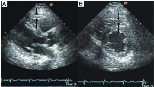
(2) 258. C. Sengul and D. Duman. centimeters) was recorded as the average of two measurements while the subject was standing at midpoint between the lowest rib and the iliac crest. After a 30-minute acclimation period, BP was measured 3 times to the nearest 2 mmHg in the sitting position using a mercury sphygmomanometer and appropriately sized cuffs. The average of 3 measurements was used to calculate systolic and diastolic BP. Blood sampling was obtained at least 12 hrs of fasting. Levels of total cholesterol, triglycerides (TG), low density lipoprotein-cholesterol (LDL), and high density lipoprotein-cholesterol (HDL), glucose, hemoglobin, and creatinine were assayed by routine laboratory techniques. Plasma concentration of insulin was measured in duplicate using commercial available kit (BIOSOURCE, Camarillo, CA). Homeostasis model assessment index of insulin resistance (HOMA-IR) was defined as fasting plasma insulin (mu/L) × fasting glucose (mg/dl)/405 by using Matthews’s equation (Matthews et al. 1985). The coefficients of variation were less than 5% for every measurement. Due to lack of definite criteria of MS in Turkey, MS was defined according to the Adult Treatment Panel III (ATP-III) criteria (Tartan et al. 2006). The subjects were diagnosed as having MS if 3 or more of the 5 criteria were met; 1) Waist circumference > 102 cm in men or > 88 cm in women, 2) triglycerides > 150 mg/dl, 3) HDL < 40 mg/dl in men or < 50 mg/dl in women, 4) BP > 130/85 mmHg, 5) fasting blood glucose > 110 mg/dl. Stress test All the subjects underwent a symptom-limited exercise stress test according to the Bruce protocol (Quinton Treadmill system, Quinton, Inc., Bothell, WA, USA). The Tango exercise BP monitor (SunTech Medical, Morrisville, North Carolina) was used to automatically measure and display a subject’s systolic, diastolic BP, and HR. BP and HR were measured before, at 2 min of each stages of exercise. Exercise was stopped when the subjects demanded cessation due to exhaustion, or if the HR achieved was more than 95% of estimated maximal HR (220-age). During the recovery phase, the subjects continued to walk for 60 sec at the speed of 1.5 mph and they sat down for 3 min with continued BP, HR, and rhythm monitoring. HRR-1 was obtained by subtracting HR at the first minute of recovery from peak HR obtained during exercise. A HRR-1 ≤ 18 beats/min was. considered blunted (Watanabe et al. 2001). Metabolic equivalents (METS) were calculated from the treadmill speed and the grade at peak exercise in accordance with previous studies (Gulati et al. 2003). Echocardiography Complete transthoracic 2-dimensional echocardiograms were obtained from the standard parasternal and apical views with the available equipment (Vivid 3 pro, GE Vingmed, Milwaukee, USA). Images were digitally stored and reviewed by a single cardiologist blinded to the subject’s information in order to avoid inter-reader variability. Left ventricular ejection fraction (EF) was measured with modified Simpson’s rule from 2-dimensional echocardiographic tracings obtained in apical 4-chamber view according to American Society of Echocardiography criteria (Lang et al. 2005). EFT was measured according to the method previously described and validated (Iacobellis et al. 2003). The epicardial fat was identified as the echo-free space between the outer wall of the myocardium and the visceral layer of the pericardium. EFT was measured perpendicularly on the free wall of the right ventricle at enddiastole in 3 cardiac cycles. The maximum EFT was measured at the point on the free wall of the right ventricle along the mid-line of the ultrasound beam, perpendicular to the aortic annulus. For the midventricular parasternal short-axis assessment, maximum EFT was measured on the free wall of the right ventricle along the mid-line of the ultrasound beam, perpendicular to the interventricular septum at mid-chordal and the tip of the papillary muscle level, as the anatomic landmark (Fig. 1). The maximum value at any site was measured, and the average value was calculated. Echocardiograms of 12 subjects were randomly selected and second measurement of EFT was performed 1 week later to assess intra-observer variability. Intraobserver variability of the EFT was 4.7%. Statistical Analysis Mean values were calculated for continuous variables and absolute and relative frequencies for discrete variables. Univariate comparisons of continuous data were carried out with the use of unpaired Student’s t-tests (or Mann-Whitney U test if applicable) and discrete variables were compared with [chi]2 tests (or Fisher’s exact tests if. Fig. 1. Measurement of epicardial fat thickness by two-dimentional transthoracic echocardiography. Shown are the images used for the measurement of epicardial fat thickness in parasternal long axis (A) and short axis (B) at end-diastole of the cardiac cycle..
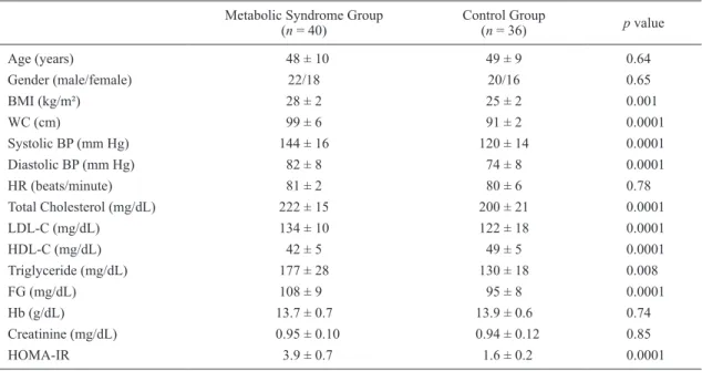
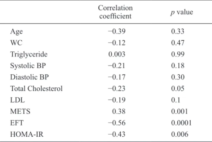
(3) 259. Epicardial Fat and Heart Rate Recovery applicable). The relationship between HRR-1 and the other variables were assessed using Pearson correlation analysis in patients with MS. MS patients were categorized into two groups according to HRR-1 cut-off value (normal HRR: HRR-1 > 18; blunted HRR: HRR-1 ≤ 18). The univariate analysis was performed as the initial step. The variables with p value ˂ 0.10 (EFT, total cholesterol, glucose, HOMA-IR, METS) were included in multivariate stepwise logistic regression analysis to assess the association with blunted HRR. LDLcholesterol was not included due to significant correlation with total cholesterol levels. The cut-off value of EFT which was related to the blunted HRR with corresponding sensitivity and specificity was estimated by receiving operator characteristic (ROC) curve analysis. All comparisons were two-sided and P ˂ 0.05 was considered to be significant. Statistical analysis was carried out with a commercially available statistical package (SPSS for Windows Version 10.0; SPSS, Inc., Chicago, Illinois, USA).. Results The baseline characteristics of the patients with MS and the controls were presented in Table 1. HOMA-IR, BMI, waist circumference, total cholesterol, LDL cholesterol, fasting glucose and triglyceride levels were significantly increased in patients with MS. Plasma HDL levels were lower in patients with MS. Age, sex, plasma hemoglobin and creatinine levels were similar for the two groups. EFT was significantly increased (7.2 ± 2 vs. 5.6 ± 1.8 mm; p = 0.001) in subjects with MS, while METS (11.1 ± 0.9 vs. 11.7 ± 0.8; p = 0.001) and HRR-1 (21 ± 8 vs. 26 ± 9; p = 0.006) were significantly lower compared to the control group. Left ventricular EF, basal HR, and peak HR were similar between two groups (Table 2). In Pearson correlation analysis, HRR-1 significantly correlated with EFT (r: −0.56, p: 0.0001), HOMA-IR (r: −0.43, p: 0.006), METS (r: 0.38, p: 0.001), and total cholesterol levels. Table 1. Demographic and laboratory variables of the patients with MS and the controls.. Age (years) Gender (male/female) BMI (kg/m²) WC (cm) Systolic BP (mm Hg) Diastolic BP (mm Hg) HR (beats/minute) Total Cholesterol (mg/dL) LDL-C (mg/dL) HDL-C (mg/dL) Triglyceride (mg/dL) FG (mg/dL) Hb (g/dL) Creatinine (mg/dL) HOMA-IR. Metabolic Syndrome Group (n = 40). Control Group (n = 36). p value. 48 ± 10 22/18 28 ± 2 99 ± 6 144 ± 16 82 ± 8 81 ± 2 222 ± 15 134 ± 10 42 ± 5 177 ± 28 108 ± 9 13.7 ± 0.7 0.95 ± 0.10 3.9 ± 0.7. 49 ± 9 20/16 25 ± 2 91 ± 2 120 ± 14 74 ± 8 80 ± 6 200 ± 21 122 ± 18 49 ± 5 130 ± 18 95 ± 8 13.9 ± 0.6 0.94 ± 0.12 1.6 ± 0.2. 0.64 0.65 0.001 0.0001 0.0001 0.0001 0.78 0.0001 0.0001 0.0001 0.008 0.0001 0.74 0.85 0.0001. MS, metabolic syndrome; WC, waist circumference; BP, blood pressure; BMI, body mass index; LDL-C, low density lipoprotein-cholesterol; HDL-C, high density lipoprotein-cholesterol; FG, fasting glucose; Hb, hemoglobin; HOMA-IR, Homeostatic model assessment of insulin resistance; HR, heart rate; NS, not significant.. Table 2. Echocardiographic and exercise stress test variables between groups.. EFT (mm) EF (%) Basal HR (beats/minute) Peak HR (beats/minute) METS HRR-1. Metabolic Syndrome Group (n = 40). Control Group (n = 36). p value. 7.2 ± 2 66 ± 3 81 ± 2 162 ± 10 11.1 ± 0.9 21 ± 8. 5.6 ± 1.8 65 ± 3 80 ± 6 164 ± 8 11.7 ± 0.8 26 ± 9. 0.001 0.84 0.78 0.25 0.001 0.006. EFT, epicardial fat thickness; EF, ejection fraction; HR, heart rate; HRR-1, heart rate recovery at 1 min after exercise; METS, Metabolic equivalents..
(4) 260. C. Sengul and D. Duman. (r: −0.23, p: 0.05) (Table 3). The MS patients were categorized into two groups according to HRR-1 value: normal HRR (HRR-1 > 18) and blunted HRR (HRR-1 ≤ 18). EFT (8.5 ± 2 vs. 5.9 ± 1.1 mm, p ˂ 0.001), total cholesterol (228.2 ± 13.5 vs 216.5 ± 16.1 mg/dL, p = 0.018), LDL cholesterol (137.6 ± 7.1 vs. 130.6 ± 11.1 mg/dL, p = 0.025), and HOMA-IR (4.2 ± 0.7 vs. 3.7 ± 0.7, p = 0.052) were higher, but glucose (104.9 ± 7.6 vs. 111.3 ± 10.1 mg/dL, p = 0.033) and METS (10.8 ± 0.8 vs. 11.5 ± 0.9, p = 0.023) were significantly lower in patients with MS and blunted HRR (Table 4). Multivariate Table 3. Correlation between HRR1 and other measured variables.. Age WC Triglyceride Systolic BP Diastolic BP Total Cholesterol LDL METS EFT HOMA-IR. Correlation coefficient. p value. −0.39 −0.12 0.003 −0.21 −0.17 −0.23 −0.19 0.38 −0.56 −0.43. 0.33 0.47 0.99 0.18 0.30 0.05 0.1 0.001 0.0001 0.006. WC, waist circumference; BP, blood pressure; LDL-C, low density lipoprotein-cholesterol; METS, Metabolic equivalents; EFT, epicardial fat thickness; HOMA-IR, Homeostatic model assessment of insulin resistance.. logistic regression analysis demonstrated that EFT was the only parameter associated with blunted HRR in patients with MS (OR = 2.34, p = 0.001) (Table 5). An EFT of ≥ 5.5 mm was associated with the blunted HRR with 84.2% sensitivity and 52.4% specificity [ROC area under curve: 0.84, 95% CI (0.70-0.96), p ˂ 0.001].. Discussion We found that EFT was the independent predictor of blunted HRR in patients with MS. This finding may help identify patients who have potentially increased cardiovascular risk during the routine echocardiographic examinations. Autonomic dysfunction characterized by either high sympathetic or low vagal activity is a marker of increased risk for all-cause mortality (Tsuji et al. 1996; Manfrini et al. 2003). MS is also associated with enhanced sympathetic neural drive and vagal impairment (Manzella et al. 2001). Previous studies have shown that hyperinsulinemia or insulin resistance, the basic pathological process in MS is associated with high sympathetic activity and may contribute to low cardiac vagal tone (Van De Borne et al. 1999; Straznicky et al. 2008). Blunted HRR after exercise, a sign of autonomic dysfunction, has been demonstrated to be a strong predictor of all-cause mortality in several studies (Imai et al. 1994; Lind and Andren 1999). Moreover, patients with MS and several of its components have been shown to have blunted HRR (Thomson et al. 2010). Cardiac autonomic dysfunction and blunted HRR may partly explain increased cardiovascular risk related to the MS. In accordance with these studies, the present study. Table 4. Univariate analysis of clinical variables in two groups of MS patients according to blunted HRR.. Age (years) Female gender (%) WC (cm) FG (mg/dL) Total cholesterol (mg/dL) LDL cholesterol (mg/dL) HDL cholesterol (mg/dL) Triglyceride (mg/dL) Systolic BP (mm Hg) Diastolic BP (mm Hg) Basal HR (beats/minute) Peak HR (beats/minute) HOMA-IR METS EFT (mm). Blunted HRR (n = 19). Normal HRR (n = 21). p value. 48 ± 9 52.6 101.2 ± 8.3 104.9 ± 7.6 228.2 ± 13.5 137.6 ± 7.1 43.3 ± 6.2 180.2 ± 34.7 146.6 ± 18 82.1 ± 8.5 81 ± 2.8 161.8 ± 9.1 4.2 ± 0.7 10.8 ± 0.8 8.5 ± 2. 48.6 ± 11.9 38.1 98.9 ± 5.1 111.3 ± 10.1 216.5 ± 16.1 130.6 ± 11.1 41.6 ± 5.1 174.6 ± 22.4 142.2 ± 13.1 82 ± 8 80.9 ± 1.9 163.1 ± 12.1 3.7 ± 0.7 11.5 ± 0.9 5.9 ± 1.1. 0.85 0.36 0.28 0.033 0.018 0.025 0.35 0.55 0.38 0.99 0.79 0.69 0.052 0.023 0.001. WC, waist circumference; BP, blood pressure; HR, heart rate; LDL-C, low density lipoprotein-cholesterol; HDL-C, high density lipoprotein-cholesterol; FG, fasting glucose; HOMA-IR, Homeostatic model assessment of insulin resistance; EFT, epicardial fat thickness; METS, Metabolic equivalents; NS, not significant..
(5) 261. Epicardial Fat and Heart Rate Recovery Table 5. Multivariate logistic regression analysis to identify the independent determinants of blunted HRR in patients with MS. Odds Ratio. 95% CI*. P value. EFT. 2.34. 1.42 - 3.87. 0.001. Glucose Total cholesterol HOMA-IR METS. 1.19 1.09 3.05 3.40. 1.01 - 1.40 0.99 - 1.19 0.34 - 27.3 0.57 - 20.23. 0.066 0.30 0.81 0.48. HOMA-IR, Homeostatic model assessment of insulin resistance; EFT, epicardial fat thickness; METS, Metabolic equivalents; *Confidence interval. demonstrated that HRR-1 was significantly lower in patients with MS. It also showed that HOMA-IR and total cholesterol were correlated with HRR-1. However, EFT was the only independent predictor of blunted HRR in patients with MS. The findings of our study suggest that EFT may have an active role in cardiovascular disorders in patients with MS. The epicardial fat is in close contact to the myocardium, and it may have local deleterious effects on the cardiac structure and function (Iacobellis et al. 2005a; Gastaldelli and Basta 2010). Recent studies suggest that epicardial fat is not only a fat depot of heart but also an extremely active organ (Brunn et al. 2003; Tan et al. 2004; Iacobellis et al. 2005b; Dick et al. 2006). Tan et al. (2004) reported that hypoadiponectinemia was associated with impaired endothelium-dependent vasodilation and they suggested adiponectin as a link between adipose tissue and the vasculature. Dick et al. (2006) showed that resistin impaired endothelium-dependent vasodilation to bradikinin in the coronary circulation. Manzella et al. (2001) showed that elevated plasma fatty acid concentrations may also stimulate cardiac autonomic nervous system activity through an increase in plasma catecholamine concentrations. These data suggest that EFT is not a bystander; but it can have an active role in the development of cardiovascular disorders. Endothelial function plays a key role in determining the clinical manifestations of established atherosclerotic lesions and further cardiovascular events (Celermajer et al. 1994). Epicardial fat seems to affect the endothelial function and increase sympathetic system activity by its paracrine effect. A recent study by Aydin et al. (2010) reported that EFT correlated with endothelial dysfunction assessed by flow-mediated dilatation at the brachial artery in patients with MS. On the other hand, increased sympathetic activation and decreased vagal activity could markedly suppress endothelial function (Hijmering et al. 2002; Huang et al. 2004). Based on these data and our findings, cardiac autonomic dysfunction and associated-endothelial dysfunction may be suggested as mechanisms of EFT for increased CVD risk in MS patients. An important limitation of this study is that the pres-. ence of coronary heart disease can not be ruled out definitely, because coronary angiography was not performed. However, it seems unlikely that a major ischemic contribution was present, because of absence of the ischemic signs during exercise stress test. We performed echocardiography for measuring EFT, although there are other methods such as cardiac computed tomography or cardiac magnetic resonance, which provide accurate data (Ueno et al. 2009). Our cross-sectional design and lack of biachemical data about cytokines do not permit any causal inference. Future research is needed with more direct measurements of EFT and epicardial fat volume.. Conclusions EFT was significantly associated with blunted HRR in patients with MS. Given that blunted HRR is an independent predictor of cardiovascular disease and all-cause mortality, the echocardiographic assessment of epicardial fat may have the potential to be a simple marker of increased cardiovascular risk in patients with MS.. Conflict of Interest The authors do not report any conflict of interest regarding this work.. References Aydin, H., Toprak, A., Deyneli, O., Yazici, D., Tarcin, O., Sancak, S., Yavuz, D. & Akalin, S. (2010) Epicardial fat tissue thickness correlates with endothelial dysfunction and other cardiovascular risk factors in patients with metabolic syndrome. Metab. Syndr. Relat. Disord., 8, 229-234. Bruun, J.M., Lihu, A.S., Verdich, C., Pedersen, S.B., Toubro, S., Astrup, A. & Richelsen, B. (2003) Regulation of adiponectin by adipose tissue-derived cytokines: in vivo and in vitro investigations in humans. Am. J Physiol. Endocrinol. Metab., 285, 527-533. Celermajer, D.S., Sorensen, K.E., Bull, C., Robinson, J. & Deanfield, J.E. (1994) Endothelium-dependent dilation in the systemic arteries of asymptomatic subjects relates to coronary risk factors and their interaction. J. Am. Coll. Cardiol., 24, 1468-1474. Cole, C.R., Blackstone, E.H., Pashkow, F.J., Snader, C.E. & Lauer, M.S. (1999) Heart-rate recovery immediately after exercise as a predictor of mortality. N. Engl. J. Med., 341, 1351-1357. Dick, G.M., Katz, P.S., Farias, M. 3rd., Morris, M., James, J., Knudson, J.D. & Tune, J.D. (2006) Resistin impairs endothelium-dependent dilation to bradykinin, but not acetylcholine, in the coronary circulation. Am. J. Physiol. Heart Circ. Physiol., 291, H2997-3002. Gastaldelli, A. & Basta, G. (2010) Ectopic fat and cardiovascular disease: What is the link? Nutr. Metab. Cardiovasc. Dis., 20, 481-490. Gulati, M., Pandey, D.K., Arnsdorf, M.F., Lauderdale, D.S., Thisted, R.A., Wicklund, R.H., Al-Hani, A.J. & Black, H.R. (2003) Exercise capacity and the risk of death in women: the St James Women Take Heart Project. Circulation, 108, 15541559. Hijmering, M.L., Stroes, E.S., Olijhoek, J., Hutten, B.A., Blankestijn, P.J. & Rabelink, T.J. (2002) Sympathetic activation markedly reduces endothelium-dependent, flow-mediated vasodilation. J. Am. Coll. Cardiol., 39, 683-688. Huang, P.H., Leu, H.B., Chen, J.W., Cheng, C.M., Huang, C.Y., Tuan, T.C., Ding, P.Y. & Lin, S.J. (2004) Usefulness of atten-.
(6) 262. C. Sengul and D. Duman. uated heart rate recovery immediately after exercise to predict endothelial dysfunction in patients with suspected coronary artery disease. Am. J. Cardiol., 93, 10-13. Iacobellis, G., Corradi, D. & Sharma, A.M. (2005a) Epicardial adipose tissue: anatomic, biomolecular and clinical relationship with the heart. Nat. Clin. Pract. Cardivasc. Med., 2, 536-543. Iacobellis, G., Pistilli, D., Gucciardo, M., Leonetti, F., Miraldi, F., Brancaccio, G., Gallo, P. & di Gioia, C.R. (2005b) Adiponectin expression in human epicardial adipose tissue in vivo is lower in patients with coronary artery disease. Cytokine, 29, 251-255. Iacobellis, G., Ribaudo, M.C., Assael, F., Vecci, E., Tiberti, C., Zappaterreno, A., Di Mario, U. & Leonetti, F. (2003) Echocardiographic epicardial adipose tissue is related to anthropometric and clinical parameters of metabolic syndrome: a new indicator of cardiovascular risk. J. Clin. Endocrinol. Metab., 88, 5163-5168. Imai, K., Sato, H., Hori, M., Kusuoka, H., Ozaki, H., Yokoyama, H., Takeda, H., Inoue, M. & Kamada, T. (1994) Vagally mediated heart rate recovery after exercise is accelerated in athletes but blunted in patients with chronic heart failure. J. Am. Coll. Cardiol., 24, 1529-1535. Lang, R.M., Bierig, M., Devereux, R.B., Flachskampf, F.A., Foster, E., Pellikka, P.A., Picard, M.H., Roman, M.J., Seward, J., Shanewise, J.S., Solomon, S.D., Spencer, K.T., Sutton, M.S. & Stewart, W.J. (2005) Recommendations for chamber quantification: a report from the American Society of Echocardiography’s Guidelines and Standards Committee and the Chamber Quantification Writing Group, developed in conjunction with the European Association of Echocardiography, a branch of the European Society of Cardiology. J. Am. Soc. Echocardiogr., 18, 1440-1463. Lind, L. & Andren, B. (1999) Heart rate recovery after exercise is related to the insulin resistance syndrome and heart rate variability in elderly men. Am. Heart J., 144, 666-672. Manfrini, O., Pizzi, C., Trerè, D., Fontana, F. & Bugiardini, R. (2003) Parasympathetic failure and risk of subsequent coronary events in unstable angina and non-ST-segment elevation myocardial infarction. Eur. Heart J., 24, 1560-1566. Manzella, D., Barbieri, M., Rizzo, M.R., Ragno, E., Passariello, N., Gambardella, A., Marfella, R., Giugliano, D. & Paolisso, G. (2001) Free fatty acids on cardiac autonomic nervous system in noninsulin-dependent diabetic patients: effects of metabolic control. J. Clin. Endocrinol. Metab., 86, 2769-2774.. Matthews, D.R., Hosker, J.P., Rudenski, A.S., Naylor, B.A., Treacher, D.F. & Turner, R.C. (1985) Homeostasis model assessment: insulin resistance and beta-cell function from fasting plasma glucose and insulin concentrations in man. Diabetologia, 28, 412-419. Straznicky, N.E., Eikelis, N., Lambert, E.A. & Esler, M.D. (2008) Mediators of sympathetic activation in metabolic syndrome obesity. Curr. Hypertens. Rep., 10, 440-447. Tan, K.C., Xu, A., Chow, W.S., Lam, M.C., Ai, V.H., Tam, S.C. & Lam, K.S. (2004) Hypoadiponectinemia is associated with impaired endothelium-dependent vasodilation. J. Clin. Endocrinol. Metab., 89, 765-769. Tartan, Z., Uyarel, H., Kasikcioglu, H., Alper, A.T., Ozay, B., Bilsel, T., Gul, M., Ozturk, R. & Cam, N. (2006) Metabolic syndrome as a predictor of non-dipping hypertension. Tohoku J. Exp. Med., 210, 57-56. Thomson, R.L., Buckley, J.D., Noakes, M., Clifton, P.M., Norman, R.J. & Brinkworth, G.D. (2010) Heart rate recovery improves after weight loss in overweight and obese women with polycyctic ovary syndrome. Fertil. Steril., 93, 1173-1178. Tsuji, H., Larson, M.G., Venditti, F.J. Jr., Manders, E.S., Evans, J.C., Feldman, C.L. & Levy, D. (1996) Impact of reduced heart rate variability on risk for cardiac events. The Framingham Heart Study. Circulation, 94, 2850-2855. Ueno, K., Anzai, T., Jinzaki, M., Yamada, M., Jo, Y., Maekawa, Y., Kawamura, A., Yoshikawa, T., Tanami, Y., Sato, K., Kuribayashi, S. & Ogawa, S. (2009) Increased epicardial fat volume quantified by 64-multidetector computed tomography is associated with coronary atherosclerosis and totally occlusive lesions. Circ. J., 73, 1927-1933. Van De Borne, P., Hausberg, M., Mark, A.L. & Anderson, E.A. (1999) Hyperinsulinemia produces cardiac vagal withdrawal and nonuniform sympathetic activation in normal subjects. Am. J. Physiol., 276, 178-183. Wang, C.P., Hsu, H.L., Hung, W.C., Yu, T.H., Chen, Y.H., Chiu, C.A., Lu, L.F., Chung, F.M., Shin, S.J. & Lee, Y.J. (2009) Increased epicardial adipose tissue (EAT) volume in type 2 diabetes mellitus and association with metabolic syndrome and severity of coronary atherosclerosis. Clin. Endocrinol., 70, 876-882. Watanabe, J., Thamilarasan, M., Blackstone, E.H., Thomas, J.D. & Lauer, M.S. (2001) Heart rate recovery immediately after treadmill exercise and left ventricular systolic dysfunction as predictors of mortality. The case of stress echocardiography. Circulation, 104, 1911-1916..
(7)
Şekil
Benzer Belgeler
The efficacy endpoints included changes in lipid profiles, including total cholesterol(TC), low-density lipoprotein cholesterol(LDL-C), high-density lipoprotein cholesterol(HDL-C)
1 Department of Cardiology, Shanghai Ninth People’s Hospital, Shanghai Jiaotong University School of Medicine; Shanghai-People's Republic of China. 2 Department of
Visit-to-visit variability in low-density lipoprotein cholesterol is associated with adverse events in non-obstructive coronary artery disease.. Anatol J Cardiol 2019;
HDL hete- rogeneity is the result of the activity of several factors that assemble and remodel HDL particles in plasma: ATP-binding cassette transporter A1 (ABCA1),
Keywords: cardiac autonomic function, polycystic ovary syndrome, heart rate turbulence, heart rate variability.. Gülay Özkeçeci, Bekir Serdar Ünlü*, Hüseyin Dursun 1 , Önder
We documented that plasma catestatin is an independent predictor of high-density lipoprotein cholesterol (HDL-C), besides male gender and waist circumference, in untreated
In men, of whom only 43 were described as healthy, an existing dif- ference of 5 mg/dl in HDL-C between the studies cannot be con- vincingly ascribed to indicating a change in levels
The most interesting point of the paper is the authors' conclusion that the average HDL-C levels of these CAD and non- CAD patients were in the 45-48 mg/dl range, values that
