Abstract
I
ntroductIonWith the methods of tissue engineering, an ideal for the design of an “artificial tympanic membrane graft” (ATMG) is evolving. An improved regenerative potential with a decreased risk of apoptosis and increased uptake rate at the perforation site with good tensile strength and conductive properties comparable to a conventional TM graft and easy availability are the criteria aimed with ATMG production.[1,2]
In our clinical practice, tragal cartilage and perichondrium are routinely used on the basis of their advantages of low rejection rates, proper tensile strength, and ideal conductive properties. Fabricating an ample amount of an ATMG with the
possessing qualities of a tragal perichondrio-chondral graft is an ideal. With this aspiration, the objective of our in vitro study is to generate an ATMG with a process of regenerating tragal chondrocytes over decellularized extracellular matrix (ECM) of the nasal septal mucosa (NSM). The aim of supplementing a novel ATMG to the diversity of TM graft alternatives will lead to a significant potential for surgical convenience and an increased success in tympanoplasty. Objective: Tympanic membrane (TM) perforations require surgical repair with graft constituents. Tissue engineering which used
cells, scaffold materials, and bioactive molecules facilitates an opportunity in otology for the synthesis of an ideal TM graft having proper mechanical possessions and high acoustic properties. The purpose of this study was to analyze the efficacy and feasibility of decellularized nasal septal mucosa (DNSM) seeded with tragus chondrocytes for in vitro regeneration of a TM graft material.
Methods: A NSM scaffold constructed with decellularization was seeded with flow cytometry-characterized tragus chondrocytes. Cells
were grown on NSM scaffold to produce an “artificial TM graft” (ATMG). Sections of untreated NSM (UNSM), DNSM, and ATMG were compared by histological examinations and immunohistochemistry analysis (i.e., total oxidant status [TOS] assay, cytokeratin K15 expression, Bcl-2, and tumour necrosis factor-alpha TNF-α). Results: Histological analysis of DNSM seeded with chondrocytes
indicated a healthy tissue formation suggesting that a cytocompatible ATMG was produced artificially. When compared with UNSM and DNSM, low TOS level, high cytokeratin K15 expression, high Bcl-2, and acceptable TNF-α levels in ATMG samples indicated the presence of healthy chondrocytes and their proper integration to the scaffold with low risk of apoptosis. Conclusion: This study
is a prospective investigation for ATMG engineering based on the patient’s own tragal chondrocytes seeded over DNSM scaffold, the employed biological niche. The aim of supplementing an ATMG to the armamentarium of graft alternatives will lead to a significant potential for surgical convenience in otology.
Keywords: Chondrocytes, grafts, tissue engineering, tympanic membrane, tympanoplasty
Address for correspondence: Dr. Özmen Öztürk,
Istanbul Medipol University, Istanbul Medipol Hospital, Haydarpasa‑ Harem yolu, 34718, Kosuyolu, Kadikoy, Istanbul, Turkey. E‑mail: [email protected]
Access this article online
Quick Response Code:
Website:
www.indianjotol.org
DOI:
10.4103/indianjotol.INDIANJOTOL_181_20
This is an open access journal, and articles are distributed under the terms of the Creative Commons Attribution‑NonCommercial‑ShareAlike 4.0 License, which allows others to remix, tweak, and build upon the work non‑commercially, as long as appropriate credit is given and the new creations are licensed under the identical terms.
For reprints contact: [email protected]
How to cite this article: Öztürk Ö, Somuncu ÖS, Özen SB,
Somuncu D, Bozkurt Z. Tissue engineering of a tympanic membrane graft using decellularized nasal septal mucosa seeded with tragus chondrocytes: a morphological In vitro study. Indian J Otol 2020;26:205-10.
Tissue Engineering of a Tympanic Membrane Graft using
Decellularized Nasal Septal Mucosa Seeded with Tragus
Chondrocytes: A Morphological In vitro Study
Özmen Öztürk, Özge Sezin Somuncu1, Sunel Bade Özen2, Doruk Somuncu2, Ziya Bozkurt3
Department of Otorhinolaryngology, School of Medicine, Istanbul Medipol University, 1Department of Medical Biology, Faculty of Medicine, Bahçeşehir University, 2Faculty of Medicine, Bahçeşehir University, 3Department of Otorhinolaryngology, Faculty of Medicine, Bahçeşehir University, Istanbul, Turkey
Submitted: 12‑Aug‑2020 Revised: 25‑Sep‑2020 Accepted: 21‑Oct‑2020 Published: 23‑Apr‑2021
M
aterIalsandM
ethodsHuman nasal septal mucosa extraction
NSM samples were obtained, with informed consent, from patients undergoing septoplasty. After removal of a severe septal spur, a roll of redundant nasal septal mucoperichondrial flap at the collapsed site was straightened with trimming. Excised mucosal fragments were transported to the laboratory within 30 min after harvesting to be used as a scaffold. All NSM samples were inspected by the attending surgeon (first author) and released to the study if found suitable.
Decellularization of nasal septal mucosa
Decellularization process was improved with respect to the related studies.[3] The decellularization protocol was
initiated with hypertonic saline solution (Polifarma, Turkey) application for 30 min. Isotonic saline solution (Polifarma, Turkey) incubation was employed for 30 min and UltraPure 20% SDS Solution (Invitrogen, Turkey) was applied for 1 h at room temperature. Following this procedure, hydrogen peroxide (H2O2) solution (Dermosept, Turkey) was applied for 20 min. The samples were transferred to absolute ethanol.
Isolation of chondrocytes from human tragal cartilage
Perichondrium attached tragal cartilage (PATC) was obtained routinely with informed consent from patients undergoing tympanoplasty. After collecting, PATC graft was trimmed and shaped properly according to the size and configuration of the TM perforation, a chondral plate of the graft was decreased in thickness to decrease mechanical stiffness, and excess tragal cartilage tissue was collected for the study. Typically, crescent or ovoid-shaped chondral samples were 10 mm in length and 8 mm in width. Samples were transported to the laboratory within 10 min for harvesting. Samples incubated with Collagenase I and Collagenase IV for 2 h at 37°C for tragal chondrocyte isolation were grown in Dulbecco’s modified Eagle’s medium (DMEM) complemented with 10% volume per volume (v/v) fetal bovine serum (FBS) and 1% (v/v) penicillin-streptomycin-amphotericin B (PSA) (10,000 units/ mL penicillin, 10,000 μg/mL streptomycin, and 25 μg/mL amphotericin B) (Invitrogen, Gibco, UK). The harvested cells began to be detected 3–4 days later and reached confluency at the end of day 10. The cells were stored in a humidified incubator at 37°C and with 5% CO2.
Flow cytometry analysis of tragus chondrocytes
Chondrocytes were suspended to a concentration of 1–5 × 10⁶ cells/mL in ice-cold phosphate-buffered saline (PBS) containing 10% fetal calf serum (FCS) and 1% sodium azide. Chondrocytes were stained in polystyrene round bottom 12 mm × 75 mm Falcon tubes and centrifuged sufficiently. The supernatant fluid was removed and 0.1–10 μg/mL of conjugated primary antibodies was added individually. Dilutions were made in 3% (v/v) FBS and 1% (v/v) PSA. Samples were incubated for 2 h in dark at 4°C. Then, the cells were washed two times by centrifugation at 400 G for 5 min. Later, they were resuspended in 500 μL of ice-cold PBS containing 10%
FCS.[4] Flow cytometry immunophenotyping was performed by
BD FACSCanto II cytometer via using a comprehensive panel of antigens, including CD 45 (ab134202), CD 73 (ab157335), CD 105 (ab53321), CD 90 (ab95700), and CD 34 (ab18227).
Seeding of tragus chondrocytes on decellularized nasal septal mucosa
Decellularized NSM (DNSM) scaffolds were placed in a 24-well plate and the cell suspensions were applied on this scaffold at concentrations ranging from 2 to 100 × 106 cells/mL in a final DMEM volume supplemented with 10% (v/v) FBS and 1% (v/v) PSA. The cells penetrated NSM scaffolds passively and vacuum suction was applied within NSM scaffolds to draw the cell suspension into the scaffold for integration. The samples were then incubated at 37°C in a humidified atmosphere of 7.5% with carbon dioxide (CO2) for 3 h to allow attachment. The samples were inverted periodically every hour to suppress gravity-induced settling of the cells within the scaffolds.[5]
Total oxidant status assay
The total oxidant status (TOS) of a sample is measured for tragus chondrocytes. In the test, oxidants present in the sample oxidize the ferrous ion-chelator complex to ferric ion. In an acidic medium, the ferric ion produces a colored complex with chromogen. The spectrophotometrically measured color intensity of a sample is related to the total amount of oxidant molecules.[6] After calibrating the assay with H
2O2, the results
are expressed in terms of micromolar H2O2 equivalent per liter (μmol H2O2 eq/L). Tragus samples were stored at −80°C for the measurement of TOS. Serum TOS was measured using Erel’s TOS method (REL Assay Diagnostics, Turkey). The parameters of the test were as follows: method, endpoint measurement; sample volume, 10 μL; R1 volume, 200 μL; R2 volume, 50 μL; reaction time, 10 min; temperature, 37°C; read points, 34; primary wavelength, 560 nm; and secondary wavelength, 800 nm. The results were expressed in μmol H2O2 eq/L.[7]
Histological analysis
Untreated NSM (UNSM), DNSM, and ATMG samples were collected. After applying distilled water to sections, hematoxylin was used to stain the nuclei of the samples. Following the rinsing of the samples in water, 0.3% acid alcohol was applied. Samples were re-washed with water and stained with eosin at a successive stage. Sections were left for dehydration in room temperature.[8] Sections were
also stained with Alizarin Red and Alcian Blue. Alizarin red was used to identify calcium deposits. The sections were incubated with 100% methanol for 20 min.[9] After adding
Alizarin red, cells were incubated for 15 min. One gram of Alcian blue dye (Sigma, USA) was dissolved in 100 mL of 3% (v/v) acetic acid to make an Alcian blue staining mixture. The sections were incubated with Alcian blue staining solution for 30 min. Samples were washed three times with PBS.[10] After washing three times with dH2O, the sections
were investigated under the light microscope (Zeiss Primo Vert, Germany).
Immunocytochemistry
Mounted paraffin sections of 5–15 μm thickness are de-waxed and re-hydrated. The antigen retrieval procedure was proceeded out in 10 mM citrate buffer. The sections were then washed and blocked with buffer containing 5% normal goat serum (Gibco, USA) and 0.3% Triton X-100 (Sigma-Aldrich) for 1 h. They were then incubated overnight at 4°C with rabbit monoclonal anti-cytokeratin (1:100), rabbit monoclonal anti-Bcl-2 (1:100), and rabbit monoclonal anti-tumor necrosis factor-alpha (TNF-α) (1:100) antibodies (Santa Cruz Biotechnology, USA). Three times washed sections were incubated with goat anti-rabbit secondary antibody (1:200) (Santa Cruz Biotechnology, USA) for 1 h at room temperature in the dark, then washed three times. Finally, the samples were investigated with an epifluorescence microscope.
Statistical analysis
Statistical analysis was performed with SigmaPlot (USA) for Windows. The significance level was defined as P < 0.05. Data were expressed as mean, median, standard deviation, and fractions. For the analysis of the relevance among the categorical variables, normality test (Shapiro–Wilk) or independent two-sample Student’s t-test was used. Brown–Forsythe test was used when the two groups were sampled from populations with equal variances. If the difference in the mean values of the two groups was greater than would be expected, Student’s t-test was performed.
Ethical approval
Samples were included in the study when the patients gave informed consent. Research ethics board approval for this study was obtained from the Researchers’ Institutional Clinical Research Ethics Board with an approval number 21.10.2018; 2018-16/01.
r
esultsPhenotypic properties of tragus chondrocytes
In initial attempts to isolate tragus chondrocytes, we obtained PATC and generated cell suspensions by mincing and without using enzymatic digestion. Isolated cells were seeded on polystyrene-coated dishes in DMEM with high glucose (DMEM High, Invitrogen, Gibco, UK). Although the resulting cells were capable of being propagated in vitro, the number of cells isolated was found low [Figure 1a]. Consequently, samples were then incubated with Collagenase I and Collagenase IV for 2 h at 37°C for chondrocyte isolation. Upon the process, cells were found regular and better proliferating [Figure 1b]. The cells exhibit a flat and fibroblast-like phenotype.
We then sought to identify the stem cell population by flow cytometry. Cells were found (11.71%) positive for CD 45; 24.87% positive for CD 73; 10.61% positive for CD 90; 5.90% positive for CD 105; and 15.80% positive for CD 34. TOS concentrations in tragus cell culture media (2.78 μmol H2O2 eq/L) was almost five times lower than the standard
solution (10 μmol H2O2 eq/L), which was found to be statistically significant (P < 0,05).
Histological staining
The tissues were sectioned and were processed with hematoxylin and eosin (H and E) [Figure 2a]. Collagen was visible in the sections due to its pink staining characteristic. The nuclei of the cells were clearly stated in UNSM samples [Figure 2a; septal mucosa (S.M.)]. NSM was treated with decellularization chemicals and a marked volume of cells was successfully eliminated. After the decellularization protocol, no signs of cellular content was found with H and E staining of the DNSM [Figure 2a; decellularized nasal septal mucosa (D.S.M.)]. The natural scaffold was then seeded with tragal chondrocytes. Ten days after the seeding, small tissue structures started to form. H and E staining of the ATMG sample showed nuclei of the cells and cell clusters integrating to the scaffold which indicated the artificial production of a healthy ATMG material [Figure 2a; A.T.M.]. The macroscopic appearance of TM graft material samples is shown in Figure 3. The Alcian blue is envisioned for the histological sorting of sulfated and carboxylated acid mucopolysaccharides and sulfated and carboxylated glycoproteins. UNSM were found highly positive for Alcian blue staining, whereas DNSM samples were detected fully negative [Figure 2b; S. M., D. S. M.). In addition, the ATMG samples were found positive, especially in specified areas [Figure 2b; A.T.M.]. We also intended to stain the sample with both Alcian blue and Alizarin red for indicating chondrogenic and osteogenic clusters [Figure 2c]. Alizarin red is a frequently used stain to pinpoint calcium comprising osteocytes in differentiated cultures. UNSM samples [Figure 2c; S.M.] were carrying positive sections for Alcian blue and Alizarin red and occasionally positive for both. On the other hand, the results were indicating insignificant staining in DNSM [Figure 2c; D.S.M.] and ATMG samples [Figure 2c; A.T.M.].
Immunocytochemistry analysis
To examine the specific intracellular dissemination of cytokeratin 15, Bcl-2, and TNF-α immunoreactivity, we Figure 1: In initial attempts to isolate tragus chondrocytes, tragal
cartilage was obtained and cell suspensions were generated by mincing and without using enzymatic digestion. Isolated cells were seeded on polystyrene‑coated dishes. (a) Although being propagated in vitro, the number of tragus chondrocytes was found to be low before incubation with Collagenase I and Collagenase IV. (b) After incubating the tragal cartilage samples with Collagenase I and Collagenase IV for 2 h at 37°C, the chondrocytes were found regular and better proliferating
b a
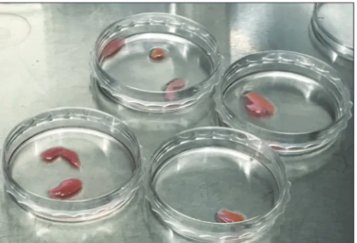
Figure 3: The macroscopic appearance of artificial tympanic membrane
graft material samples
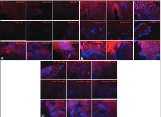
carried out fluorescent staining in UNSM, DNSM, and ATMG sections. Fluorescent microscopy confirmed the presence of cytokeratin 15 in UNSM, whereas it showed high expression in ATMG and very low expression in DNSM sections [Figure 4a]. Anti-apoptosis marker Bcl-2 expression was found significantly high in ATMG samples when compared to UNSM and DNSM samples [Figure 4b]. Interestingly, even in DNSM, there were noticeable Bcl-2 expressions [Figure 4b]. TNF-α expression profiles were detected to be almost similar to cytokeratin 15 with elevated expressions in the ATMG group and lesser expressions in UNSM group [Figure 4c].
d
IscussIonWith the notions of regenerative medicine, it is possible to acquire a cost-effective TM graft which resists the pressure gradients with advanced mechanical firmness, maintains acoustic properties, has low rejection rates, and be sufficiently available.[11]
The utilization of ECM derived from decellularized tissues is an essential field for tissue engineering.[12] Decellularized scaffolds
imitate the natural tissue structure with the conservation of ECM alignment; retain comparable concentrations of growth factors, basement membrane (BM) proteins, and glycosaminoglycans; support cell proliferation; and stimulate tissue reparation with minimized risk of rejection.[12,13] In this
study, DNSM was chosen for generating ECM scaffold instead
of ME mucosa. With its histologically confirmed similarity, the nasal mucosa is confirmed to be useful as a sheet in studies related to ME mucosal regeneration.[14]
In decellularization process, maintaining the integrity of the ECM is a key point. With decellularization, BM architecture is preserved.[15] BM, an ECM component, assists the regulation
of cell migration during tissue development, reconstruction, and differentiation.[15] In our study, with NSM decellularization,
BM showed immunofluorescent reaction with polyclonal anti-cytokeratin antibody showing marked expression in epithelial cells.
Cytoskeletal components of chondrocytes are microtubules, microfilaments, and intermediate filaments.[16] Types of
intermediate filaments may present as whorled aggregates, including vimentin and cytokeratins.[16] Since expression
profiles of vimentin and cytokeratin correlate, positive vimentin staining localization in the lamina propria of NSM in healthy controls suggests an explanation to high cytokeratin expression.[17] Our data indicated significantly
higher cytokeratin K15 expressions in ATMG samples, which is a probable evidence that the chondrocytes maintained the integrity with the scaffold [Figure 4a].
The characterization of the flow cytometry employed cells and their TOS activities were detected spectroscopically. A high TOS level would indicate an enhanced level of oxidative stress and damaged DNA of the cells.[18] TOS concentrations
in tragus cell culture media were almost five times lower than the standard solution, which was found to be statistically significant (P < 0.05) [Figure 2]. This finding indicated the presence of healthy chondrocytes.
During tissue repair, as a part of the healing process, regulation of cell populations must down-regulate the tissue repair process and avoid apoptosis with an elevated expression of Bcl-2 expression. Bcl-2 is thought to increase during periods of proliferation to prevent apoptosis.[19] Our results indicated
slightly higher expression of Bcl-2 in ATMG samples when compared with UNSM samples [Figure 4b].
Figure 2: Untreated nasal S.M., D.S.M, and A.T.M. tissue samples were
stained and analyzed. (a) With H and E staining, the nuclei of the cells were clearly stated in the “S. M.” samples. Cells were found eliminated after the decellularization protocol and no signs of cellular content were found in “D.S.M.” samples. Staining of the “A.T.M.” sample showed nuclei of the cells and cell clusters integrating to the scaffold. (b) The Alcian blue is envisioned for use in the histological visualization of sulfated and carboxylated acid mucopolysaccharides and sulfated and carboxylated sialomucins (glycoproteins). “S.M.” was found highly positive for Alcian blue staining, whereas “D.S.M.” was detected fully negative. In addition, “A.T.M.” was found positive especially in specified areas. (c) The samples were stained with both Alcian blue and Alizarin red for indicating chondrogenic and osteogenic clusters. “S.M.” was found carrying positive sections for Alcian blue or Alizarin red and occasionally positive for both. On the other hand, the results were indicating insignificant staining in “D.S.M” and “A.T.M” groups. S.M.: Septal mucosa, D.S.M.: Decellularized nasal septal mucosa, A.T.M.: Artificial tympanic membrane
c b
Figure 4: Fluorescent immunohistochemistry for untreated nasal S.M., D. S. M, and A.T.M. samples (Red: Alexa Fluor 488 conjugated rabbit secondary
antibody, Blue: DAPI: All signals were adjusted to the same scale. Scale bar equals 10 μm). (a) More CK15 was expressed in “A.T.M.” than that of “S.M.” samples. Almost no expressions of CK15 was observed in “D.S.M.” (b) A slightly more Bcl‑2 expression was observed in “A.T.M.” than “S.M.” samples. “D.S.M.” samples were also found positive for Bcl‑2 with low expressions. (c) Higher expressions of tumor necrosis factor alpha was observed for “A.T.M.,” whereas significantly lower expression was found for “S.M.” samples. S.M.: Septal mucosa, D.S.M.: Decellularized nasal septal mucosa, A.T.M.: Artificial tympanic membrane
c
b a
TNF-α has a role in the programmed cell death and is associated with hearing loss related to vibration, noise, bacterial meningitis, and autoimmune neurosensory hearing loss.[20] As expected, the expression levels of TNF-α in
NSM and ATMG samples were found inefficient to initiate programmed cell death [Figure 4c].
In our study, in contrast to utilizing the nasal septal cartilage with hyaline character, tragal (auricular) cartilage with an elastic character was preferred for obtaining human chondrocytes. It is known that maintaining the developmental characteristics by each chondrocyte type is critical in the design of a tissue-engineered cartilage model.[21,22] Elastic cartilage,
a mesenchymal derivative, has abundant chondrocytes throughout its tissue with its matrix having many elastic fibers in addition to collagen type II.[21,22]
In TM, it has been found out that type II and type III are the most abundant collagen types.[23] Auricular chondrocytes
begin synthesizing type III collagen in 24 h after they are isolated from elastic cartilage with enzymatic digestion.[24]
Due to the similarity by means of collagen compositions, auricular chondrocytes are appropriate to resemble distinctive properties of a TM in a constructed graft. By choosing tragal chondrocytes over DNSM, viability of the ATMG is ensured because chondrocytes are bradytrophic with low metabolic rates and nourished by simple diffusion from surrounding tissue.[25] In the early posttympanoplasty period, viability of
chondrocytes in a relatively avascular condition will allow time for neovascularization and fibroblasts proliferation into ATMG.[25]
ATMG has the potential to ensure advantages in particular conditions. ATMG is promising as a tool in recurrent TM perforations. If donor supplies are consumed and deficient, ATMG can be prepared in ample quantity from a patient’s own cells and be used in revision surgeries. The etiology of a recurrent perforation after tympanoplasty is due to various technical, immunological, and infectious problems adversely affecting the healing process, including angiogenesis, connective tissue proliferation, and re-epithelization.[26] Active biomolecules,
hormones, and biochemical substances (e.g. hyaluronic acid, epidermal growth factor, basic fibroblast growth factor, and pentoxifylline) applied with an ATMG may increase the regeneration potential at the perforation site, decrease the risk of rejection, and buttress its integration with TM remnant.[27]
Transplantation of ATMG as a wound-healing platform and engraftment material to promote rapid regeneration of the ME mucosa following tympanoplasty will improve the ME recovery.[14]
Latest advances in organ transplant medicine will facilitate a homograft ATMG to be used in recurrent TM perforations and other otologic defects. A tissue-engineered TM graft can be created according to three dimensional specifications (e.g., to reconstruct the mastoid cavity after mastoidectomy, to
periodontitis patients in relation to bacterial load. Front Cell Infect Microbiol 2016,5:97.
7. Esen R, Aslan M, Kucukoglu ME, Cıkman A, Yakan U, Sunnetcioglu M,
et al. Serum paraoxonase activity, total thiols levels, and oxidative
status in patients with acute brucellosis. Wien Klin Wochenschr 2015;127:427-33.
8. Liu X, Tan J, Hatem I, Smith BL. Image processing of hematoxylin and eosin-stained tissues for pathological evaluation. Toxicol Mech Methods 2004;14:301-7.
9. Kolesky DB, Homan KA, Skylar-Scott MA, Lewis JA. Three-dimensional bioprinting of thick vascularized tissues. Proc Natl Acad Sci U S A 2016;113:3179-84.
10. Ovchinnikov D. Alcian blue/ alizarin red staining of cartilage and bone in mouse. Cold Spring Harb Protoc. 2009;2009;pdb.prot5170. doi: 0.1101/pdb.prot5170.
11. Mürbe D, Zahnert T, Bornitz M, Hüttenbrink KB. Acoustic properties of different cartilage reconstruction techniques of the tympanic membrane. Laryngoscope 2002;112:1769-76.
12. Gilbert TW, Sellaro TL, Badylak SF. Decellularization of tissues and organs. Biomaterials 2006;27:3675-83.
13. Keane TJ, Londono R, Turner NJ, Badylak SF. Consequences of ineffective decellularization of biologic scaffolds on the host response. Biomaterials 2012;33:1771-81.
14. Hama T, Yamamoto K, Yaguchi Y, Murakami D, Sasaki H, Yamoto M, et al. Autologous human nasal epithelial cell sheet using temperature-responsive culture insert for transplantation after middle ear surgery. J Tissue Eng Regen Med 2017;11:1089-96.
15. Erickson AC, Couchman JR. Still more complexity in mammalian basement membranes. J Histochem Cytochem 2000;48:1291-306. 16. Benjamin M, Archer CW, Ralphs JR. Cytoskeleton of cartilage cells.
Microsc Res Tech 1994;28:372-7.
17. Onishchenko AI, Tkachenko A, Kalashnyk IM, Tkachenko VL, Nakonechna OA, Gubina-Vakulyck GI. Vimentin expression in nasal mucosa of patients with exacerbated chronic rhinosinusitis without nasal polyps. Acta Med Bulg 2019;46:39-42.
18. Kryston TB, Georgiev AB, Pissis P, Georgakilas AG. Role of oxidative stress and DNA damage in human carcinogenesis. Mutat Res 2011;711:193-201.
19. Kane CD, Greenhalgh DG. Expression and localization of p53 and bcl-2 in healing wounds in diabetic and nondiabetic mice. Wound Repair Regen 2000;8:45-58.
20. Satoh H, Firestein GS, Billings PB, Harris JP, Keithley EM. Tumor necrosis factor-alpha, an initiator, and etanercept, an inhibitor of cochlear inflammation. Laryngoscope 2002;112:1627-34.
21. Haisch A, Marzahn U, Mobasheri A, Schulze-Tanzil G, Shakibaei M. Development and phenotypic characterization of a high density in vitro model of auricular chondrocytes with applications in reconstructive plastic surgery. Histol Histopathol 2006;21:467-76.
22. Isogai N, Kusuhara H, Ikada Y, Ohtani H, Jacquet R, Hillyer J, et al. Comparison of different chondrocytes for use in tissue engineering of cartilage model structures. Tissue Eng 2006;12:691-703.
23. Stenfeldt K, Johansson C, Hellström S. The collagen structure of the tympanic membrane: Collagen types I, II, and III in the healthy tympanic membrane, during healing of a perforation, and during infection. Arch Otolaryngol Head Neck Surg 2006;132:293-8. 24. Madsen K, von der Mark K, van Menxel M, Friberg U. Analysis of
collagen types synthesized by rabbit ear cartilage chondrocytes in vivo and in vitro. Biochem J 1984;221:189-96.
25. Chhapola S, Matta I. Cartilage-perichondrium: An ideal graft material? Indian J Otolaryngol Head Neck Surg 2012;64:208-13.
26. Somers Th, Houben V, Goovaerts G, Govaerts PJ, Offeciers FE. Histology of the perforated tympanic membrane and its muco-epithelial junction. Clin Otolaryngol Allied Sci 1997;22:162-6.
27. Seonwoo H, Kim SW, Kim J, Chunjie T, Lim KT, Kim YJ, et al. Regeneration of chronic tympanic membrane perforation using an EGF-releasing chitosan patch. Tissue Eng Part A 2013;19:2097-107. 28. Bos EJ, van der Laan K, Helder MN, Mullender MG, Iannuzzi D,
van Zuijlen PP. Noninvasive measurement of ear cartilage elasticity on the cellular level: A new method to provide biomechanical information for tissue engineering. Plast Reconstr Surg Glob Open 2017;5:e1147.
support the stability of retraction pocket during cartilage tympanoplasty, and to reduce the risk of TORP/PORP prosthesis extrusion). In future, the presence of a homologous ATMG during endoscope-assisted tympanoplasty will eliminate morbidity due to incision of graft harvesting and will assist in the realization of the minimal invasive philosophy with less morbidity.
A possible disadvantage of tissue-engineered graft is the possible behavioral difference between the donor cells which may lead to early necrosis, deformation, calcification, and a possible early reperforation.[28] Further investigations have to
be carried out to utilize ATMG for routine clinical practice both as an autograft and a homograft.
c
onclusIonWith the efforts to develop an ideal ATMG for tympanoplasty, we attempted to design the exact microenvironment of a TM with the potential to permit the migration of keratinocytes and be covered with squamous epithelium. Our ATMG is promising due to its low TOS levels indicating the presence of healthy chondrocytes and high cytokeratin K15 expression showing successful integration of chondrocytes to the scaffold. High Bcl-2 and acceptable TNF-α levels of ATMG samples are indicative of a low risk of apoptosis and a possibility of increased uptake rate at the TM perforation site.
This study presents one of the initiative studies for the three-dimensional reconstruction of an ATMG. As a future prospect, in vivo investigations of ATMGs in animal models are thought to be the next step. With in vivo or in vitro studies, it will be possible to compare the mechanical strength and acoustic vibrational properties of ATMG with conventional grafts and investigate possible application fields in otology.
Financial support and sponsorship
Nil.
Conflicts of interest
There are no conflicts of interest.
r
eferences1. Hong P, Bance M, Gratzer PF. Repair of tympanic membrane perforation using novel adjuvant therapies: A contemporary review of experimental and tissue engineering studies. Int J Pediatr Otorhinolaryngol 2013;77:3-12.
2. Teh BM, Marano RJ, Shen Y, Friedland PD, Dilley RJ, Atlas MD. Tissue engineering of the tympanic membrane. Tissue Eng Part B Rev 2013;19:116-32.
3. Keane TJ, Swinehart IT, Badylak SF. Methods of tissue decellularization used for preparation of biologic scaffolds and in vivo relevance. Methods 2015;84:25-34.
4. Lo Giudice MC, Herda LM, Polo E, Dawson KA. In situ characterization of nanoparticle biomolecular interactions in complex biological media by flow cytometry. Nat Commun 2016;7:13475.
5. Faulk DM, Wildemann JD, Badylak SF. Decellularization and cell seeding of whole liver biologic scaffolds composed of extracellular matrix. J Clin Exp Hepatol 2015;5:69-80.
6. Zhang T, Andrukhov O, Haririan H, Müller-Kern M, Liu S, Liu Z,