Address for correspondence: Dr. Alpen Ortug,
Department of Anatomy, School of Medicine, Istanbul Medipol University, Istanbul, Turkey. E‑mail: [email protected]
Access this article online Website: www.jasi.org.in DOI:
10.4103/JASI.JASI_93_19 Quick Response Code: Abstract
Introduction: It is widely known that the production of knee prostheses is based on the morphometric
parameters of the distal femur and proximal tibia. Although there have been studies on this subject in different populations in the literature, such a study has not been found in the Turkish population. It is aimed to establish an index and also compare it with previous results of different populations, in this study. Material and Methods: This study was conducted on 200 patients (78 females, 122 males)
aged between 18 and 65 years who had magnetic resonance imaging retrospectively. People with previous fractures, dislocations or ruptures, tears, or tensions in the anterior cruciate ligament, history of knee surgery, and trauma were excluded from the study. Various measurements were conducted on images of the distal femur and proximal tibia morphometry. Results: Mean values of
the measurements made on the distal femur and proximal tibia were calculated, it was determined that all parameters were higher in males than females. A statistically significant difference was found between the sexes in all the parameters evaluated except for the intercondylar notch height parameter (P < 0.05). According to the results of our study, the mean intercondylar notch width index (NWI) and intercondylar notch shape index (NSI) in the total population were calculated as 0.88 ± 0.18. There was no statistically significant difference between male and female values for NWI and NSI (P > 0.05). Discussion and Conclusion: In this study, finally, intercondylar notch
classification was done. According to this, in 46.5% of cases, notch type, which defined as type A and 53.5% of the notches were identified as U type. None of the cases was classified as W type. Studies in which normal morphometric data are presented due to differences in races between skeletal structures have crucial importance. Therefore, the data obtained from our study are thought to be useful to produce specific knee prostheses.
Keywords: Distal femur, intercondylar notch width index, knee morphometry, knee radiology, proximal tibia
The Magnetic Resonance Imaging Evaluation of Morphometry of the
Distal Femur and Proximal Tibia on Adult Anatolian Population
Paria Shojaolsadati, Neslihan Yüzbasioglu1, Asrin Nalbant2, Tugrul Ormeci3, Soner Albay4, Alpen Ortug1,
Bayram Ufuk Sakul1
Department of Anatomy, School of Medicine, Okan University, Departments of 1Anatomy and 3Radiology, School of Medicine,
Istanbul Medipol University, Istanbul, 2Department of
Anatomy, School of Medicine, Bakircay University, Izmir,
4Department of Anatomy, School
of Medicine, Suleyman Demirel University, Isparta, Turkey
How to cite this article: Shojaolsadati P, Yüzbasioglu N, Nalbant A, Ormeci T, Albay S, Ortug A, et al. The magnetic resonance imaging
evaluation of morphometry of the distal femur and proximal tibia on adult anatolian population. J Anat Soc India 2020;69:9-14.
Introduction
The anatomic components forming the knee joint include the distal femur and the proximal tibia. Previous anthropometric studies were present in the literature for both on the distal femur and proximal tibia.[1‑4]
For those patients for whom total knee arthroplasty (TKA) is planned, it is extremely important to ensure size matching of the components belonging to the distal femur and the proximal tibia to maximize the bony structure and the load transmission in the proximal tibia.[5,6] Ensuring this match during TKA will extend implant stability and life. During a TKA performance, a too small tibial component might lead to loosening due to an early relocation in the
orthesis because of insufficient support by the distal bony compartment after resection.[7] Moreover in the case of a too large tibial component, soft‑tissue irritation and pain might occur when the orthesis used protrudes over the distal bony compartment after resection. Studies about the distal femur also report that anatomic structures might be variable and that such variations might lead to certain pathologies.[8]
It is emphasized in recent anthropometric studies that the components involved in the formation of knee vary depending on races and that, therefore, the orthoses used in arthroplasty should be produced in consideration of racial differences.[9]
The systematic review study conducted in 2017 compares all the English articles in the literature, revealing results about the This is an open access journal, and articles are
distributed under the terms of the Creative Commons Attribution‑NonCommercial‑ShareAlike 4.0 License, which allows others to remix, tweak, and build upon the work non‑commercially, as long as appropriate credit is given and the new creations are licensed under the identical terms. For reprints contact: [email protected]
Received: 15 July 2019 Accepted: 06 February 2020 Available online: 11 April 2020 Article Info
distal femur and proximal tibia. Accordingly, available information includes only white, Eastern Asian nations and Indians. There is still nocomparative index of the Middle Eastern and African populations.[10] The current study was planned because of a lack of morphometric data regarding the Turkish population that could be used for making prostheses for people of this region.
Magnetic resonance imaging (MRI) is noninvasive method that was utilized in previous studies for making such measurements and which enables distinguishing compartment boundaries in the clearest way possible and carrying out reliable measurements. For this reason, in our study as well, the measurements relating to the proximal tibia and the distal femur were made by MRI images retrospectively.
Material and Methods
Study groupThe approval of this study was obtained before the commencement of work from the Non‑Interventional Ethics Committee of Istanbul Medipol University. Two hundred Turkish patients were included in the study between 2016 and 2017 (122 males, 78 females; mean age: 35.37 ± 9.82) at Istanbul Medipol University Hospital Radiology Department. MRIs registered in picture archiving and communication systems (PACS) used for measurement and studied retrospectively. Those who had previous knee joint surgeries or had a knee trauma, fracture, dislocation, or a ruptured or torn anterior cruciate ligament (ACL) of any sprained ligament (sprain II and above) were not included in the study.
Imaging
The MRI was performed with the patient in the supine position and the knee in a relaxed position at a 5‑degree external rotation by 3T (TR3029, TE 30) and 4‑mm slice thickness. For image analysis, the archiving and measurement program PACS was utilized.
Measurements in each patient was based on two images; different longitudinal and angular parameters were measured on two different MRI sections including the section, in which the biepicondylar distance is visible in the widest aspect in the distal femur (first plane), and the section, in which the two condyles in the proximal tibia region are visible in the widest aspect (second plane). For standardization, the measurements were carried out by a single person by three repetitions, which were then averaged.
The parameters measured in the distal femur:
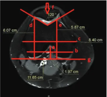
• Intercondylar notch height (NH); the longest vertical distance drawn between the postcondylar line and the notch on the first plane
• Intercondylar notch width (NW); the widest transverse distance of the notch on the first plane
• Femoral notch shape index (NSI) =notch width (NW) notch height (NH) • femoral mediolateral line (FML); longest line connecting
the medial and the lateral dimensions of the distal femur on the first plane
• femoral medial condyle anteroposterior length (FMAP); the longest FMAP length on the first plane
• femoral lateral condyle anteroposterior length (FLAP); the longest FLAP length on the first plane
• Trochlear groove angle (TGA)
• The shape of femoral notch was classified as type A, type U and type W[1,11][Figure 1].
The parameters measured in the proximal tibia:
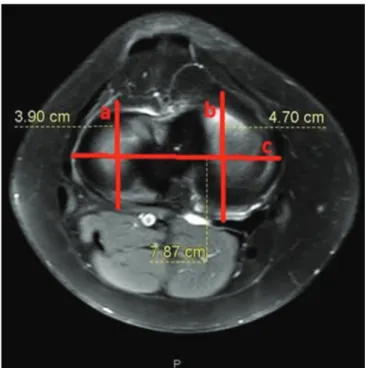
• Tibial mediolateral length (TML); the widest distance (or the widest transverse distance) of the bicondylar tibia in the axial MRI (second plane), the widest mediolateral diameter according to some references
• Tibial medial anteroposterior line (TMAP); the longest tibial medial condyle anteroposterior length on the second plane
• Tibial lateral condyle anteroposterior line (TLAP) [Figure 2]; the longest TLAP length on the second plane.
Statistics
The SPSS Version 17.0 for Windows; SPSS Inc, Chicago, Illinois, USA) was used for the analyses of all data. The Independent Student’s t‑test and Pearson’s correlation test were performed for the analysis. Those with a value of P < 0.05 were considered to be statistically significant.
Results
The mean and standard deviation values of the parameters
Figure 1: Axial magnetic resonance image of distal femur. a: NH, b: NW, c: FML, d: FLAP, e: FMAP, f: TGA, g: Reference line
as type A (46.5%), 107 recorded as type U (53.5%). A different type of intercondylar notch was not encountered except for A, U and W defined in previous studies.[1,11] The W type notch that was revealed in these previous studies was not encountered in our study. No relation was found in regard to notch type and sex.
Finally, in our study, the correlation between the parameters measured morphometrically was assessed by Pearson’s correlation test [Table 2].
Discussion
Success in TKA highly depends on the proper sizing of the product. In this purpose, anthropological information about proximal tibia has a crucial effect. As a result of many different reasons such as altering gene pools because of migrations, anthropological differences, and lack of knowledge about different racial morphometry, data used for TKA and prosthetic production secondary to TKA is not very effective. Studies evaluating different groups of Asian subpopulations indicates that knee and body size difference of these populations result with the improper fit for western products.[12] In their study on three‑dimensional knee morphology, Mahfouz et al.[13] revealed differences in the morphology of this region among races. Relevant morphological assessments of various populations have been made, but there is no clear information on the Turkish population. It is expected that this study will reveal the anatomical values of the knee in the Turkish population.
A previous study conducted in 1994, compared direct cadaveric notch measurements with radiographic and MRI Figure 3: Distribution of the population according to notch shape index
index
Table 1: Mean values and standard deviations of the measured parameters (mm) Mean±SD Gender Total Male (n=122) Female (n=78) NH 2.30±0.37* 2.31±0.37* 2.30±0.37 NW 1.99±0.26 1.72±0.27 1.88±0.30 NSI 0.88±0.18 0.76±0.16 0.84±0.18 FML 8.58±0.51 7.60±0.52 8.20±0.70 FMAP 6.28±0.54 5.80±0.64 6.10±0.62 FLAP 5.88±0.46 5.45±0.47 5.71±0.51 TGA 130.31±5.33 128.56±5.08 129.63±5.29 TML 8.10±0.62 7.09±0.72 7.70±0.82 TMAP 4.83±0.52 4.51±0.78 4.71±0.65 TLAP 3.94±0.65 3.70±0.58 3.85±0.63 Age 39.20±12.93 35.21±12.91 37.65±13.04 *Difference not found between genders (P˃0.05). SD: Standard deviation, NH: Notch height, NW: Notch width, NSI: Notch shape index, FML: Femoral mediolateral line, FMAP: Femoral medial condyle anteroposterior line, FLAP: Femoral lateral condyle anteroposterior line, TGA: Trochlear groove angle, TML: Tibial mediolateral length, TMAP: Tibial medial anteroposterior line, TLAP: Tibial lateral condyle anteroposterior line
Figure 2: Axial magnetic resonance image of proximal tibia. a: TLAP, b: TMAP, c: TML
described the distal femur and the proximal tibia are shown separately for both total population and for sexes in Table 1. Accordingly, when an assessment is made between sexes, all parameters appear to be higher in males when compared with women except for NH.
When the obtained data were assessed statistically, a significant difference was found between sexes in all the parameters assessed except for NH (P < 0.05).
A statistically significant difference was determined between the mean NSI values of males and females. The mean NSI value in the total population was calculated to be 0.84 ± 0.18 [Figure 3].
In our study, an intercondylar notch classification was also made. Accordingly, two notch types A and U were defined in our study. Ninety‑three of all knee specimens recorded
measurements and resulted in no direct difference between both measurement methods.[14] For this reason, we used MRI for our metric evaluations.
The morphometric knee values among Chinese, Koreans, Japanese, and Iranians were explored through different studies.[15‑17] For the first time, we made a complete calculation of the bony components of the knee in Turkish people in a single study including both distal femur and proximal tibia measurements also with intercondylar notch type.
Significant differences between genders in terms of the distal femur and the proximal tibia values except for the NH were noted in our study. In their study on the distal femur morphology in 2007, Conley et al. determined a significant difference in the measurements of this region in men and women.[18] Previously, it is found 33.2 ± 2.8 for males and 29.0 ± 2.6 for females also in the Turkish population, similarly on 200 people without abnormal knee.[19] Our results are lesser than their value as 23.00 ± 0.37 for males and 23.10 ± 0.37 for females. The difference was statistically not significant between genders. However, the obtained results are interesting. Yet, the presence of such different values in the same population with similar age groups and similar sample size suggests that people vary even within themselves or that there may be also regional differences in the Anatolian population.
NW is commonly related to ACL injuries. It is stated that there is still a controversy as some authors claim that ACL injured groups have narrower NW; on the other hand, some authors report nonsignificance between each other.[20] Park et al.[20] compared this value with the ACL injured and control group. The result was found for 20.33 mm and 18.52 mm in the control group for males and females, respectively. Their results have shown a significant difference between the injured and control groups. Our results revealed 19.90 ± 0.26 for males and 17.20 ± 0.27 for females for the normal knee. For an accurate comparison, this value should be studied also in the ACL injured group
in the Turkish population. Index of notch shape (NSI) served as a ratio between NW and NH. This ratio is related to ACL deformations. It is stated that this index might even be related to ACL ruptures.[1] In the presented study, the mean value for NW was found 1.88 cm and NH was found 2.30 cm for Turkish people. Hence that NSI was found (0.84 ± 0.18). Previous records indicate that this ratio was found in African as (0.60 ± 0.08) and (0.63 ± 0.09) in the European people.[21] According to Ouyang et al.[22] “The greater the ratio, the more rounded the intercondylar notch.” Hence that according to this information, Turkish people have a more rounded notch with the highest NSI ratio. According to the results of our study, the FML value was found to be 82 mm. The same value indicated in the White people as 73.5 mm, in the African‑origin as 69.1 mm, in East Asia 71.4 mm and in the Indian as 65.4 mm. Interestingly, this value revealing width of the femur was found larger than all other populations in the Turkish people.[23‑25] Hence that, the antropo‑morphologic type of the knee in Turkish people found narrower at the sagittal axis but wider at the transverse axis.
FMAP and FLAP are the measurements indicating medial and lateral anteroposterior dimensions of the knee. Previously, the mean value for FMAP was 61.7 mm for White, 64.00 mm for African‑origin and 58.00 mm for the East Asian populations and FLAP was 61.7 mm for White, 63.2 mm for African‑origin and 58.7 mm for East Asian and 57.8 for Indian people.[10] Our study reveals that the mean value of FMAP for Turkish people is 61.00 ± 0.62 and FLAP 57.1 mm. As compared to all given populations, FMAP value is similar, but FLAP value for Turkish people is the smallest.[10,24‑26] This is an interesting result, because Turkish people anthropologically considered as White‑Caucasian like many central and Eastern European populations. Hence that, the femoral anteroposterior length smaller than East Asian people is a significant result. TGA is related to dysplasia of the extensor mechanism.[27] It is stated that measurement of the angle is easy, quick, and Table 2: Correlation between measured parameters correlation
NH FML NW FMAP FLAP TGA TML TMAP TLAP
NH 1 FML −0.127 1 NW 0.009 0.482** 1 FMAP 0.077 0.502** 0.314** 1 FLAP 0.078 0.658** 0.434** 0.714** 1 0. TGA −0.205** 0.190** 0.072 −0.026 0.123 1 TML −0.088 0.748** 0.510** 0.081 0.488** 0.234** 1 TMAP 0.166* 0.349** 0.161* 0.749** 0.475** 0.086 −0.004 1 TLAP 0.163* 0.299** 0.036 0.674** 0.451** 0.029 −0.023 0.786** 1
*: P <0.001, **: P <0.01, r=1.00–0.76 very good correlation, r=0.75–0.51 good correlation, r=0.50–0.26 fair correlation, r=0.25–0.00 weak correlation. NW: Notch width, NH: Notch height, NW: Notch width, FML: Femoral mediolateral, FMAP: Femoral medial condyle anteroposterior, FLAP: Femoral lateral condyle anteroposterior, TGA: Trochlear groove angle, TML: Tibial mediolateral length, TMAP: Tibial medial anteroposterior line, TLAP: Tibial lateral condyle anteroposterior line
a reliable method for the prediction of severity.[27] Sulcus angle higher than 150° on an axial radiograph is considered to indicate femoral trochlear dysplasia.[28] Toms’s comparative work for CT, MRI and ultrasound found similar results for all three methods on patients with patellar instability.[29] It is stated that previous studies revealing the sulcus for normal knee reported that the sulcus angle was 132° and the other was 138°.[30,31] Murshed et al. conducted a study on 100 people also in the Turkish population. They found 134.00° and 132.20° for males and females, respectively. This angles are slightly larger than our results which is 130.31°, 128.56°, and 129.63° for males, females, and mean, respectively. The difference may be because of the sample size which is 200 people in our study.
TMAP, TLAP, and TML are the dimensions for the measurement of anteroposterior and transverse dimensions of the proximal tibia. The value of TMAP was found 47.1 mm in the Turkish population which was 49.8 mm in White people, 49 mm in East Asian, and 47.5 mm in Indian.[23,24] TLAP was found to be 44.3 mm in Whites, 44.4 mm in East Asia, and 43.8 mm in Indians. Our result for the Turkish people was 38.5 mm.[23] In comparison to numerical TMAP and TLAP values, results for Turkish people were found lower than all other races. The TML value was found 77 mm in the Turkish population, 74.3 mm in White, 73.2 mm in African‑origin 72.8 mm in East Asian, and 72.8 mm in Indian people. The numerical value of TML compared with the other race and in Turkish people was found to be higher than all other populations.[23,24] These results reveal that morphologically proximal tibia of the Turkish population is narrower in sagittal axis and wider in the transverse axis similar to the distal femur.
Intercondylar notch shape is divided into three as A, U, and W.[32] Even though, association between the shape and ACL injury risk is not commonly clear, some of the researchers suggest A type should be more vulnerable because of its narrow shape.[1] Similarly, Al‑Saeed found 73% of the patients with ACL tear with A type.[1] Presented study of ours, revealed 46.5% of the cases as type A and 53.5% of the cases as type U with no type W, on normal knee. In order to find the answer of “Does these results mean that nearly half of the Turkish population is more vulnerable for ACL injury?” question, a larger sample group of both normal and ACL ruptured patients should be compared.
Conclusion
According to the results of this study in Turkish people, the distal femur shape has the slightly larger mediolateral diameter and smaller or medium size anteroposterior diameter compared to all other populations. Accordingly, two femoral notch types as A and U were defined in our study. 93 of all knee specimens recorded as Type A (46.5% of total), 107 recorded as Type U (53.5% of total). The
W type notch that was revealed in previous studies was not encountered in our study.
In addition, our results revealed a wider mediolateral diameter and smaller anteroposterior diameter than other races at the proximal tibia. This study will have crucial clinical impact on the production of the specific prostheses belong to the Turkish population and populations with similar anthropologic tibial shape.
Acknowledgment
Preliminary data of this article were presented as poster at 17th National Congress of Anatomy, September 5‑9, 2016, Eskişehir, Turkey.
Financial support and sponsorship Nil.
Conflicts of interest
There are no conflicts of interest.
References
1. Al‑Saeed O, Brown M, Athyal R, Sheikh M. Association of femoral intercondylar notch morphology, width index and the risk of anterior cruciate ligament injury. Knee Surg Sports Traumatol Arthrosc 2013;21:678‑82.
2. Balcarek P, Walde TA, Frosch S, Schüttrumpf JP, Wachowski MM, Stürmer KM, et al. Patellar dislocations in children, adolescents and adults: A comparative MRI study of medial patellofemoral ligament injury patterns and trochlear groove anatomy. Eur J Radiol 2011;79:415‑20.
3. Ewe T, Ang H, Chee E, Ng W. An analysis of the relationship between the morphometry of the distal femur, and total knee arthroplasty implant design. Malay 2009;3:24‑8.
4. Havet E, Gabrion A, Leiber‑Wackenheim F, Vernois J, Olory B, Mertl P. Radiological study of the knee joint line position measured from the fibular head and proximal tibial landmarks. Surg Radiol Anat 2007;29:285‑9.
5. Berend ME, Small SR, Ritter MA, Buckley CA. The effects of bone resection depth and malalignment on strain in the proximal tibia after total knee arthroplasty. J Arthroplasty 2010;25:314‑8. 6. Innocenti B, Truyens E, Labey L, Wong P, Victor J, Bellemans J.
Can medio‑lateral baseplate position and load sharing induce asymptomatic local bone resorption of the proximal tibia? A finite element study. J Orthop Surg Res 2009;4:26.
7. Cheng FB, Ji XF, Zheng WX, Lai Y, Cheng KL, Feng JC, et al. Use of anthropometric data from the medial tibial and femoral condyles to design unicondylar knee prostheses in the Chinese population. Knee Surg Sports Traumatol Arthrosc 2010;18:352‑8. 8. Urabe K, Mahoney OM, Mabuchi K, Itoman M. Morphologic
differences of the distal femur between Caucasian and Japanese women. J Orthop Surg (Hong Kong) 2008;16:312‑5.
9. Surendran S, Kwak DS, Lee UY, Park SE, Gopinathan P, Han SH, et al. Anthropometry of the medial tibial condyle to design the tibial component for unicondylar knee arthroplasty for the Korean population. Knee Surg Sports Traumatol Arthrosc 2007;15:436‑42.
10. Kim TK, Phillips M, Bhandari M, Watson J, Malhotra R. What differences in morphologic features of the knee exist among patients of various races? A systematic review. Clin Orthop Relat Res 2017;475:170‑82.
14 Journal of the Anatomical Society of India ¦ Volume 69 ¦ Issue 1 ¦ January-March 2020 11. Chen C, Ma Y, Geng B, Tan X, Zhang B, Jayswal CK,
et al. Intercondylar notch stenosis of knee osteoarthritis and relationship between stenosis and osteoarthritis complicated with anterior cruciate ligament injury: A study in MRI. Medicine (Baltimore) 2016;95:e3439.
12. Kwak DS, Surendran S, Pengatteeri YH, Park SE, Choi KN, Gopinathan P, et al. Morphometry of the proximal tibia to design the tibial component of total knee arthroplasty for the Korean population. Knee 2007;14:295‑300.
13. Mahfouz M, Abdel Fatah EE, Bowers LS, Scuderi G. Three‑dimensional morphology of the knee reveals ethnic differences. Clin Orthop Relat Res 2012;470:172‑85.
14. Herzog RJ, Silliman JF, Hutton K, Rodkey WG, Steadman JR. Measurements of the intercondylar notch by plain film radiography and magnetic resonance imaging. Am J Sports Med 1994;22:204‑10.
15. Hosaka K, Saito S, Ishii T, Mori S, Sumino T, Tokuhashi Y. Asian‑specific total knee system: 5‑14 year follow‑up study. BMC Musculoskelet Disord 2011;12:251.
16. Hovinga KR, Lerner AL. Anatomic variations between Japanese and Caucasian populations in the healthy young adult knee joint. J Orthop Res 2009;27:1191‑6.
17. Uehara K, Kadoya Y, Kobayashi A, Ohashi H, Yamano Y. Anthropometry of the proximal tibia to design a total knee prosthesis for the Japanese population. J Arthroplasty 2002;17:1028‑32.
18. Conley S, Rosenberg A, Crowninshield R. The female knee: Anatomic variations. J Am Acad Orthop Surg 2007;15 Suppl 1:S31‑6.
19. Murshed KA, Ciçekcibaşi AE, Karabacakoğlu A, Seker M, Ziylan T. Distal femur morphometry: A gender and bilateral comparative study using magnetic resonance imaging. Surg Radiol Anat 2005;27:108‑12.
20. Park JS, Nam DC, Kim DH, Kim HK, Hwang SC. Measurement of knee morphometrics using MRI: A comparative study between ACL‑injured and non‑injured knees. Knee Surg Relat Res 2012;24:180‑5.
21. Tillman MD, Smith KR, Bauer JA, Cauraugh JH, Falsetti AB, Pattishall JL. Differences in three intercondylar notch geometry indices between males and females: A cadaver study. Knee
2002;9:41‑6.
22. Ouyang X, Wang YH, Wang J, Hong SD, Xin F, Wang L, et al. MRI measurement on intercondylar notch after anterior cruciate ligament rupture and its correlation. Exp Ther Med 2016;11:1275‑8.
23. Lim HC, Bae JH, Yoon JY, Kim SJ, Kim JG, Lee JM. Gender differences of the morphology of the distal femur and proximal tibia in a Korean population. Knee 2013;20:26‑30.
24. Chaichankul C, Tanavalee A, Itiravivong P. Anthropometric measurements of knee joints in Thai population: Correlation to the sizing of current knee prostheses. Knee 2011;18:5‑10. 25. van den Heever DJ, Scheffer C, Erasmus P, Dillon E.
Classification of gender and race in the distal femur using self organising maps. Knee 2012;19:488‑92.
26. Terzidis I, Totlis T, Papathanasiou E, Sideridis A, Vlasis K, Natsis K. Gender and side‑to‑side differences of femoral condyles morphology: Osteometric data from 360 caucasian dried femori. Anat Res Int 2012;2012:679658.
27. Davies AP, Costa ML, Shepstone L, Glasgow MM, Donell S. The sulcus angle and malalignment of the extensor mechanism of the knee. J Bone Joint Surg Br 2000;82:1162‑6.
28. Chhabra A, Subhawong TK, Carrino JA. A systematised MRI approach to evaluating the patellofemoral joint. Skeletal Radiol 2011;40:375‑87.
29. Toms AP, Cahir J, Swift L, Donell ST. Imaging the femoral sulcus with ultrasound, CT, and MRI: Reliability and generalizability in patients with patellar instability. Skeletal Radiol 2009;38:329‑38.
30. Murshed KA, Çiçekcibaşi AE, Ziylan T, Karabacakoğlu A. Femoral sulcus angle measurements: An anatomical study of magnetic resonance images and dry bones. Turk J Med Sci 2004;34:165‑9.
31. Merchant A. Patellofemoral disorders: Biomechanics, diagnosis and nonoperative treatment. Operative Arthroscopy. New York: Raven Press; 1991. p. 261‑75.
32. van Eck CF, Martins CA, Vyas SM, Celentano U, van Dijk CN, Fu FH. Femoral intercondylar notch shape and dimensions in ACL‑injured patients. Knee Surg Sports Traumatol Arthrosc 2010;18:1257‑62.