Immunohistochemical Studies on Infectious Laryngotracheitis
in the Respiratory Tract Lesions in Naturally Infected Laying Hens
[1]Orhan YAVUZ
1
Özgür ÖZDEMİR
2Zeki ARAS
3Funda TERZİ
2[1] This study was funded by Aksaray University Scientific Research Committee (ASÜBAP, Project Numbers: 2015-045 & 2016-045)
1 Aksaray University, Faculty of Veterinary Medicine, Department of Pathology, TR-68100 Aksaray - TURKEY 2 Selçuk University, Faculty of Veterinary Medicine, Department of Pathology, TR-42250 Konya - TURKEY 3 Aksaray University, Faculty of Veterinary Medicine, Department of Microbiology, TR-68100 Aksaray - TURKEY
Article Code: KVFD-2017-18805 Received: 02.10.2017 Accepted: 26.12.2017 Published Online: 26.12.2017
How to Cite This Article
Yavuz O, Özdemir Ö, Aras Z, Terzi F: Immunohistochemical studies on infectious laryngotracheitis in the respiratory tract lesions in naturally
infected laying hens. Kafkas Univ Vet Fak Derg, 24 (2): 257-264, 2018. DOI: 10.9775/kvfd.2017.18805 Abstract
In this study, naturally infected by Gallid Herpesvirus type-1 in laying hens to be diagnosed by pathological and PCR methods. Sixty pieces of hens were collected in coops from Central Anatolia region. After necropsy, routine pathological processes were applied to the trachea/ larynx, sinuses, lungs and air sacs. All organs were also stained by immunoperoxidase method, and PCR methods were applied to formalin fixed paraffin embedded (FFPE) tissues. Immunohistochemically, the positivities were seen in trachea/larynx (78.3%), sinuses (61.6%), lungs (45%) and air sacs (50%). Positive reactions were observed, in mucous and gland epithelia especially located at intracytoplasmic and rarely intranuclear. PCR positivity was observed in the trachea/larynx in 15 (25%) cases, in infraorbital sinus in 11 (18.3%) cases, in lungs in 8 (13.3%) cases and in air sacs in 6 (10%) cases following the tests performed. Following these results, it is easily concluded that histopathology and immunoperoxidase method can usable for diagnosing of the ILT. However, PCR results made by FFPE tissues showed that this method is not adequate to diagnose the ILT alone.
Keywords: Histopathology, ILT, Immunohistochemistry, Laying hens, PCR
Enfeksiyöz Laringotraheitis İle Doğal Enfekte Yumurta Tavuklarında
Solunum Kanalı Lezyonları Üzerine İmmunohistokimyasal Çalışmalar
Öz
Bu çalışmada Gallid Herpesvirus tip-1 ile doğal enfekte yumurta tavuklarında patolojik yöntemlerle teşhis konularak immunohistokimyasal ve PCR yöntemlerinin teşhiste kullanılabilirliği araştırıldı. Bu amaçla Orta Anadolu’da bulunan bazı illerdeki kümeslerden toplam 60 adet enfekte tavuk toplandı. Yapılan nekropsilerin sonrasında trake, larinks, infraorbital sinus, akciğer ve hava kesesi parçaları alınarak rutin patolojik işlemler uygulandı. Alınan tüm organlar ayrıca indirekt immunperoksidaz yöntemi ile boyandı ve organlara ait formolle fikse edilmiş parafine gömülü (FFPG) dokulara PZR testi yapıldı. İmmunohistokimyasal boyamalar sonucu trake/larinkste %78.3, sinuslarda %61.6, akciğerlerde %45 ve hava keselerinde %50 oranında pozitiflik gözlendi. Pozitif boyanmalar özellikle mukoza ve bez epitellerinde intrasitoplazmik nadiren de intranüklear olarak gözlendi. Aynı zamanda lümene dökülmüş eksudattaki hücre ve sinsityal dev hücrelerinin sitoplazmalarında da boyanmalar tipikti. Yapılan PCR testlerinin ardından trake/larinkste %25, infraorbital sinuslarda %18.3, akciğerlerde %13.3, hava keselerinde %10 oranında pozitiflik gözlendi. Bu sonuçların ardından hastalığın teşhisinde histopatolojik ve immunperoksidaz yönteminin rahatlıkla kullanılabileceği ortaya konmuştur. Ancak, FFPG dokulardan yapılan PCR yönteminin tek başına İLT’yi teşhis etmek için yeterli olmadığı gösterildi.
Anahtar sözcükler: Histopatoloji, İLT, İmmunohistokimya, Yumurta tavukları, PCR
INTRODUCTION
Infectious laryngotracheitis (ILT) is a viral disease characterized by breathing difficulty, wheezing and bloody exudate accumulation especially in the larynx, trachea, and
upper respiratory tract in chickens, turkeys and pheasants [1]. The causing agent is a Gallid alphaherpesvirus-1 (GaHV-1) belonging to Iltovirus genus in the Alphaherpesvirinae subfamily, which is in the Herpesviridae family [2]. This disease appeared firstly in the United States in 1925 [3,4].
İletişim (Correspondence)
+90 382 2882864Although it is reported that the disease is seen in chickens of all age groups, hatching chickens are more affected [5]. In the pathogenesis of the disease, the virus is primarily affinity to tracheal and larynx epithelium. Also has been emphasized that the virus is replicating in conjunctiva, sinus, air sacs and lungs. It has been reported that virus has high cytolytic activity in these tissues, as well as bleeding and damage to the epithelium [1]. In experimental studies, tracheal tissues and secretions are present on days 6-8 of virus inoculation has been reported [6]. On the other hand, there is no evidence that Infectious Laryngotracheitis Virus (ILTV) causes viremia [7]. It has been noted that the virus may be found in trigeminal ganglia, especially in subacute and latent infection period [8].
Histopathologic findings can vary according to the stage of the disease [4]. The first changes are goblet cell loss and inflammatory cell infiltrates in the tracheal mucosa. Findings such as bloating, ciliate loss and oedema can be observed in the epithelial cells. Mononuclear cell infiltrations and syncytial giant cells start to be seen in the days 2 between 3 in the beginning of the infection. In this phase, Cowdry A type intranuclear eosinophilic inclusion body may be found in the desquamated epithelial cells. These inclusion bodies can also be found in syncytial giant cells. It was emphasized that the inclusion bodies were encountered within the first 5 days of the disease and that disappeared with the progression of the sickness [9,10]. Immunohistochemically, Preis et al.[11] reported positive reactions in the cytoplasm of syncytial giant cells and epithelial layer in trachea, larynx, lung, and paranasal sinuses during field studies.
In this study, it was aimed to determine the spread of viral antigens in the respiratory system organs by using immunohistochemical and PCR methods in natural ILT infection in laying hens. In addition, it was aimed to compare macroscopic, histopathologic, immunohistochemical and PCR findings and usability of these methods.
MATERIAL and METHODS
Material
The material of the study comprised 10-90 weeks-old hens that were clinically demonstrating ILT. Hens were collected from the coops that perform egg laying and serologically ILTV positive in certain regions of the cities of Aksaray, Afyonkarahisar and Konya in Turkey. All hens were collected from September to December 2014. A total of 60 animals were used from ten different commercial hen coops. All layers were Leghorn strain. The necropsy of the hens were performed and samples were taken for histopathologic and immunohistochemical examinations. After the pathologic examinations, PCR method applied to samples which taken from paraffin blocks of each case.
Histopathological Method
Following necropsy, respiratory tract organs (Infra-orbital sinuses, trachea, larynx, lungs and air sacs) were examined grossly and fixed in 10% formalin solution. After the fixation, the tissues were subjected to alcohol, xylene and paraffin blocks, respectively. Subsequently, they were cut at 5 µm thickness of sections by microtome, glued to slides and examined under light microscope (Olympus BX51, Tokyo, Japan), after stained with Haematoxylin-Eosin (HE) [12]. The changes observed in larynx/trachea of HE-stained sections were scored as those of Guy et al.[13]. According to these, histopathologic changes were evaluated as normal (0), minimal (+1), mild (+2), moderate (+3), severe (+4) and very severe changes (+5).
Immunohistochemical Method
For immunohistochemistry, Totally 60 formalin fixed paraffin embedded (FFPE) the Infra orbital sinus, trachea, larynx, lung and air sac tissues were used. Polymer-based indirect immunoperoxidase method was applied for immuno histochemical (IHC) staining. Firstly, Proteinase-K solution was instilled for 10 minutes. Then, 3% H2O2 peroxidase block solution was dropped for 10 min. After that, the protein block was instilled and incubated for 5 min. Following this procedure, Rabbit polyclonal anti Infectious Laryngotracheitis Virus antibody (Biorbyt, orb10560) was instilled and incubated for 2 h at room temperature. Then, post-primer block solution was added to the slides and incubated for 30 min and the Polymer solution was dropped for 30 min. Slides stained by DAB (3,3’- diaminobenzidine tetrahydrochloride) for 5 min. After counter-staining with hematoxylin, slides were closed by coverslips and evaluated under a light microscope. The negative control slides were also stained according to the same procedure. However, TBS was used instead of the primer antibody.
PCR Analysis
Deparaffinization of Samples: For deparaffinization,
FFPE tissues were cut at 20 µm for 4 times then the samples were placed into the Eppendorf tubes. Eppendorf tubes were filled with 1 mL of xylol. Tubes were mixed for 2 min in the vortex and incubated in the heat block at 56°C for 5 min. Then tubes vortexed again for 2 min and centrifuged for 2 min at 13.200 rpm. Then supernatant fluid was drained out. This process was repeated twice. The steps described above were repeated. However, ethanol was used instead of xylol. The liquid remaining in the bottom of the tubes were taken with a micropipette into another tube and then these tubes were subjected to DNA extraction procedures.
DNA Extraction: DNA extraction from the samples was
done by the Vivantis Tissue DNA Extraction Kit (GF TD-50) as specified by the manufacturer’s instructions. 250 μL
of lysis solution and 20 μL of proteinase K were added to the samples and incubated at 65°C for 3 h. At the end of the period, 560 μL Buffer TB was added to the tube and incubated at 65°C for 10 min. Then, 200 μL of ethanol was added, to the filter, and centrifuged at 5.000 rpm for 1 min. The obtained DNA samples were stored at -20°C until use.
PCR Method: Polymerase chain reaction analysis of the
ICP4 gene was performed using the primers ICP4-1F (5’- CCT TGG TTC GGG AT¬G AAA CC-3’) and ICP4-1R (5’-TTC ATT ACC TCC AGC GGT TCC-3’) described by Preis et al.[14]. These primers showed a single amplicon, the size of which was 237 bp.
The extracted DNA was amplified in a total volume of 25 µL (2.5 µL 10x PCR buffer, 170 mM from each dNTPs (Vivantis), 10 µmol each of the primers (IDT), 1.5 mM MgCl2, 1.25 U Taq polymerase (Vivantis), and 2.5 µL extracted DNA). The cycling conditions with the Biorad gradient (T100) were the initiation step at 94°C for 3 min, followed by 40 cycles coupling 94°C for 30 s, 54°C for 30 s and 72°C for 2 min and a final extension at 72°C for 15 min. The PCR products (10 µL) were analyzed by electrophoresis on 1.6% agarose gel, and the gel was stained with ethidium bromide (1.5 g/mL) and photographed [15].
RESULTS
Gross Results
Lesions that seen in microscopic examinations of trachea, larynx, lung, infraorbital sinuses and air sacs are given in
Table 1. Also, the number and percentages of the gross
pathology results are given in Table 2. The most frequent lesions were observed in trachea and larynx. Bloody, mucopurulent, fibrinous exudate deposits and diphtheroid
lesions were observed in the lumen of trachea and larynx
(Fig. 1A-C). In 7 cases, both mucopurulent and fibrinous
lesions were observed together (Fig. 1D). Mucopurulent exudate deposits were observed in 16 cases in the infraorbital sinus. In these cases, swelling of the sinuses was also noticed on the gross examination. The lesions observed in the lungs consisted of congestion, and findings related to pneumonia were not observed. The air sacs had whitish colored, thickened and opaque.
Histopathological Results
The numbers and percentages of histopathological results and inclusion bodies are given in Table 2. Distribution of histopathological lesions in the organs is given in Table 3. Pathological changes were mostly detected in the trachea and larynx. Various degrees of thickening was noted due to oedema, mononuclear cell infiltrates, hyperemia, and heterophil granulocyte accumulation in the mucosa of the trachea and larynx (Fig. 2A-E). In some cases, erythrocytes and desquamated epithelial cells, as well as intranuclear inclusion bodies in syncytial giant cells were found in the lumen of the trachea (Fig. 2F). These inclusions were often determined in the nuclei of giant cells. In infraorbital sinuses, oedema, mononuclear cells, and heterophil granulocytes infiltrations were observed in the subendothelial layer, whereas in some cases only epithelial desquamation was noticed. Lymphoid hyperplasia was observed around the parabronchus in the lungs. In few cases, inclusion bodies were determined in giant cells in the exudate of bronchial lumens. In the air sacs, the propria was found to thickened due to lymphoid cells, mononuclear cells, and oedema.
Immunohistochemical Results
IHC staining results are given in Table 2. Larynx and trachea were the organs showed most positivity (47 (78.3%)) Table 1. Gross results in the respiratory tract organs
Gross Changes
(n=60)
Organs
Trachea and Larynx Infraorbital Sinus Lung Air Sac
No Lesion Observed Bloody and Mucopurulent Lesions Diphtheroid Lesions Bloody, Mucopurulent and Diphtheroid Lesions No Lesion
Observed AccumulationExudate No Lesion Observed Congestion No Lesion Observed Opaque
Numbers
of lesions 10 38 5 7 44 16 56 4 41 19
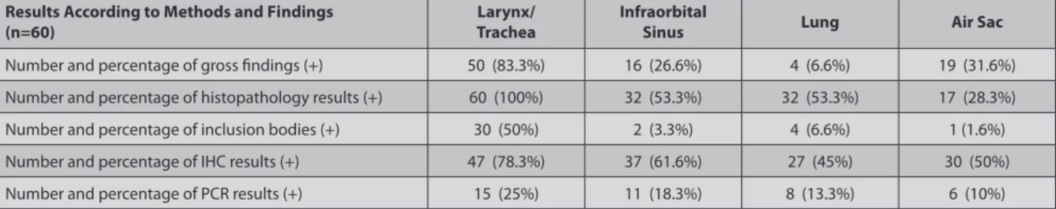
Table 2. The numbers and percentages of gross, histopathology, inclusion bodies, immunohistochemistry and PCR results
Results According to Methods and Findings
(n=60) TracheaLarynx/ Infraorbital Sinus Lung Air Sac
Number and percentage of gross findings (+) 50 (83.3%) 16 (26.6%) 4 (6.6%) 19 (31.6%) Number and percentage of histopathology results (+) 60 (100%) 32 (53.3%) 32 (53.3%) 17 (28.3%) Number and percentage of inclusion bodies (+) 30 (50%) 2 (3.3%) 4 (6.6%) 1 (1.6%) Number and percentage of IHC results (+) 47 (78.3%) 37 (61.6%) 27 (45%) 30 (50%) Number and percentage of PCR results (+) 15 (25%) 11 (18.3%) 8 (13.3%) 6 (10%)
against anti-Infectious Laryngotracheitis Virus antibody (Anti-ILTV antibody). In 37 cases (61.6%) in the infraorbital sinus, in 27 cases (45%) in lungs and in 30 cases (50%) in air sacs, positive immunoreactivities were observed. Immuno- positive staining was seen in the cytoplasm and rarely in the nuclei of macrophages, mucosal epithelial cells, gland epithelia and mononuclear cells in lamina propria and submucosa in the larynx and trachea (Fig. 3A-C). Immuno-positive reactions were also detected in the infraorbital sinuses in the mucosal epithelia and luminal desquamated epithelial cells. Immunohistochemical positivity was seen in the cell debris in the lumen of the parabronchus in the
lungs (Fig. 3D). Positive stainings were noted in inflammatory cells in the epithelium and subepithelial layer in the air sacs
(Fig. 3E). In addition, intracytoplasmic immunopositivity
was also observed in the giant cells in all examined organs
(Fig. 3F). PCR Results
PCR results from FFPE tissues are given in Table 2. The most positive reaction was observed in 15 (25%) cases in trachea and larynx. This was followed by infraorbital sinuses in 11 (18.3%) cases, in lungs in 8 (13.3%) cases and air sacs in 6 (10%) cases.
Table 3. Distribution of histopathological lesions in the organs
Organs (N=60) Histopathological Changes Score of Lesions/ Number of Lesions Degeneration, Necrosis, and Desquamation in the Epithelia Thickening of the Propria (Hyperemia, Edema, MNC Infiltration) Inclusion
Bodies Giant CellsSyncytial
Heterophil Granulocyte Infiltrations Lymphoid Cell Infiltrations Trachea and Larynx 0 0 0 0 0 0 0 0 +1 5 0 4 0 0 0 2 +2 9 5 8 3 3 0 6 +3 19 15 18 18 15 0 14 +4 18 16 11 9 8 5 7 +5 9 9 2 0 0 2 2 Infraorbital Sinus + 32 5 31 2 2 3 1 Lungs + 32 4 21 4 4 9 19 Air Sac + 17 2 17 1 0 0 2
Fig 1. A. Trachea. Hyperemic appearance in the mucosa of the trachea, and bloody exudate accumulation in
the lumen (arrows), B. Larynx. Fibrinous exudate deposits in the cavum larynx (arrow), C. Larynx and Trachea. Diphtheroid lesions on the surface of the larynx and trachea (arrows), D. Larynx and Trachea. Fibrin mass accumulation in the cavum larynx (white arrows) and mucopurulent exudate deposits in the lumen of the trachea (red arrow)
DISCUSSION
ILT is a viral disease that infects the respiratory tract of the chickens and, rarely pheasants and turkeys. It has
been reported to be diagnosed by clinical, necropsy and laboratory examinations [4,15,16]. Laboratory methods include serological methods, histopathological methods, fluorescent antibodies (FA), IHC methods, electron microscopy, virus
Fig 2. A. Trachea. +1 (minimal) lesions. Mild mononuclear cell infiltrates (arrows) in lamina propria and hyperemia in blood vessels (arrowheads), HxE, B. Trachea. +2 (mild) lesions. Mononuclear cell infiltrations (arrows), normal mucous glands (arrowheads) in the
lamina propria, and normal mucosal epithelia (red arrows), HxE, C. Trachea. +3 (moderate) lesions. Epithelial degeneration, necrosis and desquamation. Numerous giant cells and Cowdry A type intranuclear eosinophilic inclusion bodies (arrowheads) in these cells. Hyperemia in blood vessels (H), HxE, D. Trachea. +4 (severe) lesions. Mucosa thickened due to mononuclear cell infiltrates, oedema (O) and hyperemia (H). Mucosal epithelia are flattened (arrows) due to fibrinonecrotic masses, HxE, E. Trachea. +5 (Very serious) lesions. The epithelial layer is completely separated from the mucosa (black arrows) and desquamated in the lumen (red arrows). Hyperemia in the blood vessels (H) and oedema in the lamina propria (O), HxE, F. Larynx. Lumen has numerous giant cells (arrows), and eosinophilic inclusion bodies (arrowheads) in the nuclei of these cells, HxE
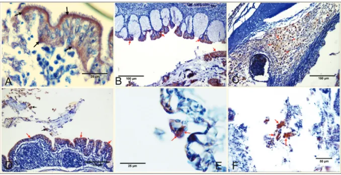
Fig 3. A. Trachea. Positive immunostaining in the mucosal epithelia (arrows), IHC, B. Larynx. Positive immunoreactions in the mucosal
epithelia (arrows), IHC, C. Trachea. Positive reactions (arrows) in the cytoplasm of macrophages in the submucosa layer, IHC, D. Lungs. Positive reactions (red arrows) in parabronchial epithelia and desquamated epithelial cells (black arrows) in the lumen, IHC, E. Air sac. Positive staining in the epithelium (arrows), IHC, F. Trachea. Positive reactions (arrows) in the cytoplasm of macrophages and epithelial cells that desquamated in the lumen, IHC
isolation, and PCR [1,16]. In a previous study, histo-pathological methods have been reported to be a more reliable method than virus isolation. However, the PCR method is more sensitive and to reduces false positives [17]. On the other hand, FA test was found to be as reliable as histopathological methods [18]. However, it needs for fluorescence microscopy has been reported as a negative aspect [9]. Williams et al.[19] compared virus isolation, electron microscopy, and PCR methods and they found that most sensitive methods were PCR, virus isolation and electron microscopy, respectively. They have also emphasized that the PCR was an advantageous method because of the other methods more costly. Abbas et al.[20] compared immunohistochemistry, virus isolation, histo-pathology, and PCR methods and they found that the most sensitive method is immunohistochemistry.
In this study, it was aimed to investigate the pathologic, immunohistochemical and PCR findings in naturally infected laying hens, and which method could be used to diagnose the disease faster and more reliably.
Although it is easy to recognize the disease by necropsy findings, it is emphasized that the advanced laboratory methods should be used, because of similar findings to some other respiratory tract infections such as Infectious Bronchitis [1,20-22]. Macroscopically; lesions are frequently seen in the laryngeal and tracheal lumen, sinuses, air sacs, and lungs [1,23-25]. The larynx, trachea, sinuses, air sacs and lungs were examined macroscopically in this study. Macroscopic changes ranged from bloody and muco-purulent contents to fibrinous and diphtheroid lesions in the larynx and trachea. Tracheal and laryngeal lesions were observed in 50 of 60 hens by grossly. Blood-mucopurulent contents were observed in thirty-eight cases, while fibrinous and diphtheroid lesions were observed in 5 cases. In seven cases, both bloody-mucopurulent and diphtheroid exudate were noted. In particular, the exudate accumulation in the tracheal lumen was similar to the findings reported by researchers [14,17,21,26]. In infraorbital sinus, mucous exudate was detected in 16 cases. Congestion was detected in only 4 cases in the lung, while thickening was observed in 19 cases at the air sacs. Mucous exudate accumulation in the sinuses and the thickening of air sacs are suggest to be that the disease is in the acute stage, as mentioned in the other studies [23,27]. Previous studies have reported that the lung is rarely affected by the disease [1]. However, only the congestions were noted in this study, while no macroscopic finding of pneumonia was found.
Histopathologically, The presence of intranuclear eosinophilic inclusion bodies and giant cells in respiratory organs such as the larynx, trachea, sinus and lung are defined as a characteristic finding for the ILT [3]. The diagnosis based on histopathologic findings may be inadequate due to the inclusion bodies are observed between the 3rd and 5th days from the beginning of the disease [1,10]. In histopathological examinations; it has been stated that
ILT mainly affects upper respiratory tracts, such as larynx and trachea, as well as lesions can be seen in organs such as air sacs and lungs [28-30]. In this study, it was revealed that the larynx, trachea, infraorbital sinus, lung and air sacs were affected at various grades from the disease. It has been found that moderate and severe changes are observed more frequently. It was noted that intranuclear inclusion bodies in epithelial cells and syncytial giant cells that are typical signs of the disease were observed in exudates, both mucosa and luminal desquamation. In addition, degeneration, necrosis and desquamation of the epithelium, thickening due to mononuclear cell infiltrates and hyperemia and oedema in lamina propria were similar to the findings previously reported by others [1,31]. Thickening due to oedema and mononuclear cell infiltrate in the subepithelial layer of infraorbital sinuses and air sacs were quite remarkable. Giant cells and inclusion bodies within the exudate and desquamated epithelial cells were also seen in these organs. Hyperplasia of the lymphoid tissue was frequently observed in the lungs. But in some cases, intranuclear inclusions in giant cells within the parabronchial lumens were similar to those reported previously [1,4]. In only 4 cases, typical inclusion bodies and giant cell accumulations were detected in the lungs. It was noticed that the lungs were less affected than the trachea, larynx and sinus.
Immunohistochemical methods have recently been used to diagnose ILT by many researchers [8,9,11,32-34]. IHC methods have been shown to be helpful for diagnosing ILT when macroscopical and histopathological findings are not possible [35]. In this study, 78.3% positive reactions observed in the larynx and trachea. In the lungs, the immunopositive staining rate was 45%. Tadese et al.[33] found 18.18% immunoactive reactions against anti-ILTV antibody in the trachea. Preis et al.[11] detected 70% positivity in the larynx and trachea tissues, and 53.8% positive results in the lungs in the field study immunohistochemically. They reported that these positive reactions were intracytoplasmic located in the mucosa and desquamated epithelial cells in the larynx and trachea, and parabronchial epithelium in the lung. At the same time, they stated that cytoplasms of giant cells found in these tissues had immunopositive reactions against anti-ILTV antibody. In an experimental study, immunopositive stainings were observed in laryngeal and tracheal tissues, especially in the cytoplasm of epithelial desquamated and unciliated cells such as goblet cells and mucous gland epithelia [36]. Guy et al.[9] reported that in their experimental study, they found positive reactions in tracheal mucosal epithelia and desquamated epithelial cells. In this study, immunopositive reactions were observed in the larynx and tracheal epithelium, luminal epithelial cells and gland epithelium. In this study, strongly positive staining was detected in the cytoplasm of the giant cells located in the lumen. These reactions were similarly observed in the sinus and lung. Immunoactive staining was observed in the cytoplasm of the macrophages in the
submucosal layer of the trachea and larynx. This suggests that the virus tends to spread to deeper layers rather than mucosa. Kirkpatrick et al.[36] support this view that different virus strains may have different tropism in the tissues. Preis et al.[11] applied PCR method in fresh larynx, trachea, paranasal sinus, and lung from ILT suspected chickens, and they found positive reactions in 63.2% of larynx/ trachea, 56% in sinus and 57.6% in the lung. However, they found FFPE positivity was 25% in tracheal tissues. In this study, which was made from FFPE tissues the rate of positive cases were 25% in the larynx/trachea tissues. The positivity of FFPE tissues is lower than fresh tissues. They explained that the long-term fixation by formalin causes DNA damage [37]. Kleter et al.[38] emphasized that liquid paraffin at high temperature during paraffin block preparation may cause damage to DNA. Sivaseelan et al.[24] obtained positive results only in the larynx, trachea and conjunctiva as a result of the PCR tests performed on the tissue specimens. In this study, positive reactions were also observed in the lungs (13.3%), infraorbital sinuses (18.3%) and air sacs (10%). This result suggests that the virus may be present in various tissues and organs at different stages of the disease. But the viral DNA is mostly found in trachea and larynx, and less in sinuses, lungs and air sacs.
In gross examination of larynx and trachea, in 50 (83.3%) cases were diagnosed as ILT. However, histopathologically 60 (100%) cases, immunohistochemically 47 (78.3%) cases were positive for ILT. Although findings were observed in all (100%) cases in histopathology, ILT specific inclusion bodies were detected in 30 (50%) cases in the larynx and trachea. Preis et al.[14] found inclusion bodies in the trachea (70%), larynx (50%), and lung (10%). This suggests that reduces the reliability of the histopathological diagnosis. Immunohistochemically, the reaction against Anti-ILTV antibody can easily be demonstrated in the tissues. However, in histopathological examinations, ILT-like findings can also be observed in other respiratory system diseases. It can be said that the IHC results are more precise than histopathologic results. In PCR analyses, there were only 15 (25%) cases positive in the trachea and larynx. The superiority of IHC over PCR, viral structures can easily be demonstrated in the tissues. In contrast, virus localization cannot be predicted by PCR. Although FFPE tissues can be stored for many years, the positive rate of PCR is lower than other methods such as histopathology and immuno-histochemistry, so it cannot be used to the diagnostic tool. In macroscopic examinations of other tissues such as infraorbital sinus and lung, there were 16 cases in the sinuses and 4 cases in the lungs, suggesting ILT findings. Thirty-two cases in the sinuses and lungs were positive in histopathology. It suggests histopathology is more sensitive against gross pathology. Histopathological (28.3%) and macroscopic (31.6%) positivity rates of air sacs were close to each other, but immunohistochemically, this ratio was 50%. Grossly, opaque appearance of the air sacs may
be regarded as a helpful finding in the diagnosis. However, IHC analyses needed to definitive diagnose.
In conclusion, lesions were evaluated by macroscopic, histopathologic, immunohistochemical and PCR methods in naturally infected with Infectious Laryngotracheitis Virus in laying hens. The larynx, trachea, infraorbital sinuses and air sacs should be carefully examined by gross examinations. In histopathological examinations of the larynx and trachea, all cases were found positive for ILT, however, a positivity rate of 78.3% was determined by IHC examinations, and it was thought that this method could be used for definite diagnosis. On the other hand, positive immunostaining was obtained in 61.6% in the sinus, 45% in the lung and 50% in the air sacs. This suggests that it may be useful to examine trachea and larynx as well as the infraorbital sinuses, lungs and air sacs to evaluate the severity/prevalence of the ILT. The location of viral antigens not only in the epithelium and lamina propria but also in the submucosa layer reveals that deeper layers should be taken into account along with mucosal layers by IHC staining, and it is also an indicator of virulence of the agents.
REFERENCES
1. Guy S, Bagust T: Laryngotracheitis. In, Saif Y (Ed): Diseases of Poultry.
11th ed., 121-134, Iowa State University Press, Ames, Iowa, 2003.
2. International Committee on Taxonomy of Viruses (ICTV): Taxonomy
history: Gallid alphaherpesvirus-1. https://talk.ictvonline.org/taxonomy/p/ taxonomyhistory?taxnode_id=201-60844. Accessed: September 10, 2015.
3. Seifried O: Histopathology of infectious laryngotracheitis in chickens. J Exp Med, 54 (6): 817-826, 1931. DOI: 10.1084/jem.54.6.817
4. Hanson L, Bagust T: Laryngotracheitis. In, Calnek B (Ed): Diseases
of Poultry. 9th ed., 485-495, Iowa State University Press, Ames, Iowa, 1991.
5. Özdemir Ö, Erer H: Tavukların önemli üst solunum yolu hastalıklarında
patolojik değişiklikler. AVKAE Derg, 2, 29-38, 2012.
6. Purcell D, McFerran J: Influence of method of infection on the
pathogenesis of infectious laryngotracheitis. J Comp Pathol, 79 (3): 285- 291, 1969. DOI: 10.1016/0021-9975(69)90041-3
7. Bagust T, Calnek B, Fahey K: Gallid-1 herpesvirus infection in the chicken.
3. Reinvestigation of the pathogenesis of infectious laryngotracheitis in acute and early post-acute respiratory disease. Avian Dis, 30 (1): 179-190, 1986. DOI: 10.2307/1590631
8. Yilmaz F, Timurkaan N, Bulut H: Detection of infectious
laryngo-tracheitis virus in trigeminal ganglia by avidin-biotin complex method in chickens. Acta Vet Hung, 52 (2): 167-171, 2004. DOI: 10.1556/AVet. 52.2004.2.5
9. Guy J, Barnes H, Smith LG: Rapid diagnosis of infectious laryngotracheitis
using a monoclonal antibody‐based immunoperoxidase procedure. Avian
Pathol, 21 (1): 77-86, 1992. DOI: 10.1080/03079459208418820
10. Reynolds H, Watrach A, Hanson L: Development of the nuclear
inclusion bodies of infectious laryngotracheitis. Avian Dis, 12 (2): 332- 347, 1968. DOI: 10.2307/1588234
11. Preis I, Fiúza A, Silva C, Braga J, Couto R, Martins NdS, Ecco R:
Pathological, immunohistochemical, and molecular findings in commercial laying hens and in backyard chickens naturally infected with the infectious laryngotracheitis virus. Braz J Poult Sci, 16 (4): 359-366, 2014. DOI: 10.1590/1516-635x1604359-366
12. Yenice G, Çelebi D, Yörük MA, Uçar Ö, Sağlam YS, Tunç MA, Altun S: Effect of kefir upon the performance, intestinal microflora and
histopathology of certain organs in laying hens. Kafkas Univ Vet Fak Derg, 20 (3): 363-370, 2014. DOI: 10.9775/kvfd.2013.10173
13. Guy JS, Barnes HJ, Morgan LM: Virulence of infectious laryngotracheitis
viruses: comparison of modified-live vaccine viruses and North Carolina field isolates. Avian Dis, 34 (1): 106-113, 1990. DOI: 10.2307/1591340
14. Preis IS, Braga JF, Couto RM, Brasil BS, Martins NR, Ecco R: Outbreak
of infectious laryngotracheitis in large multi-age egg layer chicken flocks in Minas Gerais, Brazil. Braz J Vet Res, 33 (5): 591-596, 2013. DOI: 10.1590/ S0100-736X2013000500007
15. Portz C, Beltrão N, Furian TQ, Júnior AB, Macagnan M, Griebeler J, Rosa CAVL, Colodel EM, Driemeier D, Back A: Natural infection of
turkeys by infectious laryngotracheitis virus. Vet Microbiol, 131 (1): 57-64, 2008. DOI: 10.1016/j.vetmic.2008.02.029
16. Arda M: İnfeksiyöz laringotraheitis. In, İzgür M, Akan M (Eds): Kanatlı
Hayvan Hastalıkları. 185-188, Medisan Yayınevi, Ankara, 2002.
17. Crespo R, Woolcock PR, Chin R, Shivaprasad H, García M:
Comparison of diagnostics techniques in an outbreak of infectious laryngotracheitis from meat chickens. Avian Dis, 51 (4): 858-862, 2007. DOI: 10.1637/7875-011907-REGR1.1
18. Goodwin MA, Smeltzer MA, Brown J, Resurreccion RS, Dickson TG: Comparison of histopathology to the direct immunofluorescent
antibody test for the diagnosis of infectious laryngotracheitis in chickens.
Avian Dis, 35 (2): 389-391, 1991. DOI: 10.2307/1591195
19. Williams R, Carol E, Jones R: A comparison of direct electron
microscopy, virus isolation and a DNA amplification method for the detection of avian infectious laryngotracheitis virus in field material.
Avian Pathol, 23 (4): 709-720, 1994. DOI: 10.1080/03079459408419039 20. Abbas F, Andreasen Jr JR: Comparison of diagnostic tests for
infectious laryngotracheitis. Avian Dis, 40 (2): 290-295, 1996. DOI: 10.2307/1592223
21. Linares J, Bickford A, Cooper G, Charlton B, Woolcock P: An
outbreak of infectious laryngotracheitis in California broilers. Avian Dis, 38 (1): 188-192, 1994. DOI: 10.2307/1591856
22. Wang Z, Gao S, Zheng Q, Sun M, Jia W, Ren T: Epidemiology and
evolutionary characteristics of avian infectious bronchitis virus in china.
Kafkas Univ Vet Fak Derg 21 (2): 179-186, 2015. DOI: 10.9775/kvfd.2014.11999 23. Bang BG, Bang FB: Laryngotracheitis virus in chickens. J Exp Med,
125 (3): 409-428, 1967. DOI: 10.1084/jem.125.3.409
24. Sivaseelan S, Rajan T, Malmarugan S, Balasubramaniam G, Madheswaran R: Tissue tropism and pathobiology of infectious
laryngotracheitis virus in natural cases of chickens. Israel J Vet Med, 69, 197-202, 2014.
25. Stewart-Brown BN, Van Alstine W: A case report and discussion
of Laryngotracheitis in chickens. Iowa St Uni Vet, 48 (1): 36-39, 1986.
26. Barhoom S: Outbreak of laryngotracheitis (LT) in vaccinated
commercial layer flocks in Palestine. Proceedings of the 2nd Scientific Conference of animal wealth research in the Middle East and North Africa,
Cairo International Convention Center, 24-26 October, 2009. 176-182. Massive Conferences and Trade Fairs, 2009.
27. Wang LG, Ma J, Xue CY, Wang W, Guo C, Chen F, Qin JP, Huang NH, Bi YZ, Cao YC: Dynamic distribution and tissue tropism of infectious
laryngotracheitis virus in experimentally infected chickens. Arch Virol, 158 (3): 659-666, 2013. DOI: 10.1007/s00705-012-1414-8
28. Bagust T, Jones R, Guy J: Avian infectious laryngotracheitis. Rev Sci Tech, 19 (2): 483-488, 2000.
29. Nakamura K, Imai K, Tanimura N: Comparison of the effects of
infectious bronchitis and infectious laryngotracheitis on the chicken respiratory tract. J Comp Pathol, 114 (1): 11-21, 1996. DOI: 10.1016/S0021-9975(96)80058-2
30. Russell R: Respiratory tract lesions from infectious laryngotracheitis
virus of low virulence. Vet Pathol, 20 (3): 360-369, 1983. DOI: 10.1177/ 030098588302000312
31. Riddell C: Avian Histopathology. 2 ed., Rose Printing, Tallahassee,
Florida, USA, 1996.
32. Fuchs W, Veits J, Helferich D, Granzow H, Teifke JP, Mettenleiter TC: Molecular biology of avian infectious laryngotracheitis virus. Vet Res, 38 (2): 261-279, 2007. DOI: 10.1051/vetres:200657
33. Tadese T, Potter AE, Fitzgerald S, Reed WM: Concurrent infection
in chickens with fowlpox virus and infectious laryngotracheitis virus as detected by immunohistochemistry and a multiplex polymerase chain reaction technique. Avian Dis, 51 (3): 719-724, 2007. DOI: 10.1637/ 0005-2086(2007)51[719:CIICWF]2.0.CO;2
34. Zhao Y, Kong C, Cui X, Cui H, Shi X, Zhang X, Hu S, Hao L, Wang Y: Detection of infectious laryngotracheitis virus by real-time PCR in
naturally and experimentally infected chickens. PloS One, 8 (6): e67598, 2013. DOI: 10.1371/journal.pone.0067598
35. Timurkaan N, Yilmaz F, Bulut H, Ozer H, Bolat Y: Pathological and
immunohistochemical findings in broilers inoculated with a low virulent strain of infectious laryngotracheitis virus. J Vet Sci, 4 (2): 175-180, 2003.
36. Kirkpatrick NC, Mahmoudian A, Colson CA, Devlin JM, Noormohammadi AH: Relationship between mortality, clinical signs
and tracheal pathology in infectious laryngotracheitis. Avian Pathol, 35 (6): 449-453, 2006. DOI: 10.1080/03079450601028803
37. Gilbert MTP, Haselkorn T, Bunce M, Sanchez JJ, Lucas SB, Jewell LD, Van Marck E, Worobey M: The isolation of nucleic acids from fixed,
paraffin-embedded tissues-which methods are useful when? PloS One, 2 (6): e537, 2007. DOI: 10.1371/journal.pone.0000537
38. Kleter B, van Doorn L-J, ter Schegget J, Schrauwen L, van Krimpen K, Burger M, ter Harmsel B, Quint W: Novel short-fragment PCR assay
for highly sensitive broad-spectrum detection of anogenital human papillomaviruses. Am J Pathol, 153 (6): 1731-1739, 1998. DOI: 10.1016/ S0002-9440(10)65688-X