INVESTIGATION OF THE ROLE OF cGAMP IN
DIFFERENTIATION OF T LYMPHOCYTES
A THESIS SUBMITTED TO
THE GRADUATE SCHOOL OF ENGINEERING AND SCIENCE OF BILKENT UNIVERSITY
IN PARTIAL FULFILLMENT OF THE REQUIREMENTS FOR THE DEGREE OF
MASTER OF SCIENCE IN
MOLECULAR BIOLOGY AND GENETICS
By Begüm YILDIZ
INVESTIGATION OF THE ROLE OF cGAMP IN DIFFERENTIATION OF T LYMPHOCYTES
By Begüm YILDIZ October 2016
We certify that we have read this thesis and that in our opinion it is fully adequate, in scope and in quality, as a thesis for the degree of Master of Science.
__________________________________
İhsan Gürsel (Advisor)
__________________________________
Dicle Güç
__________________________________
Ali Osmay Güre
Approved for the Graduate School of Engineering and Science:
____________________________________ Ezhan Karaşan
Abstract
INVESTIGATION OF THE ROLE OF cGAMP IN
DIFFERENTIATION OF T LYMPHOCYTES
Begüm YILDIZ
M.S. in Molecular Biology and Genetics Advisor: İhsan Gürsel
October, 2016
STING is the pivotal mediator for the recognition of host and pathogenic cytosolic dsDNA as well as cyclic di-nucleotides metabolites from microbes. STING can either recognize DNA itself or sense the presence of cGAMP, which is converted from ATP and GTP upon DNA binding to cGAS enzyme. Not only strategy against intracellular pathogens makes STING an ideal target, but also the recognition of DNA from host cells has a significant role in tumor immunity. Previous studies demonstrated that DNA released from cancerous cells are internalized by innate immune cells such as macrophages and dendritic cells in tumor microenvironment and trigger the production of IFN-β and other pro-inflammatory cytokines including IL-6, TNF-α, and IL-12 through STING triggered signaling pathway. These cytokines then enhance cytotoxic activity of CD8+ T cells by further increasing IFNγ production. Since enhanced T cell immunity is the hallmark of vaccine adjuvants, cyclic di-nucleotides such as cGAMP become an important and effective vaccine adjuvants against intracellular pathogens and malignant cells. Although STING activating cyclic di-nucleotides are envisioned as novel and
potent vaccine adjuvants, more thorough research is needed to unearth the mechanism of action of STING on different immune cells. Therefore, it will pave the way for the initiation of successful human trials. The important criteria while developing vaccine adjuvant are the magnitude, and the quality of an immune response and its toxic side effects. To identify these, members of the both innate and adaptive immune system should be taken into account. However, previous studies merely focus on the function and effect of cGAMP in innate immune cells such as macrophages, monocytes and dendritic cells. However, to date there is no explicit study investigating the effect of STING signaling cascade on T-cells. In the light of these findings, we aimed to investigate the direct effect and function of cGAMP on T lymphocytes. Since there were not any preliminary data, we firstly stimulated Pan T cells with cGAMP alone or together with various TLR ligands and then, checked the cytokine profiles and the viability of cells. Surprisingly, 2.5µg/ml dose of cGAMP had a toxic effect on T cell but not on bone marrow derived dendritic cells and macrophages. While cGAMP triggered cell death, interestingly IL-17 secretion from both CD4+ and CD8+ T cells was dramatically
increased. Beside, cGAMP stimulation drastically increased CD4+/CD8+ T cells ratio of Pan T cells population. Next, we sought to identify the source of IL-17. The IL17 inductive capacity of cGAMP was investigated on purified CD4+ T cells from mice. Unexpectedly, data revealed that cGAMP elicited apoptosis of CD4+ T cells. Moreover, there was no significant induction of IL-17 secretion. Next, we aimed to find a condition that will reduce the toxic effect of cGAMP, while maintaining IL-17 secretion. When Pan T cells were stimulated with cGAMP and R848 (a TLR7 ligand), the toxic action of cGAMP decreased while IL-17 secretion was enhanced. Lastly, the potency of T cells stimulated with cGAMP was investigated. According to our results, macrophages were activated in the presence of conditioned medium obtained from T cells stimulated with cGAMP. When taken together our findings point out that STING dependent direct activation of T-cells via cGAMP and its subsequent effect on macrophages might be utilized as an immunotherapeutic approach where IL17 induction is important and could be harnessed as vaccine adjuvants against mucosal infections or against cancer.
Özet
T LENFOSİT FARKLILAŞMASI ÜZERİNE
cGAMP`IN ROLÜNÜN İNCELENMESİ
Begüm YILDIZ
Moleküler Biyoloji ve Genetik, Yüksek Lisans Tez Yöneticisi: İhsan Gürsel
Ekim, 2016
STING, hem patojen ve konağın kendi sitozolik çift iplikli DNA (çiDNA) sının hem de mikrop kaynaklı siklik di-nükleotidlerin tanınmasında önemli bir mediyatördür. STING ya DNA’nın kendisini ya da cGAMP molekülünü tanır. cGAMP molekülü DNA’nın cGAS enzimine bağlanması sırasında ATP ve GTP’nin dönüştürülmesiyle oluşmaktadır. Hücre içi patojenlere karşı geliştirilen strateji STING’i ideal bir hedef yaptığı gibi, konağın kendi DNA’sının tanınması sonucu verilen tepki tümöre karşı geliştirilen immün yanıt açısından önemlidir. Daha önceki çalışmalar göstermiştir ki; kanser hücrelerinden salınan DNA, makrofaj ve dendiritik hücreler gibi doğal bağışıklık sistemi hücreleri tarafından tümör mikro çevresinde hücre içine alınır. Bu internalizasyon STING’in tetiklediği yolak üzerinden IFN-β ve IL-6, TNF-α, ve IL-12 gibi diğer pro-enflammatuar sitokinlerin salınımını tetikler. Daha sonra bu sitokinler IFNγ üretimini artırarak CD8+ T hücrelerinin sitotoksik aktivitelerini artırırlar. Aşı adjuvanlarının öne çıkan özelliklerinden biri de T hücre bağışıklığının artırılmasını sağlamaktır. Bu sebeple cGAMP gibi siklik di-nükleotidler hücre içi patojen ve kanser hücrelerine karşı önemli ve
etkili bir aşı adjuvanıdırlar. STING’i aktive eden siklik di-nükleotidler özgün ve potansiyel aşı adjuvanları olarak öngörülseler de, STING mekanizmasının farklı bağışıklık sistemi hücreleri üzerindeki etkilerinin açığa çıkarılması için daha kapsamlı bir çalışma gerekmektedir. Bu çalışmalar, başarılı insan deneylerinin başlamasının önünü açacaklardır. Aşı adjuvan geliştirirken, immün tepkinin niteliği, şiddeti ve toksik yan etkileri önemli kriterler arasında yer almaktadır. Bu kriterleri belirmek için doğal ve sonradan kazanılan bağışıklık sistemi hücrelerinin tümü göz önünde bulundurulmalıdır. Fakat, şu ana kadar yapılan tüm çalışmalar cGAMP etkisinin sadece doğal bağışıklık sistemi üzerine etkilerini araştırmaktadır. Bugüne dek, T hücrelerinde STING yolağının aktivasyon şelalesini açıkça gösteren hiç bir çalışma bulunmamaktadır. Bu bulgular ışığında, biz bu çalışmada cGAMP’ın T hücreleri üzerindeki direkt etkisini ve fonksiyonunu araştırmayı amaçladık. Bu alanda herhangi bir ön veri olmadığı için ilk olarak Pan T hücreleri sadece cGAMP veya cGAMP ile birlikte bazı TLR ligandlarıyla stimüle edildiler. Bu stimülasyon sonucunda, sitokin profilleri ve hücrelerin durumu kontrol edildi. Şaşırtıcı bir şekilde, cGAMP 2.5µg/ml dozu makrofaj ya da dendiritik hücreler üzerinde hiç bir etki göstermezken, T hücreleri üzerine toksik etkisi olmuştur. Hücre ölümü etkisi yanı sıra, cGAMP, Pan T hücreleri içindeki hem CD4+ hem de CD8+ T hücrelerinin IL-17 salınımı ilginç şekilde artırmıştır. Bunun yanı sıra; Pan T hücreleri içinde CD4+/CD8+ oranı ciddi şekilde artmıştır. Bu sonuçlardan sonra, IL-17 kaynağını aramayı hedefledik. cGAMP’ın IL-17 indükleme kapasitesi farelerden pürifiye eldilen CD4+ T hücreleri üzerinde araştırılmıştır. Beklenmedik bir şekilde, cGAMP CD4+ T hücreleri üzerinde toksik etki gösterip, IL-17 salınımını etkilememiştir. Bundan sonra, cGAMP’ın Pan T üzerine olan toksik etkisini IL-17 salınımını etkilemeyecek şekilde azaltan bir durum bulmayı amaçladık. Pan T hücreleri cGAMP ve R848 (TLR7 ligandı) ile stimüle edildiğinde IL-17 salınımı artarken, toksik etkinin azaldığı gözlemlenmiştir. Son olarak, cGAMP ile stimüle edilmiş T hücrelerinin potansiyeli araştırılmıştır. Sonuçlarımıza göre, cGAMP ile stimüle edilmiş T hücrelerinin salgıları makrofaj aktivasyonuna yol açmıştır. Tüm bunlar göz önüne alındığında, sonuçlarımız T hücrelerinin cGAMP kullanılarak STING’e bağlı direkt aktivasyonları ve buna bağlı makrofajlar üzerine etkilerinin immünoterapötik amaçlar üzerine kullanılmasına dikkat
çekmektedir. Ayrıca, IL-17 salınımı önemli bir bulgu olmakla beraber, mukozal enfeksiyonlara ve kansere karşı aşı adjuvanı olarak koşullanabilir.
Acknowledgement
First, I would like to express my heartfelt gratitude to my advisor Prof. İhsan Gürsel for the opportunity to work in his lab and for his continuous support, patience, motivation, guidance and encouragement.
I would like to express my sincere thanks to Assoc. Prof. Dr. Ali Osmay Güre and Prof. Dr. Dicle Güç for accepting to become members of my thesis jury and sparing time to evaluate and improve my thesis.
I am lucky to be a part of the Gürsel group. I thank my fellow lab mates: Fuat Cem Yağcı, Tamer Kahraman, Gizem Tinçer-König, Kübra Almacıoğlu, Begüm Han Horuluoğlu, Gözde Güçlüler, Defne Bayık, Muzaffer Yıldırım, Fehime Kara Eroğlu, Banu Bayyurt and particularly to Elif Senem Köksal, Hakan Köksal, Alican Savaş and Aslı Yıldırım for their support and friendship.
It was fun to be a part of the MBG family. I would like to express my gratitude to all my instructors and friends in graduate school for their companionship and assistance.
Without my family, none of the exceptional things in my life would have been possible. I would like to express my deepest love and thankfulness to my mother Betül, my father Hasan, my grandmothers Gülser and Türkan, my aunts Gülnur and Hülya for their invaluable and everlasting support. Their love is the most significant motivation for me, which makes my accomplishments meaningful. Besides, I know that my dear, sweetheart grandfather Seyhan is always watching and protecting me during my journey. I love him very much and I would like to dedicate this thesis to him.
Last but not least, I would like to thank to my dearest fiancée Tekin for supporting me throughout this period with his constant love and patience.
Table of contents
Abstract ... iii Özet ... v Acknowledgement ... ix Table of contents ... x
List of figures ... xiv
List of tables ... xvi
Abbreviations ... xvii
Chapter 1 ... 1
Introduction ... 1
1.1 The immune system ... 1
1.1.1 Innate immune system ... 2
1.1.1.1 Pattern recognition receptors (PRRs) ... 2
1.1.1.1.1 Toll-like receptors (TLRs) ... 3
1.1.1.1.2 Cytosolic DNA sensors ... 4
1.1.2 Adaptive immune system ... 6
1.2 T lymphocytes ... 7
1.2.1 CD8+ T lymphocytes ... 10
1.2.2 CD4+ T lymphocytes ... 11
1.3 Interleukin-17 (IL-17A) ... 15
1.3.1 IL-17A producing T cells ... 15
1.3.2 Mechanism of IL-17 signaling ... 15
1.3.3 IL-17 role in host defense and autoimmunity ... 17
1.4 Aim of the study ... 18
Materials and Methods ... 19
2.1 Materials ... 19
2.1.1. General laboratory & cell culture reagents and materials ... 19
2.1.2 Recombinants and other agents ... 20
2.1.3 CpG ODNs ... 20
2.1.4 PRR ligands ... 21
2.1.5 Flow cytometry ... 21
2.1.6 Determination of gene expression ... 22
2.1.6.1 Primers ... 22
2.1.7 Reagents for ELISA ... 23
2.1.8 BCA ... 25
2.1.9 Western blot ... 25
2.1.10 Determination of cell viability ... 27
2.2 Solutions, Buffers and Culture Media ... 27
2.2.1 Cell culture media ... 27
2.2.2 Flow cytometry buffers ... 27
2.2.3 Agarose gel electrophoresis ... 28
2.2.4 ELISA buffers ... 28
2.2.5 Western blot buffers ... 29
2.3 Methods ... 32
2.3.1 Maintenance of cell lines ... 32
2.3.2 Cryopreservation and thawing of cells ... 32
2.3.3 Cell counting ... 33
2.3.3.1 Haemocytometer ... 33
2.3.3.2 Flow cytometer ... 33
2.3.4 Primary murine cell suspension preparation ... 34
2.3.4.1 Mouse spleen cell suspension preparation ... 34
2.3.4.2 Isolation of bone marrow cells ... 34
2.3.5 Bone marrow derived dendritic cell generation ... 35
2.3.6 Pan T and CD4+ T cell isolation from mouse spleen ... 35
2.3.8 Cytokine ELISA ... 36
2.3.9 The measurement of cell viability ... 37
2.3.10 CFSE labeling of Pan T cells ... 37
2.3.11 Flow cytometry ... 37
2.3.11.1 Surface marker staining of cells ... 38
2.3.11.2 Fixation of cells ... 38
2.3.11.3 Intracellular cytokine staining ... 38
2.3.12 Binding & uptake and internalization assays ... 39
2.3.13 Determination of gene expression ... 39
2.3.13.1 Total RNA isolation ... 39
2.3.13.2 cDNA synthesis ... 40
2.3.13.3 PCR ... 40
2.3.13.4 qPCR ... 41
2.3.13.5 Agarose gel electrophoresis ... 42
2.3.14 BCA protein assay ... 43
2.3.15 Western blot ... 43
2.3.15.1 Preparation of protein samples ... 43
2.3.15.2 SDS-PAGE running ... 44
2.3.15.3 Transfer to the membrane ... 44
2.3.15.3 Blotting and development ... 44
2.3.16 Statistical analyses ... 45
Chapter 3 ... 46
Results ... 46
3.1 Preliminary studies to optimize the culture conditions of T cells in culture .... 46
3.2 Studies to understand the effect of cGAMP on Pan T cells ... 49
3.2.1 cGAMP has a cytotoxic effect on Pan T cells ... 51
3.2.2 cGAMP triggers IL-17 secretion from Pan T cells ... 55
3.3 Efforts to understand the effect of cGAMP on CD4+ T cells ... 61
3.3.1 cGAMP has a cytotoxic effect on CD4+ T cells ... 63
3.4 Alternative approaches to decrease cytotoxic effect of cGAMP while
maintaining IL-17 secretion ... 69
3.5 Effect of cGAMP treated Pan T cells on Macrophages ... 73
Chapter 4 ... 76
Discussion ... 76
Bibliography ... 82
List of figures
Figure 1.1 Physiological functions of toll-like receptors (TLRs) ... 4
Figure 1.2: Cytosolic DNA–sensing system ... 5
Figure 1.3: T cell development in thymus ... 8
Figure 1.4: Roles of the B7–CD28/CTLA-4 pathway in regulating T-cell activation ... 10
Figure 1.5: Summary of the CD4+ T helper cell fates ... 12
Figure 1.6: IL-17 receptor signal transduction ... 16
Figure 3.1: Isolation efficiency of Pan T cells. ... 46
Figure 3.2: The activation and proliferation of T cells were analyzed by comparing different conditions ... 49
Figure 3.3: Expression analysis of STING on Pan T cells. ... 50
Figure 3.4: Activated Pan T cells internalize cGAMP ... 51
Figure 3.5: Screening of the general effect of cGAMP on Pan T cells. ... 52
Figure 3.6: Determination of the toxic effect of cGAMP on Pan T cells ... 53
Figure 3.7: cGAMP has an apoptotic effect on PanT cells ... 55
Figure 3.8: Screening for functional analysis of cGAMP on Pan T cells ... 56
Figure 3.9 cGAMP triggers IL-17 secretion from Pan T cells. ... 57
Figure 3.10: The expression of transcription factors related with IL-17 increases in the presence of cGAMP. ... 58
Figure 3.11: cGAMP induces expression of Th17 related genes in Pan T cells. ... 59
Figure 3.12: IL-17 secretion is from both CD4+ and CD8+ T cells in the presence of cGAMP. ... 60
Figure 3.13: cGAMP increases CD4+/CD8+ ratio of Pan T cells ... 61
Figure 3.14: Isolation efficiency of CD4+ T cells ... 62
Figure 3.16: cGAMP induces apoptosis of CD4+ T cells ... 65 Figure 3.17: Screening for functional analysis of cGAMP on CD4+ T cells ... 66 Figure 3.18: cGAMP fail to induce IL-17 secretion from CD4+ T cells ... 67 Figure 3.19: cGAMP treatment fail to elicit expression levels of IL-17 and IL-17 related
transcription factors in CD4+ T cells ... 68 Figure 3.20: IL-17 secretion in CD4+ T cells by cGAMP ... 69 Figure 3.21: TLR 7/8 ligand, R848 diminishes the cytotoxic effect of cGAMP in Pan T
cells. ... 70 Figure 3.22: TLR 7/8 ligand, R848 improves IL-17 secretion from Pan T cells in the
presence of cGAMP ... 71 Figure 3.23: TLR7/8 ligand, R848 decreases the expression of Th17 related genes while
maintaining IL-17 expression ... 72 Figure 3.24: Condition media from cGAMP stimulated T cell activate macrophages ... 74 Scheme 4.1: Proposed mechanism of the effect of cGAMP stimulation in T lymphocytes
List of tables
Table 2.1: Commercial name of the CpG ODN and their properties ... 20
Table 2.2: Commercial name of the ligands and their sources ... 21
Table 2.3: Commercial name of the antibodies and their properties ... 21
Table 2.4: Primers used in PCR ... 23
Table 2.5: Antibodies used in ELISA ... 24
Table 2.6: BCA reagents ... 25
Table 2.7: Antibodies used in Western blot ... 25
Table 2.8: Sample PCR Reaction ... 40
Table 2.9: PCR Protocol ... 41
Table 2.10: Sample qPCR reaction ... 42
Abbreviations
Ab Antibody
AIM2 Absent in melanoma 2 ALR AIM2 like receptor AP-1 Activator Protein 1 APC Antigen presenting cell Bcl-2 B-cell lymphoma 2
BM Bone marrow
BSA Bovine serum albumin
C/EBPβ CCAAT/enhancer-binding protein-β
cGAMP Cyclic guanosine adenosine monophosphate cGAS Cyclic GMP-AMP Synthase
CLR C-type lectin receptor CTL Cytotoxic T cell
CTLA-4 Cytotoxic T lymphocyte antigen 4 DC Dendritic cell
ddH2O Double-distilled water DMSO Dimethyl sulfoxide
DN Double negative
DP Double positive dsRNA Double stranded RNA
EtOH Ethanol
FBS Fetal bovine serum FOXP3 Forkhead box P3
IBD Inflammatory bowel disease
IFN Interferon
Ig Immunoglobulin
IL Interleukin
IRF3 Interferon regulatory factor 3 iTreg Induced regulatory T cell JAK Janus Kinase
KIR Killer inhibitory receptor
LPS Lipopolysaccharide MAPK Mitogen-activated protein kinase
MHC Major histocompatibility complex mRNA Messenger ribonucleic acid MS Multiple sclerosis
NF- κB Nuclear factor- κB NK Natural killer
NLR Nucleotide oligomerization receptor ODN Oligonucleotide
PBS Phosphate-buffered saline PCR Polymerase chain reaction PD-1 Programmed death 1
PRR Pattern Recognition Receptor RA Rheumatoid arthritis
RLR RIG-1 like receptor RNA Ribonucleic acid
RPMI Roswell Park Memorial Institute
RT Room Temperature
SOCS Suppressor of cytokine signalling ssRNA Single stranded RNA
STAT Signal transducer and activator of transcription STING Stimulator of interferon genes
T-bet T-box transcription factor TBK1 Tank binding kinase 1 TCR T-cell receptor
TGF-β Transforming growth factor beta
Th T helper
TLR Toll-like Receptor TNF Tumor necrosis factor
TRAF TNF receptor associated factor PMA Phorbol 12-myristate 13-acetate
Chapter 1
Introduction
1.1 The immune system
The general and the main goal of the immune system are to guard the body from wide range of microorganism. During recognition and elimination of the microbes, the first discernment mechanism is its ability to distinguish self from non-self. This ability gives the immune system to demolish not only microbes but also toxins and allergens [1]. The immune system has two arms including innate and adaptive immunity. These two arms of the immune system implement the main mechanism, which is the discrimination of self from non-self to protect host organism. Although the innate and adaptive immunity are based on the same mechanism, they differ in terms of rapidity and specificity. The innate immune system is the first line of defense of the body and consists of both physical, chemical barriers and specific type of cells such as monocytes, macrophages, and neutrophils. The basic virtue of the innate immune system is an immediate defense [2]. The members of the innate immune system react against pathogens at the same extent no matter how many times they encounter. On the other hand, adaptive immune system possesses the antigen specific response, which gives the specificity to T and B-lymphocytes. In other words, the hallmark of the adaptive immunity is an increase
response in the presence of repeated infectious and the development of a memory against specific antigen [3].
1.1.1 Innate immune system
Innate immune system, which comprises of various types of cells including monocytes, dendritic cells, macrophages, neutrophils, basophils, eosinophils, mast cells and natural killer cells, provides the first and immediate response against pathogens. Although innate immune system does not have any capabilities to develop memory against specific type of antigens, the most of the infectious that the host encounter are cleared by innate immune cells without any interaction with adaptive immunity. Innate immune system has three distinct phases to eliminate pathogens. In the first phases, physical barriers and soluble factors such as antimicrobial peptides or complement system are implemented for the clearance of pathogens. After releasing soluble factors upon microbes, pathogen-sensing mechanism by innate immune cells become part of an activity. Pathogen associated molecular patterns (PAMPs) are molecules from pathogens that are sensed by the members of the innate immune system. They are recognized by pattern recognition receptors (PRRs) including Toll-like receptors (TLRs), Nucleotide oligomerization receptors (NLRs), C-type lectin receptors (CLRs) and RIG-1 like receptors (RLRs), AIM2 like receptors (ALRs) and cytosolic DNA sensors. As a third phase, specific type of innate immune cells called antigen presenting cells (APCs) present pathogen specific antigens to T lymphocytes for the expansion of antigen specific lymphocytes and long term memory [4].
1.1.1.1 Pattern recognition receptors (PRRs)
The pathogen sensing mechanism of innate immune system is mostly based on pattern recognition receptors (PRRs), which can identify the different part of pathogens. PRRs consist of different type of receptors including TLRs, NLRs, CLRs, RLRs, ALRs and cytosolic DNA sensors. They can be either at the cell surface or inside a cell [5]. When pathogen associated molecules are sensed by PRRs, pro-inflammatory cytokines or chemokines start to be secreted by innate immune cells by activating several signaling
transduction pathways. Upon secretion of several cytokines and chemokines, other immune system members are recruited the site of infection to strengthen the response [6].
1.1.1.1.1 Toll-like receptors (TLRs)
TLRs, which are the most well-known and studied receptors among PRRs, are glycoproteins characterized by extracellular ligand binding domain or Toll/interleukin-1 (IL-1) receptor homology (TIR) domain. They can be found either at the cell surface or embedded in endosomal membranes. The recruitment of the adaptor proteins upon recognition is the first step of the signaling pathway. TLRs have 2 main adaptor proteins including MYD88 and TRIF. Once TLRs bind their ligands, adaptor proteins recruit other proteins such as kinases to activate MAP kinase, NFkB or IRF pathway by providing secretion of pro-inflammatory cytokines [6]. TLRs can recognize various parts including
Figure 1.1 Physiological functions of toll-like receptors (TLRs) [11]
lipids, saccharides and nucleic acids of virus, bacteria, fungi or parasites and divided into subgroups. TLR1, 2, 4, 5, 6 and 10 which are found at the cell surface mainly recognize bacterial products, whereas TLR3, 7, 8, 9, which are found in endosomes, sense nucleic acids [7][8]. TLR1/2 and TLR4 sense triacetylated lipopeptides and lipopolysaccharides, respectively. On the other hand, as nucleic acid sensors, TLR3, TLR7/8, and TLR9 directly bind to double stranded RNA (dsRNA), single stranded RNA (ssRNA) and CpG DNA, respectively [9][10].
1.1.1.1.2 Cytosolic DNA sensors
The recognition of pathogen-associated deoxyribonucleic acids (DNA) is the essential and spectacular mechanism by which innate immune cells detect microbes to trigger the second response for the protection. However, one of the most important criteria for this mechanism is the differentiation of self-DNA and DNA from microbes. If this differentiation mechanism is interrupted, various types of autoimmune diseases arise. This is why the molecular mechanism of DNA sensors should be investigated and understood [12].
There are several cytosolic DNA sensors including TLRs, ALRs and other DNA sensors. However, the major difference between TLRs and other cytosolic DNA sensors is the specificity of the sequence. TLR9 only senses CpG motif by inducing Type I IFN response whereas other DNA sensors including AIM2, cGAS (Cyclic GMP-AMP synthase) and STING (Stimulator of interferon genes) recognize DNA in a sequence-independent manner and provide huge amount of inflammatory cytokine secretion from innate immune cells. AIM2 is one of the cytosolic DNA sensors and leads the formation and oligomerization of the inflamasome complex by triggering caspase-1 cleavage and secretion of pro-inflammatory cytokines such as IL-1b and IL-18 unlike cGAS and STING [13].
cGAS and STING are the key DNA sensors to boost both innate and adaptive immune system in the presence of virus or bacteria. cGAS which is an enzyme, recognize dsDNA in cytosol in a sequence-independent manner and leads the secretion of Type I interferon from innate immune cells. Upon activation of cGAS by binding dsDNA, it converts ATP and GTP to cyclic guanosine adenosine monophosphate (cGAMP). cGAMP activates STING which is an endoplasmic DNA sensor by leading phosphorylation of IRF3 and Type I interferon secretion [14-15].
STING (Stimulator of interferon genes; also known as TMEM173, MPYS, MITA and ERIS) has a critical and important role as a DNA sensor since it can sense various type of pathogens such as bacteria, virus and eukaryotic [17]. Moreover, recent studies showed that over-expression of STING causes severe autoimmune diseases. On the other hand, other studies showed that it triggers activation of the adaptive immune cells in response to DNA vaccines [18]. Thus, understanding DNA sensing mechanism of STING through innate and adaptive immunity is vital. STING is expressed mainly in the thymus, peripheral leukocytes, spleen, lung, heart and placenta but is poorly expressed in the brain, skeletal muscle, colon, small intestines, liver and kidney [19-20]. STING either binds DNA including cyclic dinucleotides directly or is activated through cGAS to then spark TBK1 (Tank binding kinase 1) and phosphorylate IRF3 (Interferon regulatory factor 3). Upon IRF3 phosphorylation, Type I IFN genes are up-regulated and innate immune cells such as macrophages, dendritic cells and monocytes abundantly secrete IFN-β, which is crucial for activation of adaptive immune system by inducing cytotoxic T lymphocytes [21-22].
1.1.2 Adaptive immune system
Adaptive immune system, also called acquired immune system steps in at the last phase of innate immune system by which innate immune cells cannot eliminate pathogens from the system. Unlike innate immunity, adaptive immune system has a capable of generating antigen-specific responses and long-term memory against specific type of pathogens. B and T lymphocytes are the members of the adaptive immune system and provide long lasting protection from infectious [23].
B-lymphocytes are responsible from humoral immunity as a precursor of plasma cells, which secrete antibodies specific to pathogens. Once they move to secondary lymphoid organs as a naïve form, they are activated by either antigens or T helper cells and start to release antigen specific antibodies for the elimination of microbes [24].
T-lymphocytes form the cell mediated immunity arm of the adaptive immune system. Naïve T cells which do not encounter any antigen yet are activated upon antigen recognition and differentiate into different subsets. The general mechanism including
both CD4+ and CD8+ T cells is based on the recognition of peptide part of an antigen that are already processed by antigen presenting cells (APCs). These peptide fragments were presented by binding to MHC (major histocompatibility complex) molecules. Naïve CD4+ T cells recognize peptide-MHCII complex to become effector CD4+ T cells and the major role of CD4+ T cells is to aid antibody production of B cells. On the other hand, naïve CD8+ T cells recognize peptide-MHCI complex to become effector or memory CD8+ T cells. Activated CD8+ T cells are mostly responsible from clearance of infection by killing infected cells via apoptosis [4,25].
1.2 T lymphocytes
Progenitors of T lymphocytes form in the bone marrow and then migrate to the thymus unlike other immune cells. When they reside in a sub-capsular region of the thymus, they are called double negative (DN) thymocytes due to lack of TCR, CD4 and CD8 expression. Until double positive (DP) stage, they experience 4 different subgroups of double negative stages, which differ the expression of CD44 and CD25 receptors.
Figure 1.3: T cell development in thymus [27]
At the first stage, which is called DN1, cells express kit and CD44. CD25 expression starts to take place from DN1 to DN2 stage. When expressions of Kit and CD44 reduce, cells move to DN3, which is a critical stage in terms of TCR beta rearrangement. If cells cannot complete the rearrangement of TCR beta, they undergo apoptosis rather than progressing to DN4 stage by decreasing CD25 expression. In other words, when cells reach the DN4 stage, it express rearranged TCR-beta and non-rearranging pre-TCR alpha chain. Once TCR alpha chain rearranges and forms complete TCR alpha-beta chain, cells transit from double negative to double positive stage by passing through cortex and start to express both CD4 and CD8. The cortex of the thymus composes of epithelial cells,
which highly express MHC class I and class II molecules bound with self-peptide. The destiny of double positive thymocytes are determined by strengthen of signaling during interaction of TCR and MHC-peptide complex. T cells that are only activated with an intermediate signal can survive and become mature. If TCR of the double positive tyhmocytes encounter and bind to MHC class I-self peptide complex, they become CD8+ T cells. However, an interaction between TCR and MHC Class II-self peptide complex leads generation of CD4+ T cells. When the fate of the T cells are determined and they are educated against self-antigens, they travel through secondary lymphoid organs such as spleen and lymph nodes and become ready to differentiate into effector cells [26-28]. Despite the fact that CD4+ and CD8+ T cells differ for the recognition of the MHC class
molecules, they have a common phenomenon in terms of activation. Dendritic cells as an APC have a key role for the activation of naïve T cells. They encounter with an antigen, process and present the peptide fragment with MHC class II or I at the cell surface for the interaction with CD4 or CD8+ T cells, respectively. However, TCR-MHC-peptide complexes are not enough for the activation of both CD4+ or CD8+ T cells. In addition to that, co-stimulation signal is necessary for the activation, proliferation and differentiation after TCR activation. The family of B7 proteins (B7-1 and B7-2) that are expressed at the cell surface of APCs provide co-stimulation signals to T cells by interacting with CD28 which is found only in naïve T cells. Interaction of B7 families and CD28 prevents anergy state of T cells, conducts secretion of survival factors such as IL-2 and induces the expression of anti-apoptotic BCL-2 (B cell lymphoma-2) family members. On the other hand, when T cells are activated by these 2 signals, they start to express inhibitory receptors such as cytotoxic T lymphocyte antigen 4 (CTLA-4), programmed death 1 (PD-1) and killer inhibitory receptors (KIRs) as well. CTLA-4 competes with CD28 to bind the B7-1 and B7-2 by inhibiting T cell proliferation and causes suppression of T cells by arresting cell cycle at the G1 phase [29-31].
Figure 1.4: Roles of the B7–CD28/CTLA-4 pathway in regulating T-cell activation [29]
1.2.1 CD8+ T lymphocytes
Naïve CD8+ T lymphocytes (Cytotoxic T cells) enter into secondary lymphoid organs such as lymph node to check whether there is an infection. At this stage, they highly express homing marker CD62L. However, if there is an infection, they encounter with dendritic cells, which present a non-self antigen on their MHC class I and they are activated by the interaction of TCR-MHC class I- peptide complex and lose the expression of homing ligand, CD62L [32]. Upon activation, activated CD8+ T cell become antigen specific and start to undergo clonal expansion to become effector cells. The clonal expansion of the CD8+ T cells result in reduction and obliteration of the virus.
After then, CD8+ T cells enter to contraction phase, only 5-20% of them stay alive and
become memory cells [33,34].
Cytotoxic T cells (CTLs) are of capital importance to respond to malignant cells and intracellular pathogens such as viruses. When CTLs are activated and trained against infectious such as virus, they induce apoptosis in infected cells by implementing different mechanisms. The most well known mechanism is the release of enzymes including
perforin and granzyme. These two proteins are found in the cytoplasmic granules in CTLs. Upon their activation, granzyme specifically granzyme B and perforin are released on target cell. Perforin causes pore formation on the cell membrane by facilitating the penetrance of granzyme B into the cell [35,36]. Granzyme B affects several pathways and proteins to trigger cell death. It provokes the cleavage of a BAX-like BH3 protein, BID which is a member of pro-apoptotic member of the Bcl-2 family by causing cytochrome c release into the cytosol with ensuing the activation of caspase cascade [37]. Beside, it directly acts on caspase 3 and 7 resulting in death of the target cell. The other affection mechanism of granzyme B is based on cleavage of ICAD, the inhibitor of a DNase, which can provide DNA hydrolysis by causing cell death [38]. Death mechanisms involving granzyme B and perforin are literally calcium dependent. However, still CD8+ T cells show cytotoxic activity during the depletion of calcium. This state can be explained by another cell death mechanism, which is perforin independent [39,40]. This mechanism is based on ligand-receptor interaction. CTLs express Fas ligand, which binds Fas in the target cell membrane. Fas-Fas ligand interaction promotes the activation of several cascades of caspases by leading to apoptosis in a target cell [41].
Not only the mechanism of killing but also the secretion of specific cytokines put CTLs forward in immune system. Activated CTLs secrete huge amount of IFN-γ and TNF-α to inhibit viral replication and to recruit the other members of the immune system such as macrophages [42].
1.2.2 CD4+ T lymphocytes
CD4+ T lymphocytes also called helper T cells (Ths) are of prime importance to centralize both innate and adaptive immune cells. The one of the most significant role of helper T cells is to help B cells make antibodies [43]. Beside, they improve and sustain responses of CTLs. For the innate arm, they promote the recruitment of cells such as neutrophils, basophils and eosinophils to sites of infection and inflammation through the secretion of several cytokines and chemokines. Also, they enhanced the activation status of macrophages by inducing their microbicidal activity [44]. Actually, they behave like a
maestro to control and balance the whole immune system. Hence, they have a role in elimination of various pathogens, regulation of autoimmune diseases and malignancies. Like CTLs, naïve CD4+ T cells, which exit from thymus, enter into secondary lymphoid organs such as spleen, lymph nodes or the mucosa-associated lymphoid tissue. Here, they can be activated by APCs, which present an antigen- MHCII complex on their membranes. Beside TCR-antigen-MHCII interaction, co-stimulatory signal such as CD28 as a second signal is necessary for complete activation of CD4+ T cells. Upon the interaction of TCR-antigen-MHC II complex with co-stimulatory signal, CD4+ T cells are activated and started to differentiate into effector cells [45,46]. The differentiation processes of CD4+ T cells are a little bit different from CTLs. They differentiate into
several subsets of CD4+ T cells. The cytokine milieu of the environment affects the
subset differentiation and they become Th1, Th2, Th17, iTreg or other minor subsets [47].
When naïve CD4+ T cells are exposed to IL-12 and IFNγ during activation by APCs, these cytokines trigger signaling pathways for differentiation of Th1 cells [49]. The most significant transcription factor for Th1 differentiation is T-box transcription factor (T-bet). T-bet not only promotes the activation of various genes related with Th1 differentiation but also prevent the development of other helper T cell subsets. T-bet as a transcription factor strongly augments the secretion of IFNγ by binding its promoter. On the other hand, expression of T-bet is connected with the phosphorylation of signal transducer and activator of transcription 1 (STAT1). Once, STAT1 is phosphorylated, T-bet starts to be activated which binds the promoter of IFNγ. Secreted IFNγ activates STAT1 which triggers positive feedback loop by promoting the expression of T-bet. This positive loop enhances the differentiation of helper T cells into Th1 [50-53]. In order to prevent the development of other subsets, T-bet inhibits the production of IL-4 and GATA3 which are the most important cytokine and transcription factor for development of Th2, respectively [54,55]. Beside, it binds to promoter region of RORc (RORγt) by suppressing the development of Th17 [56]. Th1 cells are crucial for the clearance of intracellular pathogens [57]. However, their unrestrained proliferation causes several autoimmune diseases [58]. They generally secrete IFNγ and IL-2. For the elimination of intracellular pathogens, IFNγ triggers the phagocytic features of cells such as macrophages and microglial cells, thereby killing infected cells [59]. Also, it enhances the response of CD8+ T cells during viral infectious. Besides its role as T cell growth factor, IL-2 is of capital importance for CD8+ T cells since it augments the proliferation of CD8+T cells and the development of CD8+ memory T cells [60-62].
During TCR-MHCII-peptide interaction, IL-4 and IL-2 in the environment lead the differentiation of naïve CD4+ T cells into Th2 [48]. The crucial transcriptional regulator of Th2 is GATA3, which is induced by signal transducer and activator of transcription 6 (STAT6). GATA3 affects the differentiation by either up regulating the production of Th2 cytokines or suppressing T-bet and STAT4 [63-65]. Beside STAT6, STAT5 also regulates the differentiation of Th2 cells by binding IL-4 together with GATA3 [66]. Th2 immunity is vital for the elimination of extracellular parasites such as helminthes,
bacteria, allergens and toxins. The elimination mechanism is mediated by cytokines including IL-4, IL-5, IL-6, IL-9, IL-13, and IL-25 [67,68]. IL-4 mostly takes part in allergic inflammation by promoting IgE switching. Beside, metabolites such as histamine and serotonin are released from mast cells and basophils as a result of 4 secretion. IL-5 provides the recruitment of eosinophils to the site of inflammation. On the other hand, IL-9 plays a critical role in asthma. One of the most significant roles of IL-13 is to fight intestinal helminthes and intracellular pathogens, especially Leishmania [69,70].
In the presence of cytokines including IL6, IL21, IL23, and TGF-β, naïve CD4+ T cells are differentiate into Th17 cells by activating the specific transcriptional factor, RORγt. Th17 differentiation is more complex compared with the differentiation of other subsets. TGF-β at low concentration with IL-6 are crucial for the differentiation step by inducing the production of IL-21 [71-73]. IL-6 and IL-21 directly activate STAT-3 while TGF- β is repressing SOCS3, which is the negative regulator of STAT3. Both, the activation of STAT3 and the expression of RORγt enhance the secretion of IL-17A, IL-17F, IL-21 and IL-22 from Th17 cells [74]. Also, RORα (RORa) takes place in Th17 differentiation process by collaborating with RORγt [75]. Th17 cells are of prime importance for the elimination of extracellular bacteria and fungi. However, they can also cause several autoimmune diseases. Since the IL-17 receptor is expressed in multiple tissues including skin, intestine, lung, hematopoietic tissue, IL-17 secreted from Th17 affects the whole system. IL-21 has an effect on T cells, B cells and Natural Killer (NK) cells in term of their activation and differentiation. On the other hand, IL-22 maintains the protection of mucosal immunity by triggering secretion of antimicrobial peptides [76,77].
Induced regulatory T cells (iTregs) are differentiated from naïve CD4+ T cells in the presence of TGF-β and IL-2. TGF-β signaling pathway induces expression of Forkhead transcription factor FOXP3 that is vital for iTreg differentiation [78]. Beside, IL-2 signaling pathway enhances the phosphorylation of STAT5, which further increases FOXP3 expression [79]. iTregs provide immunological tolerance and neutralize the inflammatory environment by secreting TGF-β, IL-10 and IL-35. IL-10 inhibits tissue damage by suppressing pro-inflammatory cytokines. IL-10 together with TGF-β prevent
production of IgE which causes allergic reactions. FOXP3 not only promotes iTreg development but also suppress Th17 differentiation by blocking RORγt [80,81].
1.3 Interleukin-17 (IL-17A)
1.3.1 IL-17A producing T cellsThe most potent IL-17 producing T cells are Th17 cells. TGF-β at low concentration with IL-6, IL-21 and IL-23 provide differentiation of naïve CD4+ T cells into Th17. The activation of transcription factor, RORγt and JAK-STAT3 pathway promote IL-17 secretion from Th17 cells [71-73]. Beside Th17 cells, a recent study showed that CD8+ T
cells can also secrete IL-17 and they are named as Tc17 cells. When naïve CD8+ T cells
are activated by TCR-MHCI-peptide under Th17 culture conditions, they start to secrete IL-17. However, they both lose their cytotoxicity and inhibit the expression of cytotoxic factors. The most important feature of Tc17 cells is that they have a positive impact on the generation of Th17 cells. Like Th17 cells, Tc17 cells can cause autoimmunity or amplify the severity of autoimmune diseases [82]. In addition to Tc17 cells, γδT cells, which compose 1-5% of T lymphocytes and reside in mucosal tissues, can secrete IL-17. In mucosal tissues, when they encounter with an antigen, they respond like innate immunity by secreting IL-17 mostly during Listeria monocytogenes and Mycobacterium infection [83]. Also, Hirota et. al. demonstrated that IL-17 producing γδT cells collaborates with Th17 cells to fight against C. albicans [84].
1.3.2 Mechanism of IL-17 signaling
IL17 that can be secreted by T cells including Th17, Tc17 and γδT cells acts on IL17 receptor [71,82,83]. Although IL17 receptor mostly express on non-hematopoietic cells such as epithelial, endothelial and fibroblast cells, both innate and adaptive immune cells such as macrophages, neutrophils and T cells express this receptor [85]. IL-17 binding to its receptor triggers the activation of several signaling pathways including NFkB, AP1, MAPK and C/EBP [86].
Figure 1.6: IL-17 receptor signal transduction [87]
Upon IL-17 binding, the adaptor protein ACT1 is recruited to site to sustain the K63-linked ubiquitylation of TRAF6, which further activates the non-canonical pathway of NFkB. In addition, IL-17 binding triggers MAPK pathways by inducing the expression of inflammatory genes. ERK from MAPK pathway phosphorylates the CCAAT/enhancer-binding protein-β (C/EBPβ) transcription factor. Alternatively, recruited protein ACT1 is phosphorylated by inducible IκB kinase and serine/threonine-protein kinase TBK1. ACT1 phosphorylation recruits TRAF2 and TRAF5 by calling HuR to stabilize mRNA
transcripts. As a result, activation of several signaling pathways enable cells to secrete inflammatory cytokines and chemokines [87].
1.3.3 IL-17 role in host defense and autoimmunity
IL-17 is the hallmark cytokine for the elimination of extracellular bacteria and the generation of autoimmunity. S. aureus, C. rodentium, and Klebsiella pneumoniae are the most sensitive strains against IL-17 which promotes the recruitment of macrophages, neutrophils to the site of infection [88,89]. Some intracellular bacteria such as L.monocytogenes , S.typhimurium can also be cleared by IL-17 production. In addition to bacterial infectious, fungal infectious are susceptible to IL-17. In fact, some fungi developed a strategy, which is based on the production of neutralizing antibody against IL-17 to escape from the effect of IL-17 [90]. The overall effects of IL-17 arise from induction of inflammatory mediators such as cytokines, chemokines and antimicrobial peptides.
Excessive production and secretion of IL-17 causes tissue damage and autoimmunity by enhancing the secretion of pro-inflammatory cytokines. Unstrained IL-17 secretion can be related with several autoimmune diseases such as rheumatoid arthritis (RA), multiple sclerosis (MS), inflammatory bowel disease (IBD). Since it affects broad spectrum of autoimmune diseases, IL-17 blockade strategy became hot topic as a treatment strategy [91].
1.4 Aim of the study
Recent studies demonstrated that STING activating cyclic di nucleotides such as cGAMP promote tumor shrinkage when they are used as vaccine adjuvants [92]. The effect of tumor reduction is based on IFN beta secretion through STING pathway by which adaptive immune members are recruited the site to secrete inflammatory cytokines such as IFNγ. By this way, cytotoxic effects of CD8+ T cells against tumor cells are enhanced.
This makes cyclic dinucleotides an important candidate for effective vaccine adjuvants. Studies investigating vaccine adjuvant therapies merely focus on the effect of cGAMP on myeloid cells such as macrophages and dendritic cells. However, the role or the effect of cGAMP in T lymphocytes is still unknown. Hence, we aimed to understand the direct effect of cGAMP in T lymphocytes. Since we do not have any preliminary data from the literature, our first approach is to stimulate Pan T cells with cGAMP alone or together with various TLR ligands for general screening. That screening strategy helped to discover the cytotoxic effect of cGAMP for Pan T cells. In addition to toxic effect, functional screening showed that IL-17 secretion is increased from Pan T cells stimulated with cGAMP. Then, all assays performed for Pan T cells were also conducted for CD4+ T cells to understand the source of 17. Surprisingly, cGAMP alone could not trigger IL-17 secretion from CD4+ T cells, unlike Pan T cells. As a second approach, we sought a way to decrease toxic effect of cGAMP while maintaining IL-17 secretion. To do that, Pan T cells were stimulated with cGAMP alone or combining with R848. According to our results, cGAMP together with R848 can be a better vaccine adjuvant rather than cGAMP. Lastly, the effects of IL-17 secretion on innate immune cells such as macrophages were investigated to understand the overall effect of cGAMP stimulation.
Chapter 2
Materials and Methods
2.1 Materials
2.1.1. General laboratory & cell culture reagents and materials
RPMI-1640 medium with PBS, FBS, L-glutamine and 2-mercaptoethanol were purchased from Gibco, USA. Zap-OGLOBIN® II Lytic reagent and Z-PAK Isoton II diluent which were used for cell counting purchased from Beckman Coulter, USA. Other cell counting reagent Trypan Blue alongside Hypure Molecular Biology Grade Water were purchased from Hyclone. Another cell culture reagents including Pen-Strep, Na-Pyruvate, HEPES, NEAA and Trypsin were purchased from Lonza, Switzerland. For PanT and CD4+ T cell isolation, mouse Pan T, CD4+ T cell negative magnetic isolation kits and other equipments including magnet, MS columns were purchased from Miltenyi Biotech, Germany. Container for freezing cells was purchased from Thermo Scientific, USA. Beside, 2 ml screw cap cryo vials and tissue culture dishes were purchased from Greiner bio-one, Austria.
2.1.2 Recombinants and other agents
Mouse IL-4, which was used for both ELISA and bone marrow, derived dendritic cell generation, alongside GM-CSF were purchased from Tonbo, USA. Beside, in vivo ready mouse anti-CD3 and anti-CD28 were purchased from Tonbo, USA for T cell activation.
The other recombinants used in ELISA such as mouse IL-10, mouse IFN-γ, mouse IL-17 and mouse IL-2, mouse IL-6 which were used for T cell survival and differentiation, as well as Brefeldin A were purchased from Biolegend, USA. PMA (phorbol 12-myristate 13-acetate) and Ionomycin were supplied from Sigma, USA.
2.1.3 CpG ODNs
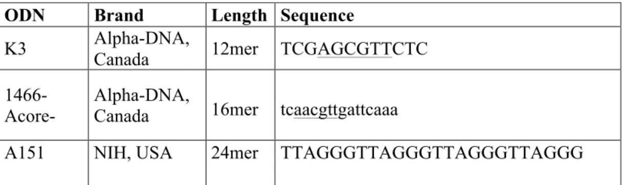
K3-PS and 1466 Acore-PO were purchased from Alpha DNA, Canada. Beside, A151 was supplied from NIH, USA. Their length and sequence are listed as the followings;
Table 2.1: Commercial name of the CpG ODN and their properties
*uppercase letters denotes phosphotothioate linkages (PS) and lowercase letters denotes phosphodiester (PO) linkages between bases. Underlined bases denote CpG motifs.
ODN Brand Length Sequence
K3 Alpha-DNA, Canada 12mer TCGAGCGTTCTC
1466-Acore-
Alpha-DNA,
Canada 16mer tcaacgttgattcaaa
2.1.4 PRR ligands
Ligands used for cell stimulations were listed in Table 2.2.
Table 2.2: Commercial name of the ligands and their sources
Ligand Source Brand
LPS E. coli Sigma, USA
R848 Synthetic Invivogen, USA
3’3’ cGAMP Synthetic Invivogen, USA
8-Fluo-AET-cGAMP(2'-5')
Synthetic Biolog, Germany
2.1.5 Flow cytometry
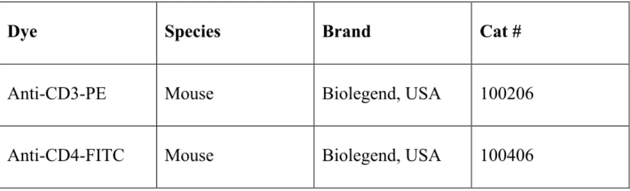
Medium A and Medium B, which were used during cell fixation and permeabilization, respectively were purchased from Invitrogen, USA. In order to perform CD4+ T cell phenotyping, mouse Th1/Th2/Th17 kit were purchased from BD, USA. Antibodies used in cell surface or intracellular staining were listed in Table 2.3.
Table 2.3: Commercial name of the antibodies and their properties
Dye Species Brand Cat #
Anti-CD3-PE Mouse Biolegend, USA 100206
Anti-CD8a-Pe/Cy5 Mouse Biolegend, USA 100710
Anti-IL17-PE Mouse Biolegend, USA 506903
Anti-CD80-FITC Mouse Biolegend, USA 104706
Anti-CD86-Pe/Cy5 Mouse Biolegend, USA 105016
Anti-I-A/I-E (MHC-II)-PE
Mouse Biolegend, USA 107608
2.1.6 Determination of gene expression
Trizol, which was used for the first step of RNA isolation, was purchased from Life Technologies, USA. In order to conversion of RNA into cDNA, ProtoScript M-MulV cDNA Synthesis Kit together with OneTaq Quick-Load 2x Master mix with Standard Buffer were purchased from NEB, USA. Reagents used for the visualization of the PCR products, 6x gel loading dye and DNA ladders were purchased from Fermentas, USA. Quantitative Real Time PCR was performed by using Light Cycler 480 Sybr Green I Master, which was purchased from Roche, USA.
2.1.6.1 Primers
Primers that were used in both PCR and qRT-PCR are listed in Table 2.4. By obtaining sequences of the specific genes from NCBI database, primers were designed with Primer3 Input v.0.4.0 program (http://frodo.wi.mit.edu/primer3/input.htm). In order to
(http://www.ncbi.nlm.nih.gov/BLAST/ or http://genome.ucsc.edu/) against the mouse genome.
Table 2.4: Primers used in PCR
Gene
Name Direction Sequence
Product size (bp) rorc F TGCAAGACTCATCGACAAGGC 86 rorc R AGCTTTTCCACATGTTGGCTG rora F GAACACCTTGCCCAGAACAT 145 rora R AGCTGCCACATCACCTCTCT il-17 F AAGGCAGCAGCGATCATCC 150 il-17 R GGAACGGTTGAGGTAGTCTGAG socs1 F ACCTTCTTGGTGCGCGAC 60 socs1 R AAGCCATCTTCACGCTGAGC sting F TGGCTGCTGATGCCATACTC 107 sting R TCGAGACTCGGGGACATCTT casp-3 F TCTACAGCACCTGGTTACTATTCCTGG 130 casp-3 R TCCTGTTAACGCGAGTGAGAATGTG bax F AACTGGTGCTCAAGGCCCTGTG 226 bax R GCCACAAAGATGGTCACTGTCTGC β-actin F GAAGATCAAGATCATTGCTCCTCCTG 120 β-actin R CTCATCGTACTCCTGCTTGCTGATCC
2HB ELISA plates were obtained from SPL Life Sciences, Korea. ELISA antibodies except IL-4 were purchased from Biolegend, USA. Mouse Anti-IL4 and IL-4 Biotinylated antibody were obtained from Endogen, USA. Beside, SA-ALP was purchased from MabTech, Sweden and PNPP Tablets and 5X Diethanolamine Buffer were purchased from Thermo Scientific, USA. The features of the antibodies including coating, biotinylated and recombinant are listed in detail on Table 2.5.
Table 2.5: Antibodies used in ELISA
Antibody Name Brand Cat # Working concentration
Anti-IFN-γ Ab Mabtech,
Sweden 3321-1a-20 1 µg/ml in PBS IFN-γ Biotinylated Ab Mabtech,
Sweden
3321-1a-20 1:1000 diluted in T cell buffer
Recombinant IFN-γ Biolegend , USA
575309 80 ng/ml in Blocker
Anti-IL-10 Ab Biolegend, USA 504902 4 µg/ml in PBS
IL-10 Biotinylated Ab Biolegend, USA 505004 1:1000 diluted in T cell buffer Recombinant IL-10 Biolegend
, USA 575804 100 ng/ml in Blocker
Anti-IL-4 Ab Endogen
, USA MM450C 4 µg/ml in PBS
IL-4 Biotinylated Ab Endogen , USA 5MM450DB 1:1000 diluted in T cell buffer Recombinant IL-4 Endogen , USA RMIL4I 1ug/ml in Blocker Anti-IL-17a Ab Biolegend, USA 506902 4 ug/ml in PBS IL-17a Biotinylated
Ab Biolegend, USA 507002 1:1000 diluted in T cell buffer Recombinant IL-17A Biolegend
, USA
576009 5 ng/ml in Blocker
Anti-IL-2 Ab Biolegend
, USA 503702 4 µg/ml in PBS IL-2 Biotinylated Ab Biolegend
, USA 503804
1:1000 diluted in T cell buffer
Recombinant IL-2 Biolegend , USA
2.1.8 BCA
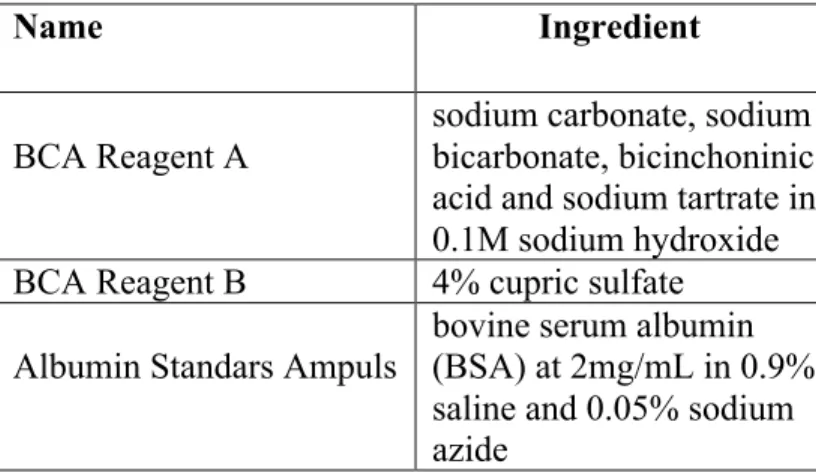
In order to measure the concentration of unknown proteins, BCA was used and purchased from Pierce, USA. The contents of the kit are listed on Table 2.6.
Table 2.6: BCA reagents
Name Ingredient
BCA Reagent A sodium carbonate, sodium bicarbonate, bicinchoninic acid and sodium tartrate in 0.1M sodium hydroxide BCA Reagent B 4% cupric sulfate
Albumin Standars Ampuls bovine serum albumin (BSA) at 2mg/mL in 0.9% saline and 0.05% sodium azide
2.1.9 Western blot
Prestained protein ladder was purchased from Fermentas, USA and PVDF transfer membranes were purchased from Thermo Scientific, USA. As a substrate, SuperSignal West Femto Kit was purchased from Thermo Scientific, USA. The visualization of the membranes was performed by Amersham Imager 600. All Western Blot antibodies were purchased from Cell Signalling, USA and listed on Table 2.7.
Table 2.7: Antibodies used in Western blot
Antibody Name Brand Cat # Working
concentration
β-actin Cell
Signaling, 3700
1:5000 diluted in 5% BSA in BSA
USA p-STAT3 (SER727) Cell Signaling, USA 9134 1:1000 diluted in 5% BSA in BSA p-STAT3(TYR705) Cell Signaling, USA 9145 1:1000 diluted in 5% BSA in BSA CASPASE-3 Cell Signaling, USA 9665 1:1000 diluted in 5% BSA in BSA CLEAVED CASPASE-3 Cell Signaling, USA 9664 1:1000 diluted in 5% BSA in BSA PARP Cell Signaling, USA 9542 1:1000 diluted in 5% BSA in BSA p-IRF3(TYR) Cell Signaling, USA 29047 1:1000 diluted in 5% BSA in BSA IRF3 Cell Signaling, USA 4947 1:1000 diluted in 5% BSA in BSA CASPASE-9 Cell Signaling, USA 9508 1:1000 diluted in 5% BSA in BSA Anti-Rabbit IgG-HRP Linked Cell Signaling, USA 7074 1:10,000 diluted in 5% milk in TBS-T Anti-Mouse IgG-HRP Linked Cell Signaling, USA 7076 1:10,000 diluted in 5% milk in TBS-T
2.1.10 Determination of cell viability
In order to determine the viabilities of cells, Cell Counting Kit-8 was used and purchased from Dojindo Molecular Technologies, Japanese.
2.2 Solutions, Buffers and Culture Media
2.2.1 Cell culture media
High Glucose RPMI-1640 (Lonza)
• 2, 5 or 10% FBS inactivated at 55 °C • 50 g/ml Penicillin/Streptomycin • 10 mM HEPES
• 0,11 mg/ml Na Pyruvate
• 1% Non-Essential Amino Acids Solution • 2 mM L-Glutamine
All ingredients were added into 500 ml medium and stored at +4 °C.
2.2.2 Flow cytometry buffers
PBS-BSA-Na azide Buffer ( FACS Buffer)
• 500 ml 1x PBS • 5g BSA (1%)
The solution was stored at +4 °C.
%0.3 Saponin Buffer
• 0.3 g Saponin
• 100 ml FACS Buffer
2.2.3 Agarose gel electrophoresis
50X TAE (Tris-Acetate-EDTA)
• 242 g Tris (C4H11NO3)
• 37.2 g Tritiplex 3 (EDTA= C10H14N2Na2O2 . 2H2O)
• 57.1 ml Glacial acetic acid
All ingredients were dissolved in 1 lt ddH2O, autoclaved and stored at RT.
1X TAE is prepared as a working concentration.
1%Agarose gel
• 1 g Agarose • 100 ml 1X TAE
The mixture was boiled at microwave and poured into tray after adding 3ul ethidium bromide.
2.2.4 ELISA buffers
Blocking buffer
• 500 ml 1x PBS • 25 g BSA (5%)
• 250 µl Tween20 (0,025%) The mixture was stored at -20°C.
T-cell buffer
• 500 ml 1x PBS • 25 ml FBS (5%)
• 250 µl Tween20 (0,025%) The mixture was stored at -20°C.
Wash buffer
• 500 ml 10x PBS • 2.5 ml Tween20 • 4.5 lt ddH2O
10X PBS (Phosphate Buffered Saline)
• 80 g NaCl • 2 g KCl
• 8,01 g Na2HPO4 . 2H2O
• 2 g KH2PO4
• 1 lt ddH2O
After adjusting pH to 6.8, it was stored at RT.
2.2.5 Western blot buffers
RIPA buffer (Radio Immuno Precipitation Assay Buffer)
• 150 mM Sodium chloride • 1.0% NP-40 or Triton X-100 • 0.5% Sodium deoxycholate • 0.1% SDS
• 50 mM Tris, pH 8.0
4X Laemmli buffer • 4% SDS • 10% 2-mercaptoehtanol • 20% Glycerol • 0.004% Bromophenol blue • 0.125 M Tris-HCl
Buffer pH was adjusted to 6.8 and stored at -20 °C.
TBS 10x
• 24.23 g Trizma HCl • 80.06 g NaCl
Solution was prepared with 1 lt ddH2O. Its pH was adjusted to 7.6 with pure HCl and
stored at RT. It was diluted to 1X before use.
TBS-T
• 100 ml of 10X TBS • 900 ml ddH2O
• 1 ml Tween20
Solution was mixed well and stored at RT.
10X Running buffer
• 25 mM Tris base • 195 mM Glycine • 0.1% SDS
Its pH was adjusted to 8.3 and stored at RT. It was diluted to 1X before use.
Transfer buffer
• 25 mM Tris base • 195 mM Glycine • 20% Methanol
Its pH was adjusted to 8.3 and stored at +4 °C.
Blocking and antibody incubation buffer
• 50 ml TBS-T
• 2.5 g Milk powder or BSA
Solution was mixed well and stored at +4 °C.
12% Resolving gel • 2.5 ml 1.5 M Tris (pH 8.8) • 100 µl 10% SDS • 3.96 ml 30% Acrylamide • 100 µl 10% APS • 10 µl TEMED • 3.44 ml ddH2O
Solution was mixed well by vortexing and used immediately.
8% Resolving gel • 2.5 ml 1.5 M Tris (pH 8.8) • 100 µl 10% SDS • 2.64 ml 30% Acrylamide • 100 µl 10% APS • 10 µl TEMED • 4.76 ml ddH2O
Solution was mixed well by vortexing and used immediately.
5% Stacking gel
• 2 ml 0.5 M Tris (pH 6.8) • 80 µl 10% SDS
• 1.3 ml 30% Acrylamide • 80 µl 10% APS
• 16 µl TEMED • 4.5 ml ddH2O
Solution was mixed well by vortexing and used immediately.
2.2.6 MACS buffer
• 0.5% BSA • 2mM EDTA
Ingredients were dissolved in 1x PBS. Buffer was freshly prepared and stored at +4 °C during isolation process.
2.3 Methods
2.3.1 Maintenance of cell lines
2.3.1.1 RAW264.7
RAW 264.7 (ATCC®, TIB-71) is an Abelson murine leukemia virus transformed mouse macrophage cell line. Cells are sustained in RMI1640 media with 10% regular FBS. For sub-culturing, when they reach around 90% confluence, cells are scraped and centrifuged at 300g for 5 min. After counting by haemocytometer, they are seeded into a new plate according to their appropriate concentration.
Cryotubes that are taken from liquid nitrogen are heated to 37 °C by using water bath. Once they are melted, cells were transferred into 15 ml falcon and additional proper medium is added until the volume reaches 15 ml to get rid of DMSO. Hereafter, cells are centrifuged at 300 g for 5 min and re-suspended with 1 ml appropriate medium. Then, cells are seeded into T-25 flask and their volume is completed to 5 ml and put into cell culture incubator, which is adjust to 37 °C with 5% CO2.
Once cells reach around 80-90% viability, they can be frozen for further experiments. When the confluency of cell is around 80%, cells are scraped and centrifuged at 300 g for 5 min. The cell pellet is re-suspended with cold FBS in such a way that 5x10⁶ cells will be in 500 ul. Then, they are transferred into cryotubes which are already labeled and 500 ul FBS with 20% DMSO is added. Cryotubes with final 10%DMSO concentration are placed into Ms. Frosty Freezing Container and stored at -80 °C. For immediate use, they can be stored at -80 °C. However; for long-term storage, they should be put into liquid nitrogen.
2.3.3 Cell counting
2.3.3.1 Haemocytometer
After cell line or primary cells are taken and centrifuged, pellet is resuspended with 1 ml appropriate medium. 10 ul from suspended pellet is mixed with 490 ul medium for 50X dilution. Afterwards, 10 ul from 50X diluted sample is mixed with 10 ul trypan blue and 10 ul from Trypan Blue-cell mixture is loaded on haemocytometer and cells are counted under light microscope. Counted cell number is multiplied with 106 to calculate actual cell number.
After cell line or primary cells are taken and centrifuged, pellet is resuspended with medium in appropriate volume (generally between 1-10 ml). 20 ul cell pellet is put into cell cuvette containing 10 ml Isoton II Diluent buffer. If necessary, 3 drops of ZAP-OGLOBIN II Lytic Reagent is added. 30 ul from the mixture is analyzed by Accuri C6 Flow Cytometry. After arranging threshold to get rid of dead cells, the numbers of gated cells are multiplied with appropriate dilution rate to obtain actual number of cells.
2.3.4 Primary murine cell suspension preparation
2.3.4.1 Mouse spleen cell suspension preparation
Spleens from C57/BL6 mice are removed by cervical dislocation. Removed spleen is put into ice in 5 ml %2 FBS supplemented complete RPMI-1640 media. Afterwards, spleen is squeezed with the backside of the 5 ml syringe plunger and squeezed cells are transferred into 15 ml falcon completed with 10 ml fresh 2% RPMI-1640 media and centrifuge at 300 g for 5 min. After centrifugation, pellet is resuspended with 1 ml ACK Lysis Buffer and incubated for 2 min at RT to remove red blood cells. Then, fresh 2% RPMI media is added and centrifuged at 300 g for 5 min. Finally, cells are resuspended in 10% RPMI media and counted in flow cytometer.
2.3.4.2 Isolation of bone marrow cells
After cervical dislocation of C57/BL6 mice, femur and tibia bones were removed. By using 26G insulin syringe, bone marrow cells were washed out from bones and washed with 2% FBS supplemented RPMI media for 3 times. Collected cells were centrifuged and suspended with 1 ml ACK lysis buffer to blow up red blood cells and incubated for 2
min. Centrifugation step was repeated and cells were resuspended with %10 FBS supplemented RPMI-1640 media to count and use further applications.
2.3.5 Bone marrow derived dendritic cell generation
After arranging concentration of bone marrow cells to 1x10⁶ cells/ml, they were seeded into 10cm2 plate in RPMI-1640 supplemented with 10% FBS including 20ng/ml mGM-CSF and 10ng/ml mIL-4 recombinant proteins. The half of the medium was aspirated and replenished with fresh 10% FBS supplemented RPMI-1640 media after three days. Six days after initial seeding, cells were collected with 1X cold PBS and centrifuged and counted by flow cytometry. For their quality control, CD11b/CD11c staining was performed before their usage.
2.3.6 Pan T and CD4+ T cell isolation from mouse spleen
After counting spleen cells with flow cytometry, centrifuged spleen cells were re-suspended in MACS buffer as 40 ul per 107 cells. Then, 10 ul Pan T or CD4+ T cell Biotin-Antibody cocktail was added per 107 cells and incubated for 10 min at 4 °C. 30 ul MACS buffer and 20 ul Pan T or CD4+ T cell Microbead Coctail were included and cells were incubated at 4 °C for an additional 15 min. In the mean time, MS column was washed with 500 ul MACS buffer. After incubation, the volume of the cells was completed to 10 ml and cells were centrifuged at 300 g for 5 min. Cell pellet was re-suspended with 500 ul MACS buffer and loaded into the MS column attached to a magnet. Flow-through was collected in a 15 ml falcon and columns were washed two times with 500 ul MACS Buffer to increase the efficiency of the isolation. Collected cells were additionally washed with 10 ml MACS buffer and centrifuged at 300 g for 10 min. Pellet was re-suspended in 1ml of RPMI-1640 supplemented with 50µM
![Figure 1.2: Cytosolic DNA–sensing system [16]](https://thumb-eu.123doks.com/thumbv2/9libnet/5845583.119851/24.918.168.827.439.949/figure-cytosolic-dna-sensing-system.webp)
![Figure 1.3: T cell development in thymus [27]](https://thumb-eu.123doks.com/thumbv2/9libnet/5845583.119851/27.918.173.813.123.677/figure-t-cell-development-thymus.webp)
![Figure 1.4: Roles of the B7–CD28/CTLA-4 pathway in regulating T-cell activation [29]](https://thumb-eu.123doks.com/thumbv2/9libnet/5845583.119851/29.918.243.645.109.502/figure-roles-cd-ctla-pathway-regulating-cell-activation.webp)
![Figure 1.5: Summary of the CD4 + T helper cell fates [48]](https://thumb-eu.123doks.com/thumbv2/9libnet/5845583.119851/31.918.161.807.569.1020/figure-summary-cd-t-helper-cell-fates.webp)
![Figure 1.6: IL-17 receptor signal transduction [87]](https://thumb-eu.123doks.com/thumbv2/9libnet/5845583.119851/35.918.167.811.106.676/figure-il-receptor-signal-transduction.webp)