137
Femoral Chondroblastic Osteosarcoma in a Kitten: A case report
Sadık YAYLA1,a,, Emin KARAKURT2,b, Uğur AYDIN3,c, Hilmi NUHOĞLU2,d, Uğur YILDIZ3,e, Serpil DAĞ2,f
1 Department of Surgery, Faculty of Veterinary Medicine, Dicle University, Diyarbakır, Turkey. 2 Department of Pathology, Faculty of Veterinary Medicine, Kafkas University, Kars, Turkey.
3Department of Surgery, Faculty of Veterinary Medicine, Kafkas University, Kars, Turkey aORCID: 0000-0001-6734-421X; bORCID: 0000-0003-2019-3690; cORCID: 0000-0001-5756-4841 dORCID: 0000-0003-2530-2542; eORCID: 0000-0002-4782-1012; fORCID: 0000-0001-7667-689X
Geliş Tarihi/Received Kabul Tarihi/Accepted Yayın Tarihi/Published
10.09.2019 13.10.2019 31.12.2019
INTRODUCTION
Bone tumors are not common in cats. They mostly occur at ages of 7-8 years and were reported to occur at a rate of 3.4% in a study scanning 100000 cases (1). Osteosarcoma (OS) is known to be the most common bone tumor with a incidence rate of 70-80% in cats. OS is usually defined as tumors consisted of malignant mesenchymal spindle cells producing bone or osteoid extracellular matrix (1).
Feline OSs can be seen throughout the skeletal sys-tem, and can also be seen in mammary gland, eye, and subcutaneous extraskeletal OS at a rate of 40% incidence (2).
Feline OSs compared to canine counterparts differ in behaviour. Metastasis in dogs was 81-90%, whereas in cats this was reported to be much lower (5-10%) (1-4). Therefo-re, treatment options in cats are controversial due to low metastasis rate. Generally wide surgical excision or
ampu-tation is recommended. In addition, radiotherapy and/or chemotherapy may prolong lifespan (1,5,6).
In this case report, we aimed to present a case of fe-moral chondroblastic osteosarcoma in a kitten with histo-pathological features and clinical results.
CASE HISTORY
A 4 month-old male kitten was brought to Veterinary Fa-culty Animal Hospital Surgery Clinics of the Kafkas Univer-sity with a complaint of severe tenderness and lameness in his left hind leg. In clinical examination, a palpable swelling and hypersensitivity were detected in the femoral region. Radiological evaluation revealed fracture of the left collum femoris, left sacroiliac separation and pubic fracture. In addition, proliferations were found to be remarkable in both collum and distal diaphyseal region of femur in radiog-ram (Figure 1A).
Dicle Üniversitesi Veteriner Fakültesi Dergisi
http://www.dicle.edu.tr/veteriner-fakultesi-dergisi
Vaka/Case Report
ISSN:1307-9972 e-ISSN:1308-0679
Abstract
Feline bone tumors are not as common as in dogs, and osteosarcomas are more common among bone tumors. Osteosarcomas in cats are not metastatic as in dogs. In this report, we aimed to present a case of femoral chondroblastic osteosarcoma with histopathological and clinical results in a kitten. Based on clinical and radiological examination, in a 4 month-old male kitten it was detected fracture of the left collum femoris, left sacroiliac separation and pubic fracture. Excision arthroplasty was performed and the caput and collum femoris were evaluated histopathologically. Following the identification of the sample as chondroblastic osteosarcoma, the related limb was amputated. As a result, a case of chondroblastic osteosarcoma in a kitten was found worthy to be survived.
Key Words: Femoral chondroblastic osteosarcoma, kitten, leg amputation
Yavru Bir Kedide Femoral Kondroblastik Osteosarkom: Olgu sunumu Öz
Kedilerde kemik tümörleri köpeklerdeki kadar yaygın olmamasına rağmen ostesarkom kemik tümörleri içerisinde daha sık görülmektedir. Ancak bu osteasarkomlar kedilerde köpeklerdeki kadar metastazik değildir. Bu raporda, yavru bir kedide karşılaşılan femoral kondroblastik osteosarkom olgusunun histopatolojik ve klinik sonuçlarıyla birlikte sunulması amaçlandı. 4 aylık erkek yavru bir kedide klinik ve radyolojik muayeneye ile sol kollum femoriste kırık, sol sakroiliak ayrılma ve pubis kırığı belirlendi. Eksizyon artroplasti ile alınan kaput ve kollum femoris histopatolojik incelemeye tabi tutuldu ve kondroblastik osteosarkom olarak tanımlanmasını takiben ilgili ekstremite ampute edildi. Sonuç olarak yavru bir kedide kondroblastik osteosarkom olgusu kedinin sağ kalmış olması bakımından sunulmaya değer bulundu.
Yayla ve ark., Dicle Üniv Vet Fak Derg 2019;12(2):137-139 Femoral Chondroblastic Osteosarcoma in a Kitten: A case report
138
Figure 1. Radiograms of the case: A. Preoperative radiogram;
prolifer-ative osteolytic areas in the distal diaphysis and collumfemoris of the left femur with collum fracture, B. Excision arthroplasty was per-formed,C. The left hind limb was amputated
Excision arthroplasty was performed for the fracture of the collum femoris (Figure 1B) and the caput femoris. The resected collum femoris were histopathologically eva-luated. Cage rest was recommended without any interven-tion for sacroiliac separainterven-tion and pubis fracture. Postopera-tive analgesics (subcutaneous ketoprofen 2 mg / kg, Keto-pet®, Teknovet, Istanbul, Turkey) and antibiotherapy (int-ramuscular cefazolin 20 mg / kg, Cefozin®, Bilimİlaç, Istan-bul, Turkey) were performed.
HISTOPATHOLOGICAL EVALUATION
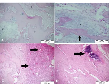
Biopsy specimen resected from collum femoris was decal-cified and routinely processed for hematoxylin and eosin staining and observed under a light microscope for evalua-tion of histopathological changes. In histopathological examination of the mass, large trabecular structures and malignant cartilage tissue formation were found in these structures. In addition to these, swirl-like structures formed by fibrosit and fibroblasts were detected (Figure 2A-B). Polygonal shaped pleomorphic tumor cells with anisocyto-sis and anisonucleoanisocyto-sis were observed. Mitotic figures and giant cells were not found in the tumoral area. In the area surrounding the mass, haemorrhage and severe cellular infiltration were observed (Figure 2C). Localized calcificati-ons were among other prominent histopathological fin-dings (Figure 2D). In the light of these finfin-dings, the mass taken from the collum femur was named as chondroblastic osteosarcoma due to the presence of irregular bone trabe-culaes in the middle of irregular atypical chondroblastic cell proliferation.
The left hind leg of the kitten was amputated 10 days after excision arthroplasty operation (Figure 1, C), upon diagnosis of chondroblastic osteosarcoma based on histo-pathological findings. Postoperative analgesic and antibiot-herapy were repeated.
Figure 2. Cat, femur, chondroblastic osteosarcoma: A. Trabecular
structures (T), chondroid tissue (K), swirl-like structures composed of fibrosit and fibroblasts (F), H&E, 200 μm. B. Trabecular structure of amorphous matrix and osteoblasts (T), malignant cartilage tissue around the trabeculaes(*), H&E, 50 μm. C. Outer surface of the mass, Hemorrhages and inflammatory cell infiltration (arrows), H&E, 200 μm. D. Calcification (arrow),H&E, 200 μm.
It was observed that the cat's condition improved in two months postoperatively and the kitten continued his life without any problem (Figure 3). In addition, there was no clinical and radiographic finding of metestasis, including the lungs.
Figure 3. Postoperative 2nd month view
DISCUSSION AND CONCLUSION
Osteosarcomas in cats have been reported to have less metastatic character than those of seen in dogs (1,7). On the other hand, cats with osteosarcoma are usually 10-12 years of age and many of them die or they are given eutha-nasia decision at the request of the owner (6). In this re-port, chondroblastic osteosarcoma was diagnosed and found to be interesting in terms of survival of the kitten that was very young being at 4 months of age.
In terms of histopathological evaluation, same types of tumors between cats and dogs may differ in their prog-nosis (1). Osteosarcomas may be classified as morphologi-cal subtypes such as osteoblastic, fibroblastic, chondroblas-tic, teleangiectachondroblas-tic, giant cell and mixed types depending on the appearance of their matrix. There is a significant relati-onship between the subtypes of tumors and survival rate.
A B C
A B
Yayla ve ark., Dicle Üniv Vet Fak Derg 2019;12(2):137-139 Femoral Chondroblastic Osteosarcoma in a Kitten: A case report
139 However, no statistical data for both humans and cats or
dogs has been reported for osteosarcomas. In addition, there is limited data on histological classification or classifi-cation system for osteosarcomas in cats unlike in dogs (1). Also, there is a significant relationship between the mitotic index and survival (8-11), however, we did not find any mitotic figures in the tumoral mass in our study. In addition to mitotic figures, the presence of multicore bizarre giant cells was not found as described in previous studies (8,9,12). Similar to the literature data (13,14), the trabecu-lar structures in the middle of irregutrabecu-lar atypical chondrob-lastic cell proliferation and the presence of inflammatory infiltration were identified and therefore the mass from the collum femoris was classified as chondroblastic osteosar-coma.
Among the localization of feline osteosarcomas, many bones such as maxilla, mandible, skull, scapula, rib, verteb-rae, pelvis, nasal cavity and tail vertebrae have been repor-ted. In addition, proximal femur, proximal tibia, radius, humerus and metatarsal/carpal bones were reported to be affected (1). In this case, the left femur was affected along the long axis, and there was a fracture at the collum level.
A wide excision or amputation of the affected area is recommended by different authors (1,2,15) for feline oste-osarcomas. In this case, in which excision arthroplasty was performed, the left hind limb was amputated upon the diagnosis of osteosarcoma. Kirpensteijn et al. (2002) repor-ted that chonrdoblastic osteosarcomas did not show me-tastasis in dogs. We haven't found enough data in cats. In our case, no clinical and radiological findings related to metastasis were found. During the postoperative 2 months follow-up period, the physical development of the cat con-tinued smoothly.
In conclusion, femoral chondroblastic osteosarcoma encountered in a very young cat is remarkable as he conti-nues to live after the related leg is amputated. Therefore, we believe that this case report will contribute to the prac-tice and literature.
REFERENCES
1. Dimopoulou M, Kirpensteijn J, Moens H, Kik M. (2008).Histologic Prognosticators in Feline Osteosarcoma: A Comparison with Phenotypically Similar Canine Osteosarcoma. Vet Surg. 37: 466-471.
2. Heldmann E, Anderson MA, Wagner-Mann C. (2000). Feline Osteosarcoma: 145 Cases (1990–1995). J Am Anim Hosp As-soc. 36:518-521.
3. Brodey RS, Riser WH. (1969). Canine osteosarcoma. A clinico-pathologic study of 194 Cases. Clin Orthop. 62:54-64. 4. Kirpensteijn J, Kik M, Rutteman GR, Teske E. (2002).The
Prog-nostic Significance of a New Histologic Grading System for Ca-nine Osteosarcoma. Vet Pathol. 39: 240-246.
5. Madewell BR, Leighton RL, Theilen GH, (1978). Amputation and Doxorubicin For Treatment of Canine and Feline Osteoge-nic Sarcoma. Eur J Cancer. 14:287-293.
6. Firat I, Bozkurt ER, Haktanir D, Ozer K, (2011). Parosteal Oste-oclastic Osteosarcoma in The Left Tarsal Joint of a Cat. Kafkas Univ Vet Fak Derg. 17 (4): 679-682.
7. Gebhard C, Fuchs-Baumgartinger A, Razzazi-Fazeli E, Miller I, Walter I. (2016). Distribution and Activity Levels of Matrix Me-talloproteinase 2 and 9 in Canine and Feline Osteosarcoma. Can J Vet Res. 80: 66-73.
8. Farjanikish G, Dezfoulian O, Mohammadi H. (2018). Metastatic Giant Cell Osteosarcoma in a Cat. Veterinary Research Forum. 9 (3): 289-292.
9. Almela R, Bomhard W, Anson A, Mayer U, (2017). Subcutane-ous Extraskeletal Osteosarcomain a Metatarsal Footpadin a Cat. Vet Dermatol. 28: 524–e129.
10. Baum JI, Skinner OT, Boston SE.(2018).Fracture-Associated Osteosarcoma of the Femur in a Cat. Can Vet J. 59: 1096-1098. 11. Breitreiter K. (2019).Late-Onset Osteosarcoma After Onychec-tomy in a Cat. JFMS Open Reports,1-6.DOI: 10.1177/2055116919842394.
12. Nakata K, Miura H, Sakai H, et al. (2017).Vertebral Replace-ment for the TreatReplace-ment of Vertebral Osteosarcoma in a Cat. JVetMedSci.79 (6): 999-1002.
13. Al Attar ASR, Kubba MA, Seham AAA, Adwak AA. (2016). Chondroblastic Osteosarcomain a Cat: Case Report. J Med Surg Pathol. 1(4): 1-3.
14. Simerdova V, Vavra M, Skoric M, Hajek I, Skor O. (2017).What is Your Diagnosis? Multilobate Nasal Massin a 5-Month-Old Sphynx Cat. Vet Clin Pathol. 46(2), 369-370.
15. Florian RLM, Walter I. (2016).Establishment and Characteriza-tion of New Canine and Feline Osteosarcoma Primary Cell Li-nes. Vet Sci. 3(9) 1-15.
Corresponding author:
Sadık YAYLA
University of Dicle, Faculty of Veterinary Medicine, Department of Surgery Diyarbakır, Turkey