Eurasian J Pulmonol 2015; 17: 119-21
Abstract
Tracheal laceration as a result of elective intubation is a rarely seen complication with a high mortality. An eighty-one years old, female patient who was followed in the intensive care unit and treated with mechanical ventilation, developed subcutaneous emphysema after intubation. Fiberoptic bronchoscopy performed via the endotracheal tube revealed laceration in the trachea 5 cm in length.. The urgent rigid bronchoscopy has been performed and one silicone straight stent and a covered metallic bifurcated stent have been inserted. The araea of laceration has been covered totally. The subcutaneous emphysema disappeared incrementally and after weaning period the patient discharged to the ward.
Keywords: Bronchoscopy, complication, intensive care, tracheal lace - ration
INTRODUCTION
Tracheal laceration can be caused by trauma or iatrogenic reasons. Subcutaneous emphysema is ob-served in almost all cases and the final diagnosis is established by identifying a laceration or rupture via bronchoscopic examination. The main treatment option in tracheal laceration is surgical, but con-servative approaches can be applied in selected cases. Considering the age, additional diseases, and the urgency of the present situation, our case was deemed unsuitable for surgery and tracheal stent was applied. Verbal and written consents were taken from the patient and relatives.
CASE PRESENTATION
An 81-year-old female patient was admitted to our emergency polyclinic with respiratory distress. Upon discovering respiratory acidosis in her arterial blood gas, she was intubated and admitted to the respiratory intensive care unit after losing consciousness following an unsuccessful noninvasive mechanical ventilation support. Her history included valvular heart disease, atrial fibrillation, hyper-tension, and chronic obstructive pulmonary disease. In her physical examination during admittance to the intensive care unit, her general condition was bad, she was unconscious, both hemithoraces participated in respiration equally, expiration in bilateral lungs was long, and breath sounds were diminished. Bilateral pretibial edema was present. Subcutaneous emphysema, more prominent in the right hemithorax, was present, extending to the neck. Arterial tension was 145/100 mmHg, car-diac apex beat was 137 per minute, and temperature was 36.8 degrees. In the arterial blood gas, the pH was 7.19, pCO2 was 81 mmHg, pO2 was 65 mmHg, oxygen saturation was 91%, and HCO3 was 32 mmoL. The patient was taken into mechanical ventilation support with the following values in pres-sure control mode: PEEP: 6 mbar, over PEEP prespres-sure: 26 mbar, respiratory rate: 15, and FiO2: 60%. In the laboratory tests, platelet was 24,000/mm3, preprandial blood glucose was 192 mg/dL, urea was
124 mg/L, creatinine was 1.7 mg/dL, and other parameters were within normal ranges. These find-ings led to the suspicion of pneumonia and sepsis in the patient. Considering that elective intuba-tion in the emergency room could lead to tracheal injury, first a chest radiography was performed. On the chest radiography that was taken lying down, severe increase in the cardiothoracic ratio in favor of the heart, increase in opacity in the bottom right area compatible with possible pneumonic
Received Date: 23.09.2013 Accepted Date: 07.12.2013 Available Online Date: 01.04.2015 Address for correspondence Levent Dalar
E-mail: leventdalar@yahoo.com
This work is licensed under a Creative Commons Attribution-NonCommercial 4.0 International License.
DOI: 10.5152/ejp.2014.48992
• Available online at www.eurasianjpulmonol.com
Case Report
Tracheal Laceration Due to Elective Intubation
and Its Treatment with Endotracheal Stenting
Aybüke Kekeçoğlu
1, Cengiz Özdemir
1, Levent Karasulu
1, Ayşe Filiz Koşar
1,
Levent Dalar
21Clinic of Chest Diseases, Yedikule Chest Diseases and Thoracic Surgery Training and Research Hospital, İstanbul 2Department of Chest Diseases, İstanbul Bilim University Faculty of Medicine, İstanbul
consolidation, and bilateral, but more prominent in the right that is observed to extend up to the neck, subcutaneous emphysema and pneumomediastinum were observed (Figure 1). After partially stabilizing the patient’s general condition, fiber optic bronchos-copy was performed in order to determine the cause of the sub-cutaneous emphysema. Prior to bronchoscopy, the platelet level was elevated to 102.000 mm3 via 8 units of platelet replacement.
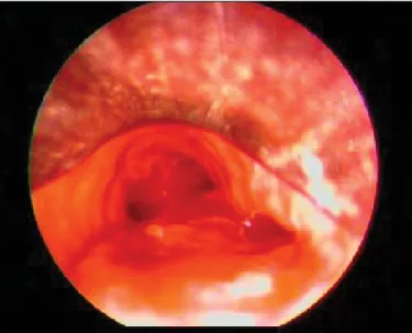
Bronchoscopy revealed normal localization of endotracheal tube, normal carina, open bilateral main bronchus, and hemorrhagic left bronchus system. When the endotracheal tube was retracted and the trachea was examined, a tear area of approximately 4-5 cm was discovered in the tracheal right posterolateral wall. The patient was scheduled for emergency rigid bronchoscopy. In the rigid broncho-scopic examination, laceration area and mediastinal fistula were observed along the line on the tracheal wall starting from a dis-tance of 5 cm from the vocal chords and continuing along the right posterolateral edge for about 5 cm (Figure 2). From this area, it was
observed that a hemorrhage in the form of a leakage had occurred and that this laceration area ended 2 cm away from the carina. In or-der to cover tracheal laceration and the fistula area, an 18 x 50 mm silicon stent was placed in the lumen. The mucosa of the laceration area was fragile and atrophic, and because it was thought that the laceration will progress from the bottom end of the stent to the dis-tal trachea, 16 x 12 x 12 mm medis-tallic Y stent was placed to the right and left primary bronchia by opening the silicon inside the stent (Figures 3, 4). Stent patency was established. The process was final-ized by placing the endotracheal tube immediately in alignment with the edge of the silicon stent following the bronchial washing. The patient was successfully extubated on the 8th day of intubation.
The weaning process was completed by intermittent noninvasive mechanical ventilation support. The patient’s general condition im-proved and mediastinal and subcutaneous emphysema completely disappeared. She was discharged into the service from the respira-tory intensive care unit (Figure 5).
Kekeçoğlu et al. Tracheal Laceration and Treatment Eurasian J Pulmonol 2015; 17: 119-21
120
Figure 1. Chest x-ray at the admission of the patient to the
in-tensive care unit
Figure 4. Bronchoscopic appearance of tracheal distal, carina,
and main bronchus entries after stent placement
Figure 2. Rigid bronchoscopy revealed a rupture area in the
right posterolateral wall
DISCUSSION
Tracheal laceration can be caused by blunt trauma, penetrating inju-ries, firearm injuinju-ries, and iatrogenic reasons.
The most commonly observed iatrogenic reason is intubation. It can develop during single or double lumen intubation. Its incidence after orotracheal intubation is 0.005% and this ratio changes to between 0.05% and 1.9% for double lumen intubation (1, 2). It has the follow-ing among its etiology: lack of experience of the personnel that intu-bate and recurring intubation attempts, over inflammation of the en-dotracheal tube cuff, changing the tube’s place when the cuff balloon is inflated, malposition of the guide wire, failure to choose an appro-priately sized endotracheal tube, and trachea anomalies (3). Although penetrating injuries are commonly observed in the cervical region, laceration and rupture more often occur in the distal trachea and pri-mary bronchia following intubation. Rupture is generally in the joint of the membranous and cartilage parts and is linear. In women and especially, in short cases, the risk is shown to be significantly higher (1, 2, 4, 5). The most commonly observed physical examination findings in tracheal rupture cases are respiratory insufficiency, subcutaneous emphysema, and hemoptysis. The first diagnostic step is radiography of the pulmonary and cervical region. The pneumothorax and pneu-momediastinum has been observed can be observed in chest X-ray and in tomography. Subcutaneous emphysema is observed in almost all tracheal laceration cases. However, in 10% of the acute period cas-es, no radiological findings are observed. Computed tomograhy can reveal the laceration in the patients with appropriate clinics. Final diagnosis of tracheal rupture is established by bronchoscopy. Rigid or flexible bronchoscopy may be used. Bronchoscopy must be per-formed as soon as possible and it must include the area under the in-tubation tube of the intubated patient. In previous studies, it is noted that 50% of the lesions can be overlooked in the first bronchoscopic examination. Therefore, if suspicion persists, the process can be re-peated, even if the first bronchoscopic findings are normal (2, 3-6). Surgical approach is the initial treatment choice in most of the cases and is recommended in order to prevent the development of medi-astinitis in the early period and of tracheal stenosis in the late period.
Although, in general, right thoracotomy is used, a transcervical ap-proach is recommended in proximal ruptures (6-8).
Although the main treatment choice is surgery, it is reported in the litera-ture that tracheal lacerations due to intubation can be treated by conser-vative methods. Conserconser-vative treatment can be applied to patients that the laceration is shorter than 2 cm and patients who are uncomplicated and stable. In the conservative treatment, the tip of the intubation tube is placed under the rupture, and it is followed up with appropriate anti-biotic treatment. Because the conservative treatment approaches have improved and their usability had increased in recent years, tracheal stent applications are reported among the conservative treatment approach-es (10, 11). In our case, considering the patient’s age, additional diseasapproach-es, her general condition not being appropriate for surgery, and the urgen-cy of the present clinic, conservative treatment is considered as a priority (3, 5, 9-11). Main steps of the treatment process are the complete cov-ering of the laceration area, prevention of the growth with a stent, and keeping the endotracheal tube in distal. The case is currently followed up in the 5th month of her treatment. Although the optimal duration for
the stent to remain in place is unknown, 6-12 months can be argued as sufficient given the experience of the center.
CONCLUSION
In conclusion, endotracheal stenting in iatrogenic trachea lacerations should be considered as an effective treatment option.
Informed Consent: Informed consent was obtained from patient who parti-cipated in this case.
Peer-review: Externally peer-reviewed.
Author contributions: Concept - A.K., C.Ö.; Design - A.K., L.D.; Supervision - L.D., F.K.; Resource - L.D., A.K.; Materials - C.Ö., L.K.; Data Collection &/or Pro-cessing - A.K., C.Ö.; Analysis &/or Interpretation - L.D.; Literature Search - A.K.; Writing - A.K., C.Ö., L.D.; Critical Reviews - L.D., F.K.
Conflict of Interest: No conflict of interest was declared by the authors. Financial Disclosure: The authors declared that this study has received no financial support.
REFERENCES
1. Borasio P, Ardissone F, Chiampo G. Post-intubation tracheal rupture. A report on ten cases. Eur J Cardiothorac Surg 1997; 12: 98-100. [CrossRef]
2. Massard G, Rougé C, Dabbagh A, Kessler R, Hentz JG, Roeslin N, et al. Tracheobronchial lacerations after intubation end tracheostomy. Ann Thorac Surg 1996; 61: 1483-7. [CrossRef]
3. Ozgulder A. Trakeobronşiyal Yaralanmalar. TTD Toraks Cerrahisi Bülteni 2010; 1: 1.
4. Conti M, Pougeoise M, Wurtz A, Porte H, Fourrier F, Ramon P, et al. Management of postintubation tracheobronchial ruptures. Chest 2006; 130: 412-8. [CrossRef]
5. Karadayı Ş, Yazıcı Ü, Gülhan E, Taştepe İ, Altınok T, Ege T. Trakeobronşiyal Ya-ralanmalarda Tedavi Yaklaşımlarımız. Solunum Hastalıkları 2007; 18: 114-7. 6. Hahn B. Tracheobronchial rupture. J Emerg Med 2007; 33: 193-4. [CrossRef]
7. Grillo HC. Tracheal and bronchial trauma. In Grillo HC ed.Surgery of the trachea and bronchi. London B.C. Hamilton Inc, 2004: 271-90.
8. Kiser AC, O’Brien SM, Detterbeck FC. Blunt tracheobronchial injuries: tre-atment and outcomes. Ann Thorac Surg 2001; 71: 2059-65. [CrossRef]
9. Karmy-Jones R, Avansino J, Stern EJ. CT of blunt tracheal rupture. AJR Am J Roentgenol 2003; 180: 1670. [CrossRef]
10. Osorio García F, Serrano Alvarez C, Martinez Sánchez A, Gallardo Medina M. Post-tracheal intubation rupture treated with a Dumon-Y stent. Rev Esp Anestesiol Reanim 2012; 59: 171-2.
11. Yopp AC, Eckstein JG, Savel RH, Abrol S. Tracheal stenting of iatrogenic trache-al injury: a novel management approach. Ann Thorac Surg 2007; 83: 1897-9.
[CrossRef]
121
Eurasian J Pulmonol 2015; 17: 119-21 Kekeçoğlu et al. Tracheal Laceration and Treatment
Figure 5. X-ray radiography at discharge from respiratory
inten-sive care unit. Complete disappearance of subcutaneous emphy-sema is seen