OTJHS
Online Turkish Journal of Health Sciences Online Türk Sağlık Bilimleri Dergisi
OTSBD
Kalp Tipi Yağ Asid Bağlayıcı Proteinin Akut Koroner Sendrom Risk Skorları ile İlişkisi
The Relationship between Cardiac Fatty Acid Binding Protein and Acute Coronary Syndrome
Risk Scores
1Uğur KÜÇÜK, 2Hakan TÜRKÖN, 3Burak ALTUN 1Sivas Numune Hospital, Department of Cardiology Specialist, Sivas, Turkey 2Çanakkale Onsekiz Mart University, Department of Medical Biochemistry, Çanakkale, Turkey 3 Istinye University Hospital VM Medical Park Florya Hospital, Department of Cardiology, Istanbul, Turkey
Uğur Küçük: https://orcid.org/0000-0003-4669-7387 Hakan Türkön: https://orcid.org/0000-0003-1888-9322
Burak Altun: https://orcid.org/0000-0002-5151-0399
Online Turkish Journal of Health Sciences 2020;5(1):165-175 Online Türk Sağlık Bilimleri Dergisi 2020;5(1):165-175
ÖZ
Amaç: Kalp tipi yağ asid bağlayıcı protein (H-FABP)
dü-şük moleküler ağırlıklı majör sitoplazmik bir proteindir ve miyokard yaralanmasına cevap olarak hızlı bir şekilde kardiyomiyositlerden salınır. H-FABP'ın akut koroner sendrom (ACS) tanısında kullanılabilirliğini ve koroner arter hastalığının (KAH) prevalansı, şiddeti ve erken teşhisi ile olan ilişkisini araştırdık.
Materyal ve Metot: Bu prospektif gözlemsel çalışmamıza,
kliniğimize Şubat 2016 ile Ocak 2017 tarihleri arasında başvuran 18 yaş ve üstü AKS tanılı 110 hasta dahil et-tik.Hastaları iki gruba ayırdık: ST yükselmeli olmayan AKS (NSTE-AKS) ve ST yükselmeli AKS (STEMI).H-FABP ve kardiyak troponin I (cTnI) için hastalardan ilk hastaneye başvuru anında ve 6 saat sonra tekrar kan örnek-leri alındı. Tüm hastalara koroner anjiografi yapıldı.
Bulgular: Başvuru anında, her iki grupta GRACE (Global
Registry of Acute Coronary Events), SYNTAX (The syn-ergy between percutaneous coronary intervention with taxus and cardiac surgery) ve Gensini risk skorları arasında istatistiksel olarak anlamlı bir ilişki bulunmadı (p=0,056, p=0,791, p=0,278). Altıncı saatte bakılan H-FABP düzeyi ile GRACE ve Gensini risk skorları H-FABP pozitif grupta istatistiksel olarak anlamlıydı (p=0,003, p=0,011). Ancak, SYNTAX risk skoru H-FABP pozitif grupta istatistiksel olarak anlamlı değildi (p=0,984).
Sonuç: Çalışmamızda H-FABP ile koroner arter hastalığı
risk skorları arasındaki ilişkiyi ve H-FABP'ın AKS tanılı hastalarda tanısında kullanılabileceğini gösterdik.
Anahtar Sözcükler: Akut koroner sendrom, inflamasyon,
ateroskleroz, kalp tipi yağ asid bağlayıcı protein, kardiyak biyobelirteç
ABSTRACT
Objective: Heart type fatty acid binding protein (H-FABP)
is a low moleculer weight major cytoplasmic protein and released quickly from cardiomyocytes in response to myo-cardial injury. We searched functionality of H-FABP in diagnosis of acute coronary syndrome (ACS) and preva-lence of coronary artery disease, relationship with its sever-ity and early diagnosis.
Materials and Methods: This was a prospective
observa-tional study. We took in 110 patients- aged 18 and above-diagnosed ACS between the dates February 2016 and Janu-ary 2017. The patient population was divided into two groups as ST segment elevation ACS (STEMI, 52 patients) and Non-ST segment elevation ACS (NSTE-ACS, 58 pa-tients). For H-FABP and cardiac troponin I (cTnI) blood samples of the patients were taken both when they first applied to the hospital and six hours later. Coronary an-giography was performed to all the patients.
Results: On admission, it wasn’t found a statistically
sig-nificant relationship in both groups, GRACE (Global Reg-istry of Acute Coronary Events), SYNTAX (The synergy between percutaneous coronary intervention with taxus and cardiac surgery) and between Gensini Risk skores (p=0.056, p=0.791, p=0.278). The H-FABP level of blood samples looked over six hours later with GRACE and Gen-sini risk scores were statistically significant in the H-FABP positive group (p=0.003, p=0.011). However SYNTAX risk score in H-FABP positive group was not statistically significant (p=0.984).
Conclusion: In our study, we demonstrated the relationship
between H-FABP and coronary artery disease risk scores and that H-FABP can be used in the diagnosis of ACS.
Keywords: Acute coronary syndrome, cardiac biomarker,
heart-type fatty acid binding protein
Sorumlu Yazar / Corresponding Author: Uğur KÜÇÜK
Addresses: Sivas Numune Hospital, Department of Cardiology Specialist, Sivas, Turkey
Tel:05345911902
e-mail: [email protected]
Yayın Bilgisi / Article Info: Gönderi Tarihi/ Received:16/05/ 2019 Kabul Tarihi/ Accepted: 29/08/2019 Online Yayın Tarihi/ Published: 31/03/2020
Atıf/ Cited: Küçük U, et al. The Relationship between Cardiac Fatty Acid Binding Protein and Acute Coronary Syndrome Risk Scores. Online Türk Sağlık Bilimleri Dergisi 2020;5(1)165-175. doi: 10.26453/otjhs.566720
INTRODUCTION
Coronary artery disease (CAD) is one of the most important causes of mortality and morbidity in the world. As with all fields, with the developing tech-nology in the field of cardiology, diagnosis and treatment modalities allow patients with Acute Coronary Syndrome (ACS) early diagnosis of dis-eases with increased survival and modification of risk factors.1
Cardiac troponins (cTn) are preferred biomarkers for the diagnosis of acute myocardial injury. Troponin is found in both skeletal muscle and cardiac muscle, but the specific versions of troponin differ between types of muscle. They are encoded by different genes and used in the diagnosis. Troponins are not used for only diagnosis; but also on risk assessment and prognosis, cTnI is the most specific.2,3 They are more sensitive than creatin kinase isoenzyme (CKMB) in cardiac damage. Fatty acid binding pro-teins (FABP) were first discovered by Ockner in 1972.4 Up to nine types are defined. H-FABP is a low molecular weight, cytosolic, soluble a protein that is not in the structure of the enzyme.5 Due to myocardial ischemia it is released from the heart tissue and its concentration in the plasma increases. It was found that 1.5 hours after the onset of symp-toms of myocardial infarction (MI), the heads peak at 6-8 hours and disappear completely within 24-36 hours.6-8
In calculating the risk of death and prognosis of pa-tients with ACS, SYNTAX (The synergy between percutaneous coronary intervention with taxus and cardiac surgery), GRACE (Global Registry of Acute Coronary Events) and Gensini scores. 9
SYNTAX score assessed for the severity of coro-nary lesions is the angiographic score system associ-ated with the lesion used. In patients with CAD mor-tality and morbidity are determined, as well as the role of revascularization. It facilitates optimal man-agement of revascularization by detecting high-risk patients in terms of adverse events that can occur after Percutaneous Coronary Intervention (PCI).10 GENSINI risk scoring is graded according to an-giography stenosis grade. GENSINI score is a pa-rameter used to assess the prevalence and severity of CAD. The GRACE risk score predicts in-hospital and first 6-month mortality with using some clinical data.11 Therefore the aim of our study is the relation-ship between H-FABP in patients with ACS and relation to the prevalence, severity, and early diag-nosis of CAD.
MATERIALS AND METHODS
We studied 110 patients who underwent coronary angiography (General Electric, Innova, 2100) due to ACS from February 2016 to January 2017. The pa-tient population was divided into two groups as ST segment elevation ACS (STEMI, 52 patients) and Non-ST segment elevation ACS (NSTE-ACS, 58 patients) based on diagnostic criteria of ACS. Pa-tients were excluded if they had a history of coro-nary artery bypass graft surgery, normal corocoro-nary arteries, impaired renal function (serum creatinine levels of >1.5 mg/dL), chest pain lasting more than 2 h before medical contact, multi organ failure, cere-bral vascular disease and missing of serum bio-marker levels and under 18 years old.
The diagnosis of AMI was based on the criteria of the Joint European Society of Cardiology/ American College of Cardiology Foundation/ American Heart Association/World Heart Federation Task Force definition.
Informed consent was obtained from all patients and the study was approved by the Ethics Committee of the Çanakkale Onsekiz Mart University,Turkey. (Date: 05/08/2015, Decision no. 2015/13).
Coronary angiography and the syntax score:
Selec-tive coronary angiography was performed in all pa-tients using the Judkins technique. Left and right coronary angiographies were performed at various projections. Assessment of coronary stenosis by coronary angiography was by 2 experienced cardi-ologists who used the SYNTAX score algorithm.12
Gensini score: Gensini score was calculated for
each patient according to coronary angiography re-sults. The score was computed by assigning a sever-ity score to each coronary stenosis according to the degree of luminal narrowing and its geographic im-portance. Reduction in the lumen diameter, and the roentgenographic appearance of concentric lesions and eccentric plaques were evaluated (reductions of 25%, 50%, 75%, 90%, 99%,and complete occlusion, were given Gensini scores of 1, 2, 4, 8, 16,and 32, respectively). Each principal vascular segment was assigned a multiplier in accordance with the func-tional significance of the myocardial area supplied by that segment: the left main coronary artery × 5; the proximal segment of left anterior descending coronary artery (LAD) × 2.5; the proximal segment of the circumflex artery × 2.5; the mid-segment of the LAD × 1.5; the right coronary artery, the distal segment of the LAD, the posterolateral artery, and the obtuse marginal artery × 1; and others × 0.5.Stenosis reducing the intraarterial diameter of
greater than 50%was regarded as hemodynamically significant. Noncritical coronary artery disease was defined as a < 50% stenosis in coronary arteries. Single-vessel disease was defined as a ≥50% steno-sis in only one major epicardial artery or an impor-tant branch of a major epicardial coronary artery. Multivessel coronary disease defined as the presence of a > 50% stenosis in two or more major epicardial arteries or left main coronary artery disease.
All patients underwent emergency, early or elective coronary angiography within 5 days of admission (mean 3 days).13
Grace score: GRACE consists of the following
eight vari-ables at admission: age, heart rate, systolic blood pressure, plasma creatinine, Killip class, ST-segment deviation, elevated myocardial necrosis marker, and cardiac arrest at admission. In-hospital percutaneous coronary intervention (PCI), coronary artery bypass grafting surgery, and past MI history are also added to the score when calculating the dis-charge GRACE score. For calculating the score elec-trocardiographic records of the first 6 hours, the first plasma creatinine value and the troponin mea-sures for the first 12 hours were used. For each patient, GRACE score was calculated by using specific vari-ables collected at admission. Patients were classified into 3categories low (1–108), intermediate (109– 140), and high (>140), according to the GRACE score.14
Collection of Samples and Laboratory Analysis: In
the first 10 minutes following the patient's admission to the triage, all participants were evaluated by using electrocardiogram (ECG) tracings, and the serum levels of H-FABP, troponin-I, lipids and the basic routine biochemical parameters.
Venous blood samples were drawn from the antecu-bital vein at initial presentation and 6 h later. EDTA tubes were used for the hematological test. H-FABP samples were centrifuged at 4000 rpm for 10 min-utes.
The serum and the supernatant of the H-FABP samples were transferred into eppendorf tubes and stored at -80°C till further analysis. Biochemical analyses (glucose, plasma creatinine, uric acid, total cholesterol, low-density lipoprotein, low-density lipoprotein, triglyceride levels) were performed with a Beckman Coulter LH-780 device (Beckman Coul-ter Ireland Inc Mervue, Galway, Ireland). The analy-sis of H-FABP was performed by an enzyme linked immunosorbent assay (ELISA) using assay kit (Cat No: HK401, Hycult Biotechnology, The Nether-lands).
The results were determined by reading on the ELX808 IU microplate reader. The cv (coefficient of variation) values for H-FABP are <10% <12.
Statistical analysis: Continuous data were expressed
as mean ± standard and categorical data were ex-pressed as number and percentage. The variables were compared using the chi-square test. The sig-nificance of relation between the H-FABP, troponin I and cardiac risk scores were evaluated by Mann-Whitney U and Kruskal-Wallis tests. A ROC (Receive Operating Curve) analysis was performed to definite the sensitivity and specificity of the meas-urement of the H-FABP levels to diagnose the acute coronary syndrome. Data were statistically analyzed using SPSS statistical package version 21 (SPSS Inc., Chicago, IL, USA). A p value of less than 0.05 was considered statistically significant.
RESULTS
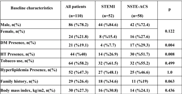
We assessed 110 patients (mean age 60.07±10.57 years, 78.2% male) for the study. Selective coronary angiography was performed in all patients. In our study, the cut-off value for troponin I value was 0.6 ng / ml and 1700 pg / ml for H-FABP. Twenty-one patients (19.1%) had diabetes mellitus, 44 patients (40%) were hypertensive, 64 patients (58.2%) were current smokers, 52 patients (47.3%) had dyslipide-mia. The time from onset of chest pain to medical contact ranged from 30 to 120 min with an average of 59.14±27.49 min. The baseline demographic/ clinical characteristics and laboratory findings of patients included in our study are summarized in Table 1 and Table 2. There were no statistically sig-nificant differences between the groups with respect to sex, body mass index (BMI), smoking status, or hemoglobin levels.
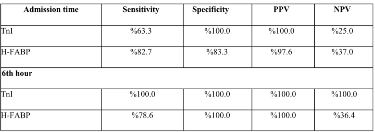
A number of patients had H-FABP positive on ad-mission 30 patients versus 9 patients with cTnI. At the time of admission, H-FABP had the highest sensitivity of 82.7% within the cardiac markers. The specificity was 83.3%, the positive predictive value was 97.6%, the negative predictive value was 37.0% and the test reliability was 82.7%.
For troponin I, the sensitivity was 63.3%, specificity was 100.0%, positive predictive value was 100.0%, negative predictive value was 25.0% and test reli-ability was 67.3% for diagnosis of ACS. We found that H-FABP within 6 h of chest pain onset had a sensitivity of 78.6%, a specificity of 100%, a posi-tive predicposi-tive value of 100%, a negaposi-tive predicposi-tive value of 36.4% for diagnosis of ACS. And we found that Troponin I within 6 h of chest pain onset had a
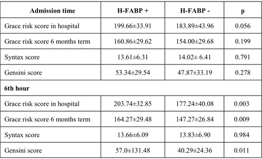
sensitivity of 100%, a specificity of 100%, a positive predictive value of 100%, a negative predictive value of 100% for diagnosis of ACS (Table 3). Based on the SYNTAX risk score, 96 patients (87.3%) were in the low-risk (<22) group, 14 (12.7%) were in the intermediate-risk (22-33) group, and no patients were in the high-risk (>33) group. On admission when the patients were classified ac-cording to the positive or negative H-FABP, there was not a statistically significant relationship be-tween the cardiac risk scores in patients with ACS. Although the GRACE risk score and GENSINI risk score were higher in the H-FABP positive group; SYNTAX risk score was higher in H-FABP nega-tive patient group. Even though at the 6th hour in the H-FABP-positive group, we found a statistically significant relationship between the GRACE and GENSINI risk scores; there was no statistically sig-nificant correlation with SYNTAX risk score (Table 4).
A ROC curve analysis of H-FABP and Troponin in the diagnosis of the acute coronary syndrome be-tween 0-6 hours (Figure 1). While the area under the curve (AUC) value of H-FABP was 0.86 (95% con-fidence interval (CI): 0.76–0.97, p<0.001), the AUC value of Tropoin I was 0.84 (95% CI: 0.76-0.97, p<0.001).
DISCUSSION AND CONCLUSION
When we evaluated the results, we showed the rela-tionship between H-fabp and cardiac risk scores and showed that it can be used to evaluate the diagnosis and prognosis of patients with ACS. The main pathophysiological mechanism is the reduction of coronary flow due to thrombus formation on the plaque in ACS. Plaque erosion is responsible for 25% of patients.
It is a dynamic process and requires early diagnosis and urgent treatment. As a result of distal emboliza-tion of the thrombus, cardiac markers become posi-tive and help us to make a diagnosis. It is important to determine mortality and morbidity in patients with ACS.
GRACE, SYNTAX and GENSINI scores currently used are available for this purpose. Syntax score is angiographic scoring used to determine how com-plex the coronary arteries are. It is helpful to deter-mine the manner of the revascularization (CABG or PCI) according to the difficulty of coronary anatomy of the patients. The GRACE risk score provides im-portant information for predicting mortality both in the hospital and within the first 6 months. As a result
of the scoring, patients are divided into low, medium and high-risk categories and performing coronary angiopraphy in appropriate time zones is recom-mended accordingly.15-18
The GENSINI score, which is investigated by the severity of CAD lesion, is a method that shows the relationship of atherosclerotic plaque when used in conjunction with simple, catheter calibration analy-sis.19,20
The frequency of ACS is effected by factors such as region, gender and race. McManus et al.21and Eren et al.22 also had male gender dominance at their studies. In our study, male gender dominance was present in STEMI and NSTE-ACS groups. The pa-tients in the STEMI group were of older. There was no statistically significant difference in BMI cate-gory in both groups. It is important to diagnose chest pain whether it is ischemic in patients who admitted to the emergency department. Chest pain does not always lead us to the diagnosis, and neither does the ECG, hence additional assistive tests are required. Here, cardiac markers play a major role. Most stud-ies have showed that diagnosis of ACS within the first 6 hours by troponins is superior to CKMB. However, troponin values cannot be detected at the blood level in the early hours. As a result, new car-diac markers were needed in early diagnosis. In our study, in order to find a new cardiac marker we investigated H-FABP may associate with the prevalence and severity of the disease in the early diagnosis of ACS patients. Of the cardiac markers examined at the time of admission, Troponin I was positivity in 9 patients, but H-FABP was positivity in 30 patients. Initially H-FABP had higher sensitiv-ity and specificsensitiv-ity than troponin.
There was no statistically significant relationship between risk scores in both groups when we classi-fied the patients according to whether H-FABP was positive or negative. Although Grace risk score and Gensini risk score were higher in H-FABP positive group; Syntax risk score was higher in H-FABP negative patient group. Risk scores were developed in early diagnosis and prognosis in patients. In our study, we have showed statistically significant corre-lations both H-FABP, Troponin I values at the 6th hour and GRACE, GENSINI risk scores. As a result of the studies in the literature, the use of GRACE risk score has become widespread.23 Because of the inadequacy of a thick plate with axial lesions in ACS patients, as a result other stenosis scoring was needed. For example, GENSINI score was found to be associated with the major cardiac event (MKO) in
the long and short term.24 Our study shows that H-FABP may be associated with major cardiac events. Cakar et al. showed a statistically significant rela-tionship between the GENSINI and GRACE risk scores in ACS patients.25 In our study, we found a significant relationship between GRACE risk score and GENSINI score (Table 4).
As a result of our study, we found the sensitivity of H-FABP as 82.7% and the specificity as 83.3% on admission. When we look at data at the 6th hour, we found the sensitivity of H-FABP as 78.6% and the specificity as 100.0%.
H-FABP was found to most sensitive cardiac marker than CKMB and cTnI at the admission to hospital. Specificity of H-FABP as 100.0% and specificity of 100.0% were found at the 6-hour, likewise Tp-I. Umut Cavus et al.26 they found the specificity of H-FABP as 88.5% at the 4th hour. In the multicentered study of Valle et al.27 the sensitivity and the specific-ity of H-FABP were found 44% and 94%, respec-tively. In the study of Kenji Inoue et al.28 the sensi-tivity of H-FABP was 78.5% and specificity was 78.2% in patients with ACS in the emergency de-partment. In the study conducted by Ruzgar et al.29 the specificity of H-FABP as 95.2%, 91% and 27.3%, respectively in ACS patients. Priya Gurura-jan et al.30 reported that the sensitivity and specific-ity of H-FABP as 87% and 93%, respectively in ACS patients. The sensitivity of H-FABP was corre-lated or higher than many studies. Sensitivity and specificity of H-FABP can vary according to the onset of symptoms in ACS patients as shown in the literature.
In conclusion, our study demonstrates that the H-FABP has significant value for assessment of sever-ity, precense and early diagnosis of coronary artery disease in patients with ACS. In addition, we showed that a significant relationship between H-FABP and GRACE risk score at admission may be significant in predicting patient prognosis.
Ethics Committee Approval: Our study was
ap-proved by the Çanakkale Onsekiz Mart University Ethics Committee (Date: 05/08/2015, Decision no: 2015/13).
Conflict of Interest: No conflict of interest was
de-clared by the authors.
Author Contributions: Concept - U.K., B.A.;
Super-vision - U.K., B.A.; Materials - U.K., H.T.; Data Collection and/or Processing - U.K., B.A., H.T.; Analysis and/ or Interpretation - U.K., B.A., H.T.; Writing - U.K.
Peer-review: Externally peer-reviewed.
REFERENCES
1. Van Domburg RT, Miltenburg-van Zijl AJ, Veer-hoek RJ, et al. Unstable angina: good long-term outcome after a complicated early course. J Am Coll Cardiol. 1998;31:1534–9.
2. Thygesen K, Alpert JS, White HD. Universal definition of myocardial infarction. Eur Heart J. 2007;28:2525-38.
3. Cohen M, Antman EM, Murphy SA, et al. Mode and timing of treatment fail-ure (recurrent ischemic events) after hospital admission for non - ST segment elevation acute coronary syn-dromes. Am Heart J. 2002;143:63–9.
4. Jaffe AS, Babuin L, Apple FS. Biomarkers in acute cardiac disease. J Am Coll Cardiol. 2006;48:1–11.
5. Hamm CW, Katus HA. New biochemical mark-ers for myocardial cell injury. Curr Opin Cardiol. 1995;10:355-60.
6. Glatz JF, van Bilsen M, Paulussen RJ, et al. Re-lease of fatty acid-binding protein from isolated rat heart subjected to ischemia and reperfusion or to the calcium paradox. Biochim Biophys Acta. 1988;961:148-52.
7. Offner GD, Brecher P, Sawlivich WB, et al. Characterization and amino acid sequence of a fatty acid binding protein from human heart. Bio-chem J. 1998;252:191-8.
8. Lindholm D, James SK, Bertilsson M, et al. Bio-markers and Coronary Lesions Predict Outcomes after Revascularization in Non-ST-Elevation Acute Coronary Syndrome. Clin Chem. 2017;63:573-84.
9. Bawamia, B, Mehran, R, Qiu, W. Risk scores in acute coronary syndrome and percutaneous coro-nary intervention: a review. Am Heart J. 2013;165:441–50.
10. Tanboga IH, Ekinci M, Isik T, et al. Reproduci-bility of syntax score: from core lab to real world. J Interv Cardiol. 2011;24:302-6.
11. Fox KA, Dabbous OH, GoldbERG RJ, et al. Pre-diction of risk of death and myocardial infarction in the six months after presentation with acute coronary syndrome: prospective multinational
observational study (GRACE). BMJ.
2006;333:1091.
12. Serruys PW, Onuma Y, Garg S, et al. Assess-ment of the SYNTAX score in the Syntax study. Euro Intervention. 2009;5:50-56.
relationship between glucose tolerance and severityof coronary artery disease using the Gen-sini score. Angiology. 2010;61:751–5.
14. Widera C, Pencina MJ, Meisner A, et al. Adjust-ment of the GRACE score by growth differentia-tion factor 15 enables a more accurate apprecia-tion of risk in non-ST-elevaapprecia-tionacute coronary syndrome. Eur Heart J. 2012;33:1095–104. 15. Lloyd-Jones D, Adams RJ, Brown TM, et al.
Executive summary: heart disease and stroke statistics--2010 update: a report from the Ameri-can Heart Association. Circulation. 2010;121:480 -86.
16. Dong C, Crawford LE, Goldschmidt-Clermon PJ. Endothelial progenitor obsolescense and atherosckerotic inflammation. J Am Coll Cardiol. 2005;45:1458-60.
17. Hagensen MK, Shim J, Thim T, et al. Circulating endothelial progenitor cells do not contribute to plaque endothelium in murine atherosclerosis. Circulation. 2010;121:898-905.
18. Libby P, Shi GP. Mast cells as mediators and modulators of atherogenesis. Circulation. 2007;115:2471-3.
19. Granger CB, Goldberg RJ, Dabbous O, et al. Predictors of hospital mortality in the global reg-istry of acute coronary events. Arch Intern Med. 2003;163:2345-53.
20. Nurkalem Z, Hasdemir H, Ergelen M, et al. The relationship between glucose tolerance and se-verity of coronary artery disease using the Gen-sini score. Angiology. 2010;61:751–5.
21. McManus DD, Gore J, Yarzebski J, et al. Recent trends in the incidence, treatment, and outcomes of patients with STEMI and NSTEMI. Am J Med. 2011;124:40-7
22. Şevki Hakan EREN, Kerim YILMAZ, İlhan KORKMAZ, ve ark. Acil Serviste Akut Miyo-kard Enfarktüsü Tanısı Almış Hastalarda Trom-bolitik Tedavi Uygulanmasını Etkileyen Faktör-ler Fırat Tıp Dergisi. 2006;11:163-5
23. Mehta SR, Grange6r CB, Boden WE, et al. Early versus delayed invasive intervention in acute coro-nary syndromes. N Engl J Med. 2009;360:2165–75.
24. Huang G, Zhao JL, Du H, et al. Coronary score adds prognostic information for patients with acute coronary syndrome. Circ J. 2010;74:490-5. 25. Cakar MA, Sahinkus S, Aydin E, et al. Relation
between the GRACE score and severity of atherosclerosis in acute coronary syndrome Jour-nal of Cardiology. 2014;63:24–8.
26. Umut Cavus, Figen Coskun, Bunyamin Yavuz, et al. Heart-type, fatty-acid binding protein can be a diagnostic marker in acute coronary syndromes. J Natl Med Assoc. 2006;98:1067–1070.
27. Valle HA, Riesgo LG, Bel MS, et al. Clinical assessment of heart-type fatty acid binding pro-tein in early diagnosis of acute coronary syn-drome. Eur J Emerg Med 2008;15:140-4. 28. 28Inoue K, Suwa S, Ohta H, et al. Heart Fatty
Acid-Binding Protein Offers Similar Diagnostic Performance to High-Sensitivity Troponin T in Emergency Room Patients Presenting With Chest Pain. Circ J. 2011;75:2813-20.
29. Ruzgar O, Bilge AK, Bugra Z, et al. The use of human heart-type fatty acid-binding protein as an early diagnostic biochemical marker of myocar-dial necrosis in patients with acute coronary syn-drome, and its comparison with troponin-T and creatine kinase-myocardial band. Heart Vessels. 2006;21:309-14.
30. Gururajan P, Gurumurthy P, Nayar P, et al. Heart fatty acid binding protein (HFABP) as a diagnos-tic biomarker in patients with acute coronary syndrome. Heart Lung Circ. 2010;19:660-4.
Table 1. Clinical characteristics of patients.
Baseline characteristics All patients (n=110) STEMI (n=52) NSTE-ACS (n=58) p Male, n(%) 86 (%78.2) 44 (%84.6) 42 (%72.4) 0.122 Female, n(%) 24 (%21.8) 8 (%15.4) 16 (%27.6) DM Presence, n(%) 21 (%19.1) 4 (%7.7) 17 (%29.3) 0.004 HT Presence, n(%) 44 (%40) 14 (%26.9) 30 (%51.7) 0.008 Tobacco use, n(%) 64 (%58.2) 32 (%61.5) 32 (%55.2) 0.499 Hyperlipidemia Presence, n(%) 52 (%47.3) 27 (%48.1) 25 (%46.6) 1.0 Family history, n(%) 29 (%26.4) 18 (%34.6) 11 (%19) 0.063
Body mass index, kg/m2, n(%) 30 (%27.3) 16 (%30.8) 14 (%24.1) 0.436
Table 2. Laboratory findings of patients.
All patients
STEMI NSTE-ACS
Mean± SD Mean± SD Mean± SD p
Glucose mmol/L 154.02±74.28 149.98±74.59 157.64±74.46 0.886 Urea mmol/L 34.00±9.78 34.08±8.26 33.92±11.05 0.933 Creatinine mmol/L 0.83±0.17 0.83±0.16 0.83±0.18 1.0 Sodium 139.49±3.17 140.12±3.21 138.93±3.06 0.132 Potassium 4.26±0.51 4.20±0.58 4.32±0.44 0.21 Wbc x 109/mL 14.90±22.03 15.52±18.76 14.35±24.75 0.005 Hgb g/dL 14.28±1.79 14.59±1.66 14.01±1.88 0.108 Plt 239.46±66.29 243.36±58.99 235.97±72.54 0.56 Ldl mmol/L 132.79±35.23 133.90±37.44 131.73±33.34 0.756 Hdl mmol/L 44.05±11.88 44.53±11.72 43.60±12.13 0.695 Total-cholesterol mmol/L 190.84±41.98 194.00±39.09 187.93±44.65 0.468 Triglyceride mmol/L 159.35±98.43 149.98±90.27 168.17±105.66 0.272 Grace 109.10±23.32 109.96±22.41 108.32±24.28 0.551 Syntax 13.7±6.31 12.57±5.50 14.72±6.84 0.981 H-fabp 6166.46±5858.75 8822.16±6660.81 3783.59±3705.52 0.004 Gensini 51.99±30.40 56.74±30.69 47.74±29.77 0.024 TnI 1.6±1.9 1.11±1.47 2.03±2.27 0.008
All values are presented mean±standard deviation (SD). Abbreviations: HDL: high density lipoprotein; LDL: low density lipoprotein; Hb: Hemoglobin; Wbc: white blood cell; Plt: Platelets; Grace: Global Registry of Acute Coro-nary Events; Syntax: The synergy between percutaneous coroCoro-nary intervention with taxus and cardiac surgery; H-fabp: Heart type fatty acid binding protein; TnI: Troponin I
Table 3. The sensitivity and specificity of the cardiac markers at the time of admission and 6th hour.
Abbreviations: TnI: Troponin I; H-FABP: Heart type fatty acid binding protein; PPV: Positive Predictive Value; NPV: Negative Predictive Value
Admission time Sensitivity Specificity PPV NPV
TnI %63.3 %100.0 %100.0 %25.0
H-FABP %82.7 %83.3 %97.6 %37.0
6th hour
TnI %100.0 %100.0 %100.0 %100.0
Table 4. The relationship between cardiac risk scores and H-FABP on admission and 6th hour.
Abbreviations: H-FABP: Heart type fatty acid binding protein
Admission time H-FABP + H-FABP - p
Grace risk score in hospital 199.66±33.91 183.89±43.96 0.056
Grace risk score 6 months term 160.86±29.62 154.00±29.68 0.199
Syntax score 13.61±6.31 14.02± 6.41 0.791
Gensini score 53.34±29.54 47.87±33.19 0.278
6th hour
Grace risk score in hospital 203.74±32.85 177.24±40.08 0.003
Grace risk score 6 months term 164.27±29.48 147.27±26.84 0.009
Syntax score 13.66±6.09 13.83±6.90 0.984
Figure 1. ROC curve analysis of H-FABP and troponin in the diagnosis of the acute coronary syndrome between 0-6 hours.