Arch Clin Exp Med 2018;3(3):186-189. e-ISSN: 2564-6567
DOI:10.25000/acem.464304 Araştırma makalesi / Research article
Atıf yazım şekli:
How to cite: Karabay Ö, Bostancı Ö. Biliary fistula after liver hydatid cyst surgery: Is it a predictable complication? Arch Clin Exp Med. 2018;3(3):186-189.
Abstract
Aim: The biliary fistula development after liver hydatid cyst surgery is a common complication. The aim of this study is to examine the factors affecting the development of postoperative biliary fistula in patients operated for liver hydatid disease.
Methods: The study was retrospectively performed in patients treated surgically for liver hydatid cyst between 1999 and 2010. The data of 53 patients operated for hydatid csyt were reviewed with hospital records. Patients were divided into two groups as biliary fistulas with (Group A) and without biliary fistula (Group B). The demographic data (age, sex), cyst diameter, cyst localization, laboratory tests and length of hospital stay were recorded. These parameters were compared with the groups.
Results: The groups were similar in terms of age and sex (p = 0.790 and p=1.0, respectively). In group A, the mean cyst diameter was significantly higher than group B (p=0.001). The mean duration of hospitalization was longer in group A than group B (p=0.001). There was no difference between the groups considering cyst localization, AST, ALT, total bilirubin and direct bilirubin (p>0.05 for all).
Conclusion: Preoperative cyst diameter may be a valuable parameter for predicting biliary fistula preoperatively. However, larger prospective studies are needed on this subject.
Key words: Hydatid cyst, liver, biliary fistula, ERCP
Öz
Amaç: Karaciğer hidatik kisti ameliyatı sonrası safra fistülü gelişimi sık bir komplikasyondur. Bu çalışmanın amacı, karaciğer hidatik hastalığı nedeniyle opere edilen hastalarda postoperatif bilyer fistül gelişimini etkileyen faktörleri incelemektir.
Yöntemler: Çalışma, 1999 ve 2010 yılları arasında karaciğer hidatik kisti nedeniyle cerrahi olarak tedavi edilen hastalarda retrospektif olarak yapıldı. Hidatik kist için ameliyat edilen 53 hastanın verileri hastane kayıtları ile gözden geçirildi. Hastalar safra fistülü olan (Grup A) ve safra fistülü olmayan (Grup B) olmak üzere iki gruba ayrıldı. Yaş, cinsiyet, kist çapı, kist yerleşimi, laboratuvar testleri ve hastanede kalış süresi kaydedildi. Bu parametreler gruplar arasında karşılaştırıldı.
Bulgular: Gruplar yaş ve cinsiyet açısından benzer özellikteydi (p = 0,790 ve p=1,0 sırasıyla). Grup A’daki ortalama kist çapı grup B’den anlamlı olarak büyüktü (p=0,001). Ortalama hastanede kalış süresi grup A’da grup B’den uzundu (p=0,001). Kist lokalizasyonu ve AST, ALT, total bilirubin, direk bilirubin değerleri açısından gruplar arasında fark yoktu (hepsi için p>0.05).
Sonuç: Ameliyat öncesi kist çapı, safra fistülünü tahmin etmek için faydalı bir parametre olabilir. Bununla beraber, bu konuda daha geniş prospektif serilere ihtiyaç vardır.
Anahtar Kelimeler: Kist hidatik, karaciğer, safra fistülü, ERCP
Ethics Committee Approval: The study wass approved by the local ethical authority.
Etik Kurul Onayı: Çalışma lokal etik komite tarafından onaylanmıştır.
Conflict of Interest: No conflict of interest was declared by the author.
Çıkar Çatışması: Yazar çıkar çatışması bildirmemiştir.
Finansal Destek: Yazarlar bu çalışma için finansal destek almadıklarını beyan etmişlerdir.
Geliş Tarihi / Received: 26.09.2018 Kabul Tarihi / Accepted: 15.11.2018 Yayın Tarihi / Published: 30.11.2018
Sorumlu yazar / Corresponding author: Onder Karabay
Adres/Address: Fatih Medical Park Hospital, General Surgery Department, Iskenderpasa Mah., Horhor Str., No:4 Fatih,Istanbul, Turkey.
e-posta: [email protected] Tel/Phone: +905057330343
Copyright © ACEM
1 Istinye University, Faculty of Health Sciences,
Istanbul, Turkey.
2 Sisli Hamidiye Etfal Research and Education
Hospital General Surgery Clinic, Istanbul, Turkey.
Financial Disclosure: The authors declared that this study has received no financial support.
Biliary fistula after liver hydatid cyst surgery: Is it a predictable
complication?
Karaciğer hidatik kist cerrahisi sonrası safra fistülü: Tahmin edilebilir bir komplikasyon mu?
Arch Clin Exp Med 2018;3(3):186-189. Hydatid cyst and biliary fistula
187
Introduction
Liver hydatid disease is a common zoonotic disease in Turkey and the Mediterranean countries [1]. If it is not treated, it can lead to serious health problems. Although there are many innovations in treatment, surgical procedures continue to be widespread.
No matter how precisely surgical treatment is done, development of postoperative biliary fistula is still one of the most important complications. The causes of biliary fistulae are an association between the cyst and the biliary tract, an injury to the biliary tract, or a high intrabiliary system pressure due to the presence of scolexes in the main bile duct. In order to prevent this, magnetic resonance cholangiopancreatography (MRCP) scanning should be performed in suspicious cases before surgical treatment and especially in the presence of large cysts. If postoperative biliary fistulas persist for a long time or the drain output is high, it can rarely result in mortality. In appropriate patients, fistulas can be controlled by endoscopic procedures or percutaneous drainage [2]. Additionally, there isn’t still a gold standard blood test for prevention of biliary fistula after liver hydatid cyst surgery.
The aim of this study is to examine the factors affecting the development of postoperative biliary fistula in patients operated for liver hydatid disease.
Material and methods
This retrospective study was performed in Sisli Etfal Education and Research Hospital in Istanbul, Turkey. Institutional review board approval was obtained. Informed consent could not be obtained from all participants for being included in this retrospective study. All of the procedures were in accordance with the World Medical Association Helsinki Declaration of 1964 and later versions.
Patients operated for liver hydatid cyst between January 1999 and December 2010, were evaluated retrospectively. There were a total of 64 patients. Eight patients with lack of clinical data and three patients with lack of follow up data were excluded. Therefore, 53 patients were included in the study.
Patients were analyzed for age, gender, laboratory values, imaging findings with regard to number, diameter, and location of the cysts, operation type, and length of hospital stay.
In the preoperative diagnosis, the patient's anamnesis, physical examination findings, and abdominal USG were used. All patients underwent a pre-operative CT scan. The location of the cyst in the liver, its size, number and its relation with surrounding tissues were examined. Laboratory analysis including alanine transaminase (ALT) (U/l), aspartate transaminase (AST) (U/l), total and direct bilirubin (mg/dl), were recorded in the preoperative period.
In the preoperative period, 10 mg/kg albendazole (Andazol, 200 mg, tablet, Biofarma, Turkey) was given for 3 weeks when the liver enzymes were normal. All the patients were informed about the surgery to be performed and the possible complications, and an informed consent form was obtained. Open or laparoscopic cystotomy and drainage were performed for all patients.
Surgical technique
Right subcostal incision was preferred in open surgical technique. Four trocars were used in the laparoscopic method. According to the placement of the cysts, trocar locations were regulated. The cyst was aspirated to reduce the pressure inside the cyst. A 0.09% NaCl solution was added into the cyst. After waiting for five minutes, the cyst was opened over, the germinative membrane and the female vesicles were removed
from the cyst. The cyst was washed with saline and aspirated. The presence of bile in the cyst was assessed. If there was an open bile duct in the cyst, it was sutured. The drain was inserted into the cyst in all patients. The oral regime started the same day after surgery. In the postoperative period, 10 mg/kg albendazole was started again. Albendazole treatment was planned for three weeks usage and one week rest for six months period.
In the postoperative period, the presence of bile fluid in the drain tube was evaluated as a biliary fistula. Patients were evaluated in two groups: postoperative biliary fistula (Group A) and no fistula group (Group B).
The drain outputs were followed in patients with post-operative biliary fistula (Group A). If the drain output is low and biliary fistula is closed spontaneously, no intervention was made.
If there was a long-standing bile fistula on day 5 or a high-output biliary fistula (>500 ml), ERCP was performed. Nasobiliary drainage or sphincterotomy was performed during ERCP. The patients were followed up for 1 year after discharge. At 1st year follow-up, CT scan was performed.
Statistical analysis
Statistical analysis was performed using SPSS ver. 20 (SPSS, Chicago, IL, USA). All continuous data were presented as means ± standard deviations. Statistical significance of the findings was analyzed using the two-tailed Student's t-test, Pearson chi-square test, and Mann-Whitney test. A p-value less than 0.05 was considered to be statistically significant.
Results
Fifty-three patients who underwent hepatic hydatid cyst surgery were retrospectively reviewed. Twenty-nine of the patients were male (55%) and 24 were female (45%). Mean symptom duration was 10.7 months (range 1-120 months).
There were multiple cysts in 12 patients (23%) and single cysts in 41 patients (77%). The mean cyst size was 81.75 mm (range 30-140 mm). When we analyzed the cyst localization, in five patients (9.44%) were in both lobes, in 30 patients (56.60%) were in the right lobe and in 18 (33.96%) patients were in the left lobe.
Three patients (5.6%) were operated by the laparoscopic method and 50 patients (94.4%) were operated by the open technique. The mean duration of hospitalization was 6.6 days (2-36) days.
14 patients (26.41%) had biliary fistula after operation (Group A). The biliary fistula was seen in 13 patients (24.52%) who underwent open surgery and one case (33.3%) under laparoscopic surgery. Solitary cysts (85.72%) were present in 12 of the patients in Group A, and multiple cysts were present in two (14.28%).
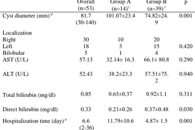
The groups were similar in terms of age and sex (p = 0.790 and p=1.0, respectively). The mean cyst diameter was 101.07±23.4 mm in group A and it was significantly higher than group B (p=0.001). The mean duration of hospitalization was 11.79±10.6 (4-36) days in group A and it was significantly longer than group B (p=0.001). Comparison of the groups for cyst localization revealed no significant difference (p=0.422).
There was no difference between the groups considering AST, ALT, total bilirubin and direct bilirubin (p>0.05 for all) (Table 1).
In 14 patients (Group A), the mean daily drainage was 341.07±284 ml. Seven patients in Group A (50%) underwent ERCP (nasobiliary stent in two) and the remaining seven patients (50%) were followed up. The mean daily fistula drainage was 500 ml (range 200-950 ml) and 175 ml (range 75-350 ml), respectively. The mean drainage of two patients with nasobiliary stents was 800 ml (range 650-950 ml) (Table 2).
Arch Clin Exp Med 2018;3(3):186-189. Hydatid cyst and biliary fistula
188 The median length of fistula closure was 11 days (range
6-60 days). In the group of patients who were followed up with fistulas, the median length of close time was 10 days (range 6-21 days). The median closure time of patients who was performed ERCP was 12 days (range 7-60 days). The mean follow up time of patients was one year and none of the patients had biliary
fistula recurrence.
Table 1. Comparison of preoperative features of the groups.
µ: mean (min-max), :mean±standard deviation
Table 2. The drainage output of the patients with fistula.
µ
: mean (min-max)
Discussion
Liver hydatid cyst disease is an important parasitic disease in terms of human and animal health. As it is nowadays in the world, it continues to be a serious health problem in our country as well. Many modalities were tried like medical treatment or percutaneous drainage with USG for hydatid disease. However, surgical treatments still maintain the efficacy and necessity. Biliary fistulas and recurrences are the most common complications after liver hydatid cyst surgery [2]. Factors that may be effective in the development of biliary fistulas; the high pressure in the cyst, a large relationship between the cyst and biliary system, an iatrogenic injury at the biliary system, ductus choledochus obstruction by germinative membranes and scolexes. It is stated that 80-90% of the small cystobiliary connections are present but clinically the connection is seen in 13-37% [3, 4]. The cystobiliary connection was presented with the appearance of bile leakage into the cyst during surgery or by imaging the connection in the ERCP due to jaundice or biliary fistula in the postoperative period [3].
In literature, there were many different biliary fistula rates. In a study with 117 patients, Patkowski et al reported biliary fistula in three patient (2.5%) [5]. Furthermore, Nooghabi et al reported 27.3% biliary fistula rate in 73 patients [6]. In large number of studies; Baraket et al reported 8 biliary fistulas in 120 patients (10.4%), Surmelioglu et al reported 36 fistulas in 186
patients (19.4%) and Nakeeb et al reported 13 fistulas in 123 patients (10.5%) [7, 8, 9]. In our study, we found postoperative biliary fistula in 14 patients (26.4%). The rate of biliary fistula in our study is higher than literature. The low number of patients and the presence of large diameter cysts may be the reason for the high fistula rate in our study.
There is not a gold standard surgery technique in liver hydatid disease for decreasing the biliary fistula rate. So, the biliary fistula is still a common complication after surgery. Although, there are many ideas to prevent the fistula and none of them are effective at 100%. In literature, there were a few studies which researched on the parameters to prevent the biliary fistula [1, 2]. The study of Kilic et al, liver enzymes (ALT, AST, GGT, ALP) and cyst type-diameter were evaluated and the difference was statistically significant about only cyst diameter [1]. Atahan et al reported that GGT can be a predictive test for preventing the biliary fistula. Furthermore, they didn't find a difference for cyst diameter in groups [2]. Liver enzymes and cyst features were examined in this study and only cyst diameter was significantly associated with biliary fistula.
Patients with biliary fistulas are also more likely to stay in the hospital than other patients. In a study conducted by Agarwal et al. [10], the mean duration of hospital stay in patients with fistulas was 18 days, while the mean duration of fistula-free patients was seven days. In our study, the mean hospital stay of biliary fistula group was significantly longer than fistula-free group.
There are several modalities in the treatment of biliary fistulas. It was observed that some of the biliary fistulas were spontaneously closed during follow-up. Balik et al reported 10 biliary fistulas in 304 patients and they were spontaneously closed during the follow-up period of 2-4 months [11]. In the study of Vagioanos et al, 7 of 12 biliary fistulas were spontaneously closed in 38 days [12]. In our study, 7 of 14 biliary fistulas were spontaneously closed in follow up. The others needed endoscopic intervention. Although some biliary fistulas can be closed spontaneously, there may be persistent fistulas. Most of the fistulas don't require re-surgery and can be treated by endoscopic or percutaneous interventions [10]. Today, endoscopic interventions are the mainstay of treatment in patients with unclosed or high-flow fistulas. It is believed that endoscopic sphincterotomy causes the early closing of the fistula by lowering intrabiliary pressure [13]. There is no definite conclusion about the timing of the endoscopy. There are different opinions between the next few days and a few months [14, 15]. In our study, there were not required a second surgery in patients with fistulas. Seven patients who have persistent fistulas were treated with endoscopic sphincterotomy (five patients) or nasobiliary stenting (two patients). All of the patients with biliary fistula were treated successfully.
Limitations of this study are the small number of patients and tests, and to be retrospective. In a prospective study with high volume series, it may be more effective and guiding.
In conclusion, preoperative cyst diameter may be a valuable parameter for predicting biliary fistula preoperatively. However, larger prospective studies are needed on this subject.
References
1. Kilic M, Yoldas O, Koc M, Keskek M, Karakose N, Ertan T, et al. Can biliary-cyst communication be predicted before surgery for hepatic hydatid disease: does size matter? Am J Surg. 2008;196:732-5. 2. Atahan K, Küpeli H, Deniz M, Gür S, Cökmez A, Tarcan E. Can occult
cystobiliary fistulas in hepatic hydatid disease be predicted before surgery? Int J Med Sci. 2011;8:315-20.
3. Kayaalp C, Bostanci B, Yol S, Akoglu M. Distribution of hydatid cysts into the liver with reference to cystobiliary communications and cavity-related complications. Am J Surg. 2003;185:175-9.
Overall (n=53) Group A (n=14) Group B (n=39) p Cyst diameter (mm) µ 81.7 (30-140) 101.07±23.4 74.82±24. 9 0.001 Localization Right Left Bilobular 30 18 5 10 3 1 20 15 4 0.420 AST (U/L) 57.13 32.14± 16.3 66.1± 80.8 0.290 ALT (U/L) 52.43 38.2±23.3 57.51±75. 2 0.940 Total bilirubin (mg/dl) 0.85 0.65±0.37 0.92±1.1 0.311 Direct bilirubin (mg/dl) 0.33 0.21±0.26 0.37±0.48 0.030 Hospitalization time (day) µ 6.6
(2-36) 11.79±10.6 4.87± 1.5 0.001 Number (n) Drainage (ml) µ Biliary fistula No 39 - Yes 14 341.07 (75-950) Treatment Conservative 7 175 (75-350) ERCP 7 500 (200-950) Nasobiliary stenting 2 800 ( 650-950)
Arch Clin Exp Med 2018;3(3):186-189. Hydatid cyst and biliary fistula
189
4. Ozaslan E, Bayraktar Y. Endoscopic therapy in the management of hepatobiliary hydatid disease. J Clin Gastroenterol. 2002;35:160-74. 5. Patkowski W, Krasnodębski M, Grąt M, Masior Ł, Krawczyk M.
Surgical treatment of hepatic Echinococcus granulosus. Prz Gastroenterol. 2017;12:199-202.
6. Jabbari Nooghabi A, Mehrabi Bahar M, Asadi M, Jabbari Nooghabi M, Jangjoo A. Evaluation and Comparison of the Early Outcomes of Open and Laparoscopic Surgery of Liver Hydatid Cyst. Surg Laparosc Endosc Percutan Tech. 2015;25:403-7.
7. Baraket O, Moussa M, Ayed K, Kort B, Bouchoucha S. Predictive factors of morbidity after surgical treatment of hydatid cyst of the liver. Arab J Gastroenterol. 2014;15:119-22.
8. Surmelioglu A, Ozer I, Reyhan E, Dalgic T, Ozdemir Y, Ulas M, et al. Risk Factors for Development of Biliary Complications after Surgery for Solitary Liver Hydatid Cyst. Am Surg. 2017;83:30-5.
9. El Nakeeb A, Salem A, El Sorogy M, Mahdy Y, Ellatif MA, Moneer A, et al. Cystobiliary communication in hepatic hydatid cyst: predictors and outcome. Turk J Gastroenterol. 2017;28:125-30.
10. Agarwal S, Sikora SS, Kumar A, Saxena R, Kapoor VK. Bile leaks following surgery for hepatic hydatid disease. Indian J Gastroenterol. 2005;24:55-8.
11. Balık AA, Başoğlu M, Celebi F, Oren D, Polat KY, Atamanalp SS, et al. Surgical treatment of hydatid disease of the liver: review of 304 cases. Arch Surg. 1999;134:166-9.
12. Vagioanos C, Androulakis JA. Capsulectomy and drainage in hepatic hydatidosis. Dig Surg. 1997;14:241-4.
13. Akaydin M, Erozgen F, Ersoy YE, Birol S, Kaplan R. Treatment of hepatic hydatid disease complications using endoscopic retrograde cholangiopancreatography procedures. Can J Surg. 2012;55:244-8. 14. Dolay K, Akçakaya A, Soybir G, Cabioğlu N, Müslümanoğlu M, Iğci
A, Topuzlu C. Endoscopic sphincterotomy in the management of postoperative biliary fistula A complication of hepatic hydatid disease. Surg Endosc. 2002;16:985-8.
15. Bilsel Y, Bulut T, Yamaner S, Buyukuncu Y, Bugra D, Akyuz A, et al. ERCP in the diagnosis and management of complications after surgery for hepatic echinococcosis. Gastrointest Endosc. 2003;57:210-3.