Comment
In previous reports [4, 5] we have demonstrated that endoscopic gluing is a valid procedure for closure of postresectional bronchopleural fistulas. On the basis of these encouraging results, we applied the technique in the present case of TEF in a ventilated patient in whom surgery was contraindicated and spontaneous closure by conservative treatment was unlikely to occur.
According to our experience, the closure of the fistula by endoscopic gluing occurs very rapidly (⬍ 24 hours), resulting from both a mechanical occlusion by the solid-ified glue and the associated reactive tissue reaction. In the present case we employed a fibrin glue, which solidifies more slowly, resulting in a more homogeneous spread over the lesion.
In conclusion, endoscopic gluing successfully closed a small iatrogenic TEF in a mechanically ventilated patient. The technique is safe, repeatable, and does not exclude the use of subsequent surgical correction in case of failure, so that it may be proposed as an alternative to surgery in high-risk patients or as a first-line attempt.
References
1. Willets IE, Dudley NE, Tam PKH. Endoscopic treatment of recurrent tracheo-esophageal fistulae: long-term results. Pe-diatr Surg Int 1998;13(4):256 –8.
2. Hoelzer DJ, Luft JD. Successful long-term endoscopic closure of a recurrent tracheoesophageal fistula with fibrin glue in a child. Int J Pediatr Otorhinolaryngology 1999;48(3):259 –63. 3. Raijman I. Endoscopic management of esophagorespiratory
fistulas: expanding our options with expandable stents. Am J Gastroenterol 1998;93(4):496 –9.
4. Scappaticci E, Ardissone F, Ruffini E, Baldi S, Mancuso M.
Postoperative bronchopleural fistula: endoscopic closure in 12 patients. Ann Thorac Surg 1994;57:119 –122.
5. Scappaticci E, Ardissone F, Ruffini E, Baldi S, Revello F, Coni F. As originally published in 1994: Postoperative broncho-pleural fistula: endoscopic closure in 12 patients. Updated in 2000. Ann Thorac Surg 2000;69:1629 –1630.
Solitary Eosinophilic Granuloma of
Sternum
Atilla Eroglu, MD, Ibrahim Can Ku¨rkc¸u¨oglu, MD, and Nurettin Karaoglanoglu, MD
Department of Thoracic Surgery, Atatu¨rk University, Medical Faculty, Erzurum, Turkey
Primary bone tumors of the chest wall are uncommon, although a wide variety of both benign and malignant tumors arise within the chest wall. Among those tumors, sternal tumors are rare and usually malignant. We report an extremely rare case of eosinophilic granuloma devel-oped in the sternum in a 30-year-old woman. She pre-sented anterior chest pain and somewhat tender mass over the sternum. Chest roentgenogram, computed to-mography scanning of the thorax, and total body bone scintigraphy revealed an isolated lytic lesion in the corpus sterni. A tru-cut biopsy of the mass exhibited the typical histologic appearance of eosinophilic granuloma. The solitary lesion was removed completely surgically. This rare condition should be kept in mind in differential diagnosis of sternal lesions.
(Ann Thorac Surg 2004;77:329 –31) © 2004 by The Society of Thoracic Surgeons
E
osinophilic granuloma (EG) or benign focal histiocy-tosis-X is one of the three variants of a clinical entity, which involves the reticuloendothelial system known as Histiocytosis-X, and was reported in 1953 [1]. The other two are Letterer–Siewe and Hand–Schuller–Christian diseases. There have been some discussion about the term EG and the pathology of EG. Lieberman and col-leagues [2] think that the term EG should not be included in the term Histiocytosis-X. EG usually has better prog-nosis, which is characterized by solitary, or multiple lytic bone lesions. EG frequently occurs in the flat bones such as ribs, pelvis, scapula, and the skull [3]. Although EG bone lesions have been discussed extensively in the literature, only a few cases of solitary EG of the sternum have been published until now [4 – 6]. An unusual pre-sentation of solitary EG of the sternum is discussed with the literature review.Accepted for publication April 9, 2003.
Address reprint requests to Dr Eroglu, Department of Thoracic Surgery, Faculty of Medicine, Atatu¨rk University, 25240 Erzurum, Turkey; e-mail: [email protected].
Fig 2. Endoscopic image of the tracheo-esophageal fistula.
329
Ann Thorac Surg CASE REPORT EROGLU ET AL
2004;77:329 –31 SOLITARY EOSINOPHILIC GRANULOMA OF STERNUM
© 2004 by The Society of Thoracic Surgeons 0003-4975/04/$30.00
Published by Elsevier Inc doi:10.1016/S0003-4975(03)01051-8
CASE
We report a 30-year-old woman patient presenting with a 1-year history of presternal swelling and pain. On physical examination this region was tender on palpa-tion. On admission laboratory investigations showed normal values for the erythrocyte sedimentation rate (ESR), fibrinogen and C-reactive protein (CRP), and the white blood cell count was minimally elevated (12,300 cells/mm3
).
Chest radiographs were normal. A contrast-enhanced computed tomographic (CT) scan was carried out and confirmed the existence of a partially sclerotic delineated osteolytic lesion with a focal interruption of the cortex in the corpus sterni (Fig 1). No contrast enhancement was illustrated. No invasion of the tumor into the thoracic cavity was found. A skeletal survey was normal and a technetium bone scan revealed only pathologic increased uptake in the corpus sterni (Fig 2). A CT scan guided tru-cut biopsy was performed. Histopathologic examina-tion revealed a mixed cellular infiltrate made up of lymphocytes, macrophages, eosinophilic granulocytes, Langerhans cells, and multinucleated cells. The histology was quite characteristic of EG.
The patient underwent an operation through a vertical incision on sternum. Chondral parts of third and fourth ribs with the adjacent segment of sternum and associated pectoralis major muscle adherent to the chondrosternal junction appeared to be involved. Cartilages of the right third and fourth ribs, involved sternum were removed completely. Prolene mesh was used for closure of the thorax wall defect. Pathologic examination revealed EG with positive immunostaining for S-100 protein and CD-1a antigen, confirming the diagnosis and surgical ends were microscopically free of tumor.
The hospital course was uncomplicated, and the pa-tient was discharged on the eighth postoperative day. Chemotherapy or radiotherapy was not given. At 1-year after the operation, no local recurrence has been found,
and no abnormal findings such as enlargement of the bone lesion on plain radiogram have been obtained.
Comment
Eosinophilic granuloma represents the localized form of a spectrum of pathologic proliferations of tissue macro-phages called Langerhans’ cells. The cause of this disease is unknown. Many factors have been considered as the cause, such as abnormal immunity, viral infection, injury, metabolic disturbance, and genetic predisposition. How-ever, none of them have been substantiated as yet. In the majority of patients EG is a solitary osteolytic lesion with a predilection for the calvarium and rarely seen in the skull base, but it can affect the femur, ribs, and the brain, too [3]. Eosinophilic granuloma is uncommon, and rep-resents less than 1% of tumorlike lesions of bone [7]. About 90% of patients with EG of bone present between 5 and 15 years of age; there is a slight male predominance [8]. Sternal EG in an adult is extremely rare.
Pain and the presence of a mass are the two most common complaints of patients with sternal tumors, and many patients experienced both. Differential diagnosis of lesions involving the sternum includes condroma, osteo-condroma, bone cyst, and malignant entities primary bone and soft tissue tumors and metastases.
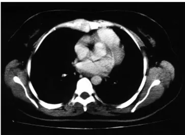
Radiographic appearances of osseous EG depend on the site of involvement and the phase of the disease. The radiographic diagnosis is speculative because the ap-pearance is nonspecific. Solitary EG appear as an osteo-lytic mass with sharp borders and a characteristic “punched-out” appearance on standard radiographs. The value of plain films is limited and the extent of the disease is better defined by CT scanning. Magnetic Fig 1. Contrast-enhanced computed tomographic scan of the thorax
reveals a large lytic and destructive lesion in the corpus sterni.
Fig 2. The technetium 99m methylenediphosphonate scintigraphy illustrates a focal zone of increased tracer uptake on the corpus sterni.
330 CASE REPORT EROGLU ET AL Ann Thorac Surg
SOLITARY EOSINOPHILIC GRANULOMA OF STERNUM 2004;77:329 –31
CASE
resonance imaging (MRI) presents low intensity on T1
-weighed image and high intensity on T2-weighed image
with heterogeneous enhancement. There are no specific CT or MRI characteristics, which allow a more specific diagnosis of EG. A complete scintigraphic bone survey was made because the lesion might have been present in other bones. Radionuclide bone scan can also be used to detect the bone involvement as well as to rule out recurrence or regrowth after surgery.
A diagnosis of localized EG can be made with certainty only by histologic examination of a truly representative specimen of tissue. Definitive diagnosis of EG is made by histopathologic means and immunohistochemical detec-tion of S-100 and CD1 antigens in the tissue samples. In our patient tru-cut biopsy was used to provide adequate tissue for exact histologic diagnosis.
Treatment of EG depends on the extent of the disease. The therapeutic modalities include radiotherapy, sur-gery, and chemotherapy alone or in combination. Specif-ically, monosystemic patients are treated by most centers with surgery, radiation, or local steroid injection. Surgery is generally indicated for isolated lesions in which an appropriate curettage may lead to complete removal of the lesion. Wide resection with tumor-free margins is required in order to provide the best chance for cure in EG. The use of radiotherapy is generally limited to those lesions not accessible to local steroid infiltration or not resectable. Chemotherapy is generally used for patients with multisystemic EG, in combination with local steroid injection whenever possible. The cytotoxic drugs used are etoposide and vinblastine. We treated this patient with only surgery due to solitary lesion. Further fol-low-up is necessary for future reference.
Although EG has only rarely been reported to occur in this location, at this age, and as a solitary lesion, this entity has to be considered in the differential diagnosis of patients who present with a sharp delineated osteolytic mass in the sternum.
References
1. Lichtenstein L. Histiocytosis X. Integration of eosinophilic granuloma of bone, Letterer–Siwe disease and Hand– Schuller–Christian disease as related manifestation of a sin-gle nosologic entity. Arch Pathol 1953;56:84 –102.
2. Lieberman PH, Jones CR, Steinman RM, Erlandson RA, Smith J, Gee T, Huvos A, Garin-Chesa P, Filippa DA, Urma-cher C, Gangi MD, Sperber M. Langerhans cell (eosinophilic) granulomatosis. A clinicopathologic study encompassing 50 years. Am J Surg Pathol 1996;20:519 –52.
3. Zinkham WH. Multifocal eosinophilic granuloma: nature, history ethiology and management. Am J Med 1976;60:457–63. 4. Chiau JH, Chang TK, Hwang B, Tiu CM. Eosinophilic gran-uloma of sternum. Report of one case. Zhonghua Min Guo Xiao Er Ke Yi Xue Hui Za Zhi 1990;31:35–9.
5. Martinelli C, Reggiani GC, Bucci D. Eosinophilic granuloma located in the sternum. Radiol Med 1989;77:270 –1.
6. Peer A, Witz E, Yona E, Manor H, Reif R. Solitary eosinophilic granuloma of sternum: case report with review of the litera-ture. Br J Radiol 1985;58:1173–6.
7. Wells CPO. The button sequestrum of eosinophilic granu-loma of the skull. Radiology 1956;67:746 –7.
8. Stull MA, Kransdorf MJ, Devaney KO. Langerhans cell histi-ocytosis of bone. Radiographics 1992;12:801–23.
Primary Chondromatous
Osteosarcoma of the Pulmonary
Artery
Yoshio Tsunezuka, MD, PhD, Makoto Oda, MD, PhD, Masao Takahashi, MD, PhD, Hiroshi Minato, MD, PhD, and Go Watanabe, MD, PhD
Departments of General and Cardiothoracic Surgery, Pathology, Kanazawa University School of Medicine, Kanazawa, Japan
Primary pulmonary sarcoma is an extremely rare malig-nancy, pulmonary osteogenic sarcoma is even more rare and has a poor prognosis. We present a report of a 58-year-old woman with primary pulmonary osteosar-coma occupying the right main pulmonary artery and extending to the main pulmonary artery. Right pneumo-nectomy was performed with total right main pulmonary artery resection under cardiopulmonary bypass. Micro-scopic examination demonstrated that tumor cells con-tained mainly chondromatous components and were atypical cells that became osteoid. There is no sign of recurrence 24 months postoperatively. The present case is the only case yet reported with even an intermediate-term survival without recurrence.
(Ann Thorac Surg 2004;77:331– 4) © 2004 by The Society of Thoracic Surgeons
P
rimary pulmonary sarcoma is an extremely rare ma-lignancy and osteogenic sarcoma is even more rare. Extraskeletal osteosarcoma usually arises in somatic soft tissues and paraenchymal organs such as soft tissue, kidney, and thyroid gland. We report here a surgically respectable case of primary chondromatous osteosar-coma of the pulmonary artery, and discuss the case and previous case reports.A 58-year-old woman was admitted to Kanazawa Univer-sity Hospital on February 19, 2001 for evaluation and treatment of dyspnea on effort and transient chest pain that began 5 months before admission. She had been treated with heparin at another hospital because the diagnosis of pulmonary emboli was made by chest com-puted tomography (CT) in September 2000, but the emboli gradually enlarged in spite of treatment. She underwent total resection of the uterus for myoma uteri in 1991, and had a history of hypertension since 1993. Physical examination revealed vital signs within normal limits and bilateral breath sounds were equal without adventitious sounds. Significant laboratory findings in-cluded C-reactive protein 0.6 U and serum alkaline Accepted for publication April 29, 2003.
Address reprint requests to Dr Tsunezuka, Department of General and Cardiothoracic Surgery, Kanazawa University School of Medicine, Takaramachi 13-1, Kanazawa 920-8530, Japan; e-mail: [email protected]. 331
Ann Thorac Surg CASE REPORT TSUNEZUKA ET AL
2004;77:331– 4 PRIMARY CHONDROMATOUS OSTEOSARCOMA OF THE PULMONARY ARTERY
© 2004 by The Society of Thoracic Surgeons 0003-4975/04/$30.00
Published by Elsevier Inc doi:10.1016/S0003-4975(03)00761-6
CASE