An Anatomical Consideration Of The Internal
Mammary (Thoracic) Artery And Vein
ARTERIA VE VENA THORACICA INTERNA’NIN ANATOMIK ÖNEMI
Çiğdem İÇKE, Mustafa GÜVENÇER, Esin KORMAN, İpek ERGÜR Dokuz Eylül University Faculty of Medicine, Department of Anatomy
Çiğdem İÇKE Department of Anatomy Faculty of Medicine Dokuz Eylül University 35340 İnciraltı, Izmir – TURKİYE Tel: (232) 4124357 Faks: (232) 2590541 e-posta: [email protected] ÖZET
Giriş: Arteria thoracica interna (ATI) ve vena thoracica interna (VTI) anatomik yerlesimleri nedeniyle koroner by-pass cerrahisinde ve meme rekonstrüksiyonlarında önemli yere sahiptir. Çalışmamızın amacı, cerrahi öneme sahip olan ATI ve VTI`nin anatomik olarak incelenmesidir.
Gereç ve yöntem: Çalışmamız formaldehit ile fikse edilmiş 10 erişkin insan kadavrasında gerçekleştirildi. Disseksiyon ile sternum`un her iki yaninda ATI ve VTI görünür hale getirilerek ölçümler yapıldı.
Bulgular: ATI`nın olguların %88,89`unda (16/18) iki dala ayrıldığı gözlendi. Ayrılma yeri 4. kostanın üst ucu (%12,50), 4. kostanın alt ucu (%6,25), 5. kostanın üst ucu (%6,25), 5. kostanın alt ucu (%43,75), 6. kostanın üst ucu (%31,75) olarak belirlendi. ATI`nin ortalama çapı 2,58 mm (min-max: 2,00-3,20 mm), 5. interkostal aralık (IA)`da sternum`a ortalama uzaklığı 13,11 mm (min-max: 6,00-22,00 mm) olarak ölçüldü. VTI´nın olguların %94,44`ünde (17/18) iki dala ayrıldığı gözlendi. Ayrılma yeri 1. kostanın alt ucu (%5,90), 2. kostanın üst ucu (%5,90), 2. kostanın alt ucu (%5,90), 2. IA (%17,65), 3. kostanın üst ucu (%23,50), 3. kostanın alt ucu (% 5,90), 3. IA (%5,90), 4. kostanın üst ucu ( %11,72), 4. kostanın alt ucu (%5,90), 5. kostanın üst ucu ( %11,72) olarak belirlendi. VTI`nin ortalama çapı 2. IA`da 3.12 mm (min-max: 1,90-5,00 mm), 3. IA` da 2,59 mm (min-max: 1,8-3,9 mm) olarak ölçüldü.
Sonuç: ATI ve VTI hem proksimal hem de distal yönde gerçekleştirilecek anastomozlar icin uygun özelliklere sahip damarlardır. Bu nedenle anatomik olarak iyi tanımlanmalarının klinik önemi büyüktür.
Anahtar sözcükler: Internal thorasik arter, internal thorasik ven, flep, myokart revaskülarizasyonu, meme rekonstrüksiyonu
SUMMARY
Introduction: Anatomic position of the internal mammary vessels is important in breast reconstruction and coronary artery myocardial revascularisation. Objective of this study was to describe detailed anatomy of the internal mammary vessels. Material and method: Dissection was performed in 10 formalin fixed human cadavers . After both of the clavicles were removed the anterior mammary wall was released by cutting it on both sides of the cadavers following the midaxillary line. The parietal pleura and endothoracic fascia were stripped on the posterior surface of the anterior thoracic wall and the transversus thoracis muscle was removed. The internal thoracic vessels were dissected. The diameter and distance of the vessels from the sternum were measured from the first intercostal space (ICS) to sixth ICS.
An anatomical consideration of the internal mammary artery and vein
The number, relationships and bifurcation points of the vessels
were recorded. Results: The internal mammary (thoracic) artery IMA was divided into twobranches in 88.89% of all cases. The point of division occured at cranial edge of 4th rib, at caudal edge of 4th rib, at the cranial edge of 5th rib, at caudal edge of 5th rib, at caudal edge of 6th rib as 12.50%, 6.25%, 6.25%, 43.75%, 31.75% respectively. The mean diameter of the IMA was mesaured as 2.58 mm (range 2.00-3.20 mm), and the mean distance of the IMA to the sternum was measured as 13.11 mm (range 6.00-22 mm) at the 5th ICS. The IMV was divided into two branches in 94.44% (17/18) of the cases. The point of division occured at the caudal edge of 1st rib (5.90%), at the cranial edge of 2nd rib (5.90%), at caudal edge of 2nd rib (5.90%), at 2nd ICS (17.65%), at cranial edge of 3rd rib (23.50%), at the caudal edge of 3rd rib (5.90%), at 3rd ICS (5.90%), at cranial edge of 4th rib (11.72%), at caudal edge of 4th rib (5.90%) and at cranial edge of 5th rib (%11.72). The mean diameter of the medial IMV was mesaured as 2.48 mm (range 1.9-3.4 mm) at the 2nd ICS, and 2.59 mm (range 1.8-3.9 mm) at the 3rd ICS.
Conclusion: The internal mammary vessels can provide double recipient vessels (proximal and distal ends) for anastomoses to both the deep inferior epigastric artery and vein.
Key words: Internal mammary artery, internal mammary vein, flap, myocardial revascularization, breast reconstruction
The internal thoracic or internal mammary arteries (IMA) are the first inferiorly directed branches of the subclavian vessels. The arteries run behind the brachiocephalic vein at the sternoclavicular joint, then they continue to costal cartilages. In the superior tho-rax the artery and committant veins (which empty into the brachiocephalc vein on each side) run extra-pleurally, deep to the costal and internal intercostal muscles. From the third intercostal space (ICS) downward, the artery and committant veins run be-tween the transversus thoracic muscle and intercostal muscles. By this well-defined muscular layer they are separated from the pleura. IMA gives off branches at each ICS. At the level of the sixth ICS or the seventh costal cartilage the IMA divides into the superior epi-gastric and musculophrenic arteries (1-3).
Owing to their anatomic position internal thoracic vessels are important in breast reconstruction and coronary artery myocardial revascularisation (2-10). In coronary by-pass surgery IMA grefts have the most widespread usage among the other ones depending on their good outcome on postoperative mortality and morbidity. The resistance of these vessels to athero-sclerosis and size congruity with the coronary vessels are the additional benefits of these grafts (10). For free
microsurgical tissue transfer in thoracic region it is difficult to choose the best recipient vessels. In breast reconstruction with free flaps, especially with the transverse rectus abdominis myocutaneous (TRAM) flap, the vessels in the axilla are the most common recipient vessels. After radiotherapy these vessels tend to be small, heavily scarred and vulnerable. Therefore, dissection of these vessels is more difficult. However, internal mammary artery (IMA) and internal mammary vein(s) IMV(s) particularly offer themselves as recipient vessels because they are centrally located and usually only minimally affected by radiotherapy for breast cancer (2,11). Internal thoracic vessels have been utilized as recipient site in breast reconstruction because of their ideal location (2,4,12).
Objective of this study was to describe the location and course of the internal thoracic vessels using a dif-ferent method to permit for the total exploration of the vessels. We thought that the total exploration of the internal thoracic vessels could give a different per-spective to a surgeon depending on their importance in microsurgery.
MATERIAL AND METHODS
Dissection was performed on 18 sides of sternum in 10 formalin fixed human cadavers (male 9; female 1). After both of the clavicles were removed, the ante-rior thoracic wall was released by cutting it on both sides of the cadavers following the midaxillary line. The parietal pleura and the endothoracic fascia were stripped on the posterior surface of the anterior tho-racic wall and the transversus thoracis muscle was re-moved. The internal thoracic vessels were dissected.
The anatomical features of the internal thoracic vessels were evaluated according to different parame-ters. These parameters included the branching per-centage of the IMA, location of the branching point of the IMA, mean diameter of the IMA over the ICSs’, mean distance between the branching point of the IMA and sternal border, branching percentage of the IMV; location of the branching point of the IMV; types of the venous anatomy; and mean diameter of the IMV over the ICSs’.
The diameter and distance of the vessels from the sternum were measured from the first ICS to sixth ICS. The number, course, relationships and bifurca-tion points of the vessels were recorded. The result was evaluated statistically; Mann-Whitney U test was used to compare the measurements between left and right side vessels.
RESULTS
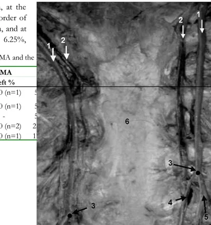
The IMA was divided into two branches in 88.89 % (16/18) of all cases (Fig 1). The point of division occured at the upper border of fourth costa, at the lower border of fourth costa, at the upper border of fifth costa, at the lower border of fifth costa, and at the lower border of sixth costa as 12.50%, 6.25%,
6.25%, 43.75%, 31.25% respectively (Fig 1, Table l). Branching occurred at the lower border of the fifth costa and below this level in majority of the cases.
The mean diameter of the IMA was measured between the first and fifth ICSs. Mean diameter was found to be placed larger on the right side than on the left side. On both sides it became smaller through the lower ICSs depending on branching of the vessels. Results are presented in the Table II (p>0.05).
The mean distance between the IMA and sternal border was measured over the ICSs. The IMA were found to be placed from medial to lateral through the lower ICSs. This displacement was silently more significant on the left side through the lover ICSs. Results are presented in the Table III (p>0.05).
The IMA had branched into medial (superior epigastric artery) (SEA) and lateral branches (musculophrenic artery) (MPA) in 88.89 % of all cases and the division point occured at the lower ICSs. The mean diameter of the SEA-MPA and the mean distance between the branches and sternal border was measured over the ICSs. There was not significant difference between the left and right sides statistically (p>0.05). The results are presented in the Table II and III (p>0.05).
Figure 1. The sample of the IMA has two branches and different bifurcation levels on both sides (1: ima, 2: imv, 3: bifurcation point, 4: SEA, 5: MPA, 6: sternum)
Table I. Distrubution of bifurcation level of the IMA and the IMV
IMA IMV
Right % Left % Total % Right % Left % Total %
Lower border of the 1st rib - 11.10 (n=1) 5.90 (n=1) - -
-Upper border of the 2nd rib - 11.10 (n=1) 5.90 (n=1) - -
-Lower border of the 2nd rib 12.50 (n=1) - 5.90 (n=1) - -
-2nd ICS 25.00 (n=2) 22.20 (n=2) 23.50 (n=4) - -
-An anatomical consideration of the internal mammary artery and vein
Lower border of the 3rd rib - 11.10 (n=1) 5.90 (n=1) - -
-3rd ICS 12.50 (n=1) - 5.90 (n=1) - -
-Upper border of the 4th rib 12.50 (n=1) 11.10 (n=1) 11.80 (n=2) - 25.00 (n=2) 12.50 (n=2)
Lower border of the 4th rib - 11.10 (n=1) 5.90 (n=1) - 12.50 (n=1) 6.25 (n=1)
Upper border of the 5th rib 12.50 (n=1) 11.10 (n=1) 11.80 (n=2) 12.50 (n=1) - 6.25 (n=1)
Lower border of the 5th rib - - - 50.00 (n=4) 37.50 (n=3) 43.75 (n=7)
Lower border of the 6th rib - - - 37.50 (n=3) 25.00 (n=2) 31.25 (n=5) IMA: Internal mammary artery
IMV: Internal mammary vein
Table II. Diameters of internal thoracic vessels (mm)
ICS: Intercostal space, IMA: Internal mammary artery, SEA: Superior epigastric artery, MPA: Musculophrenic artry, IMV: Internal mammary vein, MPV: Musculophrenic vein
Table III. Distances (mm) between the internal thoracic vessels and the sternal border
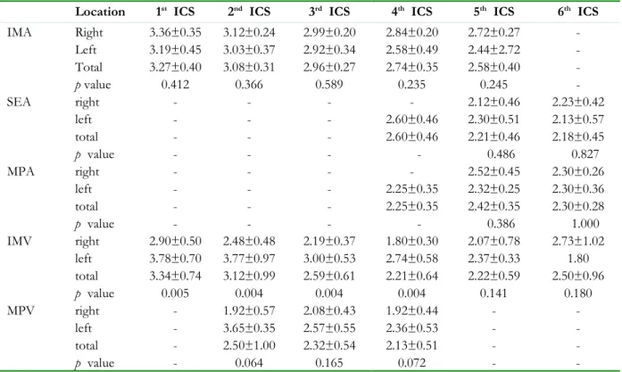
Location 1st ICS 2nd ICS 3rd ICS 4th ICS 5th ICS 6th ICS
IMA Right 3.36±0.35 3.12±0.24 2.99±0.20 2.84±0.20 2.72±0.27 -Left 3.19±0.45 3.03±0.37 2.92±0.34 2.58±0.49 2.44±2.72 -Total 3.27±0.40 3.08±0.31 2.96±0.27 2.74±0.35 2.58±0.40 -p value 0.412 0.366 0.589 0.235 0.245 -SEA right - - - - 2.12±0.46 2.23±0.42 left - - - 2.60±0.46 2.30±0.51 2.13±0.57 total - - - 2.60±0.46 2.21±0.46 2.18±0.45 p value - - - - 0.486 0.827 MPA right - - - - 2.52±0.45 2.30±0.26 left - - - 2.25±0.35 2.32±0.25 2.30±0.36 total - - - 2.25±0.35 2.42±0.35 2.30±0.28 p value - - - - 0.386 1.000 IMV right 2.90±0.50 2.48±0.48 2.19±0.37 1.80±0.30 2.07±0.78 2.73±1.02 left 3.78±0.70 3.77±0.97 3.00±0.53 2.74±0.58 2.37±0.33 1.80 total 3.34±0.74 3.12±0.99 2.59±0.61 2.21±0.64 2.22±0.59 2.50±0.96 p value 0.005 0.004 0.004 0.004 0.141 0.180 MPV right - 1.92±0.57 2.08±0.43 1.92±0.44 - -left - 3.65±0.35 2.57±0.55 2.36±0.53 - -total - 2.50±1.00 2.32±0.54 2.13±0.51 - -p value - 0.064 0.165 0.072 - -10 fig1
ICS: Intercostal space, IMA: Internal mammary artery, SEA: Superior epigastric artery, MPA: Musculophrenic artry, IMV: Internal mammary vein, MPV: Musculophrenic vein
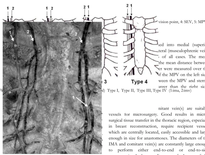
The venous anatomy in this region has beeen varied. Four different types of venous anatomy were identified by Arnez et al (13) (Fig 2). In our study we found Type I, Type II and Type III as 88.89 %, 5.56 % and 5.56 % respectively and there was no Type IV.
We also identified the presence of multiple venous junctions in the specimens. These junctions were mostly located at the neighbourhood of the bifurcation points of the IMA (Fig 3).
Location 1st ICS 2nd ICS 3rd ICS 4th ICS 5th ICS 6th ICS
IMA Right 8.00±3.47 11.24±2.47 11.93±2.94 11.49±3.64 11.24±4.60 14.83±3.33 Left 8.82±4.93 12.26±4.52 13.03±2.85 12.71±4.27 14.67±6.11 21.00 Total 8.41±4.16 11.75±3.57 12.48±2.86 12.02±3.84 13.11±5.51 16.37±4.11 P Value 0.791 0.790 0.330 0.490 0.273 0.180 SEA Right - - - - 8.95±5.38 7.55±3.18 Left - - - 8.70±6.65 6.70±9.47 8.00±4.24 Total - - - 8.70±6.65 8.20±6.06 7.77±3.07 P Value - - - - 0.643 1.000 MPA Right - - - - 18.10±7.88 16.15±3.46 Left - - - 14.00±5.66 17.00±4.24 15.50±3.54 Total - - - 14.00±5.66 17.73±6.41 15.82±2.88 P Value - - - - 0.643 0.439 IMV Right 4.92±3.74 8.80±1.83 8.17±2.48 8.82±3.47 8.11±4.92 9.25±5.56 Left 4.04±3.95 7.32±3.91 8.90±2.49 10.11±4.33 12.20±5.01 9.50 Total 4.48±3.76 8.06±3.06 8.53±2.44 9.39±3.79 10.0±5.20 9.30±4.81 P Value 0.562 0.144 0.769 0.396 0.166 1.000 MPV Right - 11.25±1.71 14.55±2.63 14.52±6.04 - -Left - 15.50±3.54 15.28±3.51 16.75±3.31 - -Total - 12.67±3.01 14.92±2.99 15.48±5.02 - -P Value - 0.100 0.936 0.475 -
-An anatomical consideration of the internal mammary artery and vein
Figure 3. The close localization of the venous junctions to the branching point of the IMA (1: ima, 2: imv)
A lateral branch joining the IMV has been found in 94.44% of the subjects (17/18)(Fig 4). The level where the lateral branch joined had a wide range of dispersion between the lower border of the first costa and the lower border of the sixth costa. The most frequent participation was seen in the second ICS at the rate of 23.50%, and at the upper border of the third costa at the rate of 17.60% (Fig 4, Table I). The branching point of the IMV was located at higher intercostal levels than the branching point of the IMA. The mean diameter of the IMV was measured over the first costa and between the first and sixth ICSs. The difference between the right and left sides was found to be significant . Results were presented in the Table II.
The mean distance between the IMV and sternal border was measured over the ICSs. At the first ICS, both on the right and left sides, IMV had its closest distance to the sternum. While it was significantly moving laterally at the second ICS and keeping the distance nearly the same through the lower ICSs on the right, it showed a lateral placement at the second ICS and descended through the lower ICSs becoming more lateral on the left. Results are presented in the Table III (p>0.05).
Figure 4. The division point of the IMV into the SEV
and MPV (1: ima, 2: imv, 3: division point, 4: SEV, 5: MPV)
The IMV had branched into medial (superior epigastric vein) SEV and lateral (musculophrenic vein) (MPV) veins in 94.44 % of all cases. The mean diameter of the MPV and the mean distance between the MPV and sternal border were measured over the ICSs. The mean diameter of the MPV on the left side, and the mean distance between the MPV and sternal border were sigificantly larger than the right side. Results are presented in the Table III (p>0.05).
DISCUSSION
The IMA and its comitant vein(s) are suitable vessels for microsurgery. Good results in micro-surgical tissue transfer in the thoracic region, especially in breast reconstruction, require recipient vessels which are centrally located, easily accessible and large enough in size for anastomoses. The diameters of the IMA and comitant vein(s) are constantly large enough to perform either end-to-end or end-to-side anastomosis. At least a diameter of 1 mm of the artery and a single vein is needed for sufficient flap perfusion. The frequent presence of two comitant veins on both sides can make two venous anastomoses possible in order to reduce venous congestion in the transferred tissue.
In contrast to the detailed anatomical information about the origin, path and branching of the IMA, little information is available about vessel diameters, the number of comitant veins and the level of the venous junction below the second costa (11). The level of the cranial edge of the fourth costa is of great importance in microsurgical tissue transfer depending on its central location in the anterior thoracic wall. And also at this level both of the IMA and IMV(s) are having accessible positions and enough diameters for the surgery (2,11). In the Study of Feng the mean diameter fig3
12
of the IMA used for the breast reconstruction was measured as 2.52 ± 0.51 mm on the right and 2.30 ± 0.55 mm on the left at the third ICS. In the same study the mean diameter of the IMV (at the third ICS) was measured as 2.89 ± 0.56 mm on the right and 2.31 ± 0.48 mm on the left (4). Han et al measured the mean diameter of the IMA and IMV in 60 breast cancer patients by sonographic 2-dimentional computed tomography. At the 2., 3., and 4. ICS’s the
mean diameter of the IMA was 2.35 ± 0.80, 2.60 ± 0.68, 1.79 ± 0.44 mm on the right and 2.07 ± 0.56, 1.91 ± 0.52, 1.76 ± 0.40 mm on the left respectively; and the mean diameter of the IMV was 3.53 ± 1.04, 3.09 ± 0.91, 2.59 ± 0.85 mm on the right and 2.30 ± 0.72, 1.85 ± 0.73, 1.70 ± 0.57 mm on the left respectively (14).
In their study Yazıcıoğlu et al was measured the mean diameter of the IMA by colored Doppler USG in 50 patients and the results for the first ICS are 2.65±0.7 mm on the right and 2.80 ± 0.6mm on the left; for the 5th ICS are 1.62 ± 0.3mm on the right and
1.85 ± 0.3 mm on the left (9).
In their studies Zenn and Heitmann measured the diameter of the IMA as 2.5 mm (2.1-2.8) on the right, 2.3 mm (1.9-2.6) on the left at the third ICS and 2.2 mm (1.8-2.5) on the right, 2.0 mm (1.5-2.3) on the left
at the fifth ICS (15). Arnez et al measured the mean diameter of the IMA at the third, fourth, fifth ICSs as 2.80 mm at the third ICS, 2.60 mm at the fourth ICS, and 2.60 mm at the fifth ICS, in our study these values were 2.96, 2.74, 2.58 mm respectively (13). While the results of our study showed differences from the results of Feng, Yazıcıoğlu et al, Han et al, Zenn and Heitmann; they were in accordance with the results of Arnez et al (4,9,10,13-15) (Graph 1).
Henriquez-Pino et al observed that the IMA had two branches in 93%, and three branches in 7% of all cases in their 100 cadaveric series. Similarly in 88.89 % of our cases the IMA had two terminal branches but as a diference from this study we did not observed that the IMA divided into three branches in any case (3).
In the study of Henriquez-Pino et al the point of
division occurred at the 5th costa, at the 5th ICS, at the
6th costa, at the 6th ICS, at the 7th costa, at the 7th ICS
as %2, %5, %21, %56, %10, %6 respectivly on the right side and as %5, %7, %23, %49, %11, %5 respectivly on the left side (3). When the division point occurred at the lower border of fifth costa, and at the lower border of sixth costa in most of our cases (Table l), it occurred mostly at the 6th costa and at the
6th ICS in the study of Henriquez-Pino et al (3).
An anatomical consideration of the internal mammary artery and vein 0 0,5 1 1,5 2 2,5 3
Arnez et al Zenn&Heitmann Feng et al İçke et al
A 3.ICS (R) B 3.ICS (L) C 4.ICS (R) D 4.ICS (L) E 5.ICS (R) F 5.ICS (L)
Graph 1. Comparison of the mean diameter of the IMA (mm)
The point of division occured at the upper border of fourth costa, at the lower border of fourth costa, at the upper border of fifth costa, at the lower border of fifth costa, and at the lower border of sixth costa as 12.50%, 6.25%, 6.25%, 43.75%, 31.25% respectively in our study (Fig 1, Table l). Branching occurred at the lower border of the fifth costa and below this level in majority of the cases.
The location of the IMA and IMV(s), on the both sides of the sternum in the thoracal region, allows a wide range of movement for the surgery. Because of that the width of the distance between the IMA and sternum is of great importance for surgeons. Several descriptions of the distance between the IMA and the sternal border can be found in the literature. Williams and Warwick notes that the IMA is about 12.5 mm
distant from the sternal border, whereas Last describes the artery passing vertically downwards a finger’s breadth from the sternal border (16,17). Kremer and Platzer noted that the IMA is normally found 11-13 mm lateral to the sternal border in the first ICS and about 15 mm at the second and third ICS (18). Soyluoğlu et al measured the mean distance between the IMA and the lateral border of the sternum over the 1st, 2nd, 3rd and 4th ICSs as 6.9 ± 2.27,
8.9 ± 1.30, 9.7 ± 2.97, 9.9 ± 3.20 mm respectively (8). Zenn and Heitmann, in their studies, reported that the distance increased from the third ICS to the sixth ICS and it was 13.9 mm on the right and 14.2 mm on the left, at the third 20.8 mm on the right and 21.3 mm on the left at the sixth ICS (15). Henriquez-Pino et al had measured the distance between artery and lateral border of the sternum from the first costal level to the 14
sixth and found that 10.1 ± 3.3, 13.5 ± 2.6, 12.8 ± 2.8, 13.2 ± 3.4, 15.9 ± 4.5 ve 18.4 ± 5.2 mm on the right and 10.5 ± 3.2, 13.6 ± 2.9, 13.4 ± 2.8, 14.5 ± 3.1, 16.4 ± 4.9, 20.0 ± 6.7 mm on the left. Similar to the literature data we observed that the distance between the artery and sternal border has increased from upper ICSs to lower ICSs (3). Also in accordance with the results of Henriquez-Pino et al, Zenn and Heitmann, we found that mean distance between artery and sternal border was always larger on left side than on right side (3,15) (Table III).
In our study the mean distance between the IMA and sternal border was measured over the ICSs (from the first to the sixth ICS) as 8.41 ± 4.16, 11.75 ± 3.57, 12.48 ± 2.86, 12.02 ± 3.84, 13.11 ± 5.51, 16.37 ± 4.11 mm respectively. On the contrary, we found that mean distance between artery and sternal border was always larger on left side than on right side (Table III). Ninkovic et al, studied the diameter of IMA and IMV, the distance between IMA and sternal border at cranial edge of fourth costa on cadavers (2). Schwabegger et al evaluated the diameter of IMA and IMV at third ICS intraoperatively (19). Ninkovic et al stated that the mean diameter of IMA and IMV; the mean distance between IMA and sternum had larger values on right side than left side (2). Contrary to Ninkovic et als’ results we found that mean distance between IMA and sternal border, mean diameter of IMV had greater values on left side than right side (2). While our mean diamater values were found to be greater than both of the studies Ninkovic et al and Schwabegger et al, distance between IMA and sternum was smaller than the results of Ninkovic et als’ (2,19). Our results were compared by the results of these authors in the Graph 2.
In the study of Feng the mean diameter of the IMV (at the third ICS) was measured as 2.89 ± 0.56mm on the right and 2.31 ± 0.48 mm on the left (4).
Graph 2. Comparison of the mean diameter and distance values in 3.1 ICS (mm)
An anatomical consideration of the internal mammary artery and vein
0 2 4 6 8 10 12 14 16
Ninkovich et al Schabbeger et al İçke et al
A IMA-sternum distance (R) B IMA-sternum distance (L) C IMA diameter (R) D IMA diameter (L) E IMV diameter (R) F IMV diameter (L)
According to results of Han et al the mean diameter of the IMV (at the 2.,3.,4. ICS was 3.53 ± 1.04, 3.09 ± 0.91, 2.59 ± 0.85 mm on the right and 2.30 ± 0.72, 1.85 ± 0.73, 1.70 ± 0.57 mm on the left respectively (14).
On the contrary, we found that mean IMV diameter was 2.48 ± 0.48, 2.19 ± 0.37, 1.80 ± 0.30 on the right and 3.77 ± 0.97, 3.00 ± 0.53, 2.74 ± 0.58 on the left. The difference between the results could be depended on the usage of different methods for measuring or location of the branching point of the IMV (Graph 3).
In their study Clark et al groupped the veins according to their diameter values which had diameter less than 3 mm and 3 mm or greater (20). The results of their study presented that veins with diameter of 3mm or larger were localized mostly at the level of
second costa. In our study they were localized mostly at the level of first ICS and had greater percentages on the left side each of the ICSs’ as the difference. The results were compared in the Graph 4.
Arnez et al identified four different types of venous anatomy (Fig 2) (13). In their study Type 1 (69%) and 2 (26%) were common, Type 3 (3%) and 4 (2%) were rare. In 95% of cases (Type 1 and 2) the IMV was found medial to the IMA throughout its course; in 5% of cases the IMVwas lateral to the IMA (Type 3 and 4). In our study we observed Type 1 (88.89 %), Type 2 (5.56 %), Type 3 (5.56 %); together with the Type 1 and Type 2 consisted 94.45% of all cases. This data was in accordance with the results of Arnez et al. As the difference Type 2 consisted only 5.56% of our cases and there was not seen any sample for the Type 4. Arnez et al studied the level of division in Types 1 and 3 (13). They reported that veins were 16
never divided above the upper border of the second costa. There were few divisions below the fifth costa; 84 % of the veins had divided before the lower border of the fourth costa. In our study the IMV was found
to be divided above the upper border of the second costa in 11.8% of cases; 82.4% of cases divided before the lower border of the fourth costa (Graph 5).
3 .I C S -R 3 .I C S -L 2 .I C S -R 2 .I C S -L 3 .I C S -R 3 .I C S -L 4 .I C S -R 4 .I C S -L 2 .I C S -R 2 .I C S -L 3 .I C S -R 3 .I C S -L 4 .I C S -R 4 .I C S -L 0 0,5 1 1,5 2 2,5 3 3,5 4
Feng Han İçke et al
2.ICS-R 2.ICS-L 3.ICS-R 3.ICS-L 4.ICS-R 4.ICS-L
An anatomical consideration of the internal mammary artery and vein C D E F G A B C D F H 0 1 0 2 0 3 0 4 0 5 0 6 0 7 0 8 0 9 0 C la r k e t a l İç k e e t a l A 1 .IC S (R ) B 1 .IC S (L ) C 2 .IC S (R ) D 2 .IC S (L ) E 3 .IC S (R ) F 3 .IC S (L ) G 4 .IC S (R ) H 4 .IC S (L )
Graph 4 . The localization rates of the veins having diameter 3 mm or greater (%)
C E F H I J A B C D E F G H I J 0 5 10 15 20 25 30 35 40 45 Arnez et al İçke et al
A lower border of 1.costa B upper border of 2.costa C lower border of 2.costa D 2.ICS
E upper border of 3.costa F lower border of 3.costa G 3.ICS
H upper border of 4.costa I lower border of 4.costa J upper border of 5.costa
Graph 5. Comparison of the division level of the IMV (%) 18
Hefel et al (11) observed the frequent presence of two comitant veins on both sides at the level of the cranial edge of the fourth costa and Arnez et al (13) emphasized the division point of the IMV mostly located at the upper border of the fourth costa (41.0%) in their series (5,6). In majority of our cases the division point of the IMV was located at the second ICS (23.5%) and upper border of the third costa (17.6%). The most frequent division point of the IMV was located one costal level in our study which was found to be above than found in the literature.
Arnez et al measured the mean diameter of SEV and MPV, and mean distance between SEV-MPV and sternal border at third, fourth, fifth ICSs (13). Both of the results of Arnez et al and our study showed that the SEV had greater diameters than the MPV at third, fourth and fifth ICSs. That result indicates that the SEV is the main vein and the MPV is a branch of it. In our study the distance between the SEV-MPV and the sternum did not show any consistancy like the results of Arnez et al and became wider through the lower ICSs. The comparison of the results are presented in Graph 6. A B C D E F G H I J K L A B C D E F G H I J K L 0,00 2,00 4,00 6,00 8,00 10,00 12,00 14,00 16,00 18,00 Arnez et al İçke et al
A MIMV diameter 3.ICS B MIMV diameter 4.ICS C MIMV diameter 5.ICS
D MIMV distance to sternum 3.ICS E MIMV distance to sternum 4.ICS F MIMV distance to sternum 5.ICS G LIMV diameter 3.ICS
H LIMV diameter 4.ICS I LIMV diameter 5.ICS
J LIMV distance to sternum 3.ICS K LIMV distance to sternum 4.ICS L LIMV distance to sternum 5.ICS
An anatomical consideration of the internal mammary artery and vein
CONCLUSION
We found differences between the results of our study and literature findings. These differences could be depended on the genetic properties of the Turkish population. On the other hand, in our study we used a method different from the other studies in the litera-ture. The most important benefit of this method was to expose the exact route of the internal thoracic ves-sels. We think that the reliability of this method is higher than the other methods used in the literature. We wanted to give a different point of view to the in-ternal thoracic vessels surgey with our findings.
The results of this study provide detailed knowl-edge about the diameter, course, branching points and types of internal thoracic vessels to the surgeon de-pending on their importance in microsurgery.
REFERENCES
1. Coleman JJ, Bostwick J. Rectus abdominis muscle-mus-culocutaneous flap in chest-wall reconstruction. Surg Clin North Am 1989; 69: 1007- 1027.
2. Ninkovic MM, Schwabegger AH, Anderl H. Internal mammary vessels as a recipient site. Clin Plast Surg 1998; 25: 213-221.
3. Henriquez-Pino JA, Gomes WJ, Prates JC, Buffolo E. Surgical anatomy of the internal thoracic artery. Ann Thorac Surg 1997;64:1041-1045.
4. Feng LJ. Recipient vessels in free-flap breast reconst-ruction: a study of the internal mammary and thoraco-dorsal vessels. Plast Reconstr Surg 1997;99: 405-416. 5. Li S, Mu L, Li Y, et al. Breast reconstruction with the
free bipedicled inferior TRAM flap by anastomosis to the proximal and distal ends of the internal mammary vessels. J Reconstr Microsurg 2002;18: 161-168. 6. Demirkılıç U, Bolcal C, Küçükarslan N, et al. Çok genç
hastalarda (20-29 yaş) koroner arter by-pass greft cerra-hisi orta ve geç dönem sonuçları. Anadolu Kardiyoloji Dergisi 2004; 4:25-29.
7. Ekim H, Kutay V, Demirbağ R. İnternal torasik arterin anatomisi ve diseksiyon tekniklerinin sternal dolaşıma ve solunum fonksiyonuna etkisi. T Klin J
Cardiovas-cular Surg 2003;4:27-32.
8. Soyluoğlu Aİ, Tanyeli E, Marur T, Ertem AD, Özkuş K. Türkiye’de yaşayan insanlarda arteria thoracica in-terna’nın sternum kenarına uzaklığı. Karadeniz Tıp Dergisi 1996;9:44-45.
9. Yazıcıoğlu L, Aral A. Koroner arter by-pass cerrahisin-de internal mammarian arter greftlerincerrahisin-de uygun anas-tomoz bölgesi. Klinik Bilimler&Doktor 1999;5:493-496 10. Yazıcıoğlu L, Elalmış AÖ, Kaya K, Aral A, Özyurda Ü.
Koroner arter by-pass cerrahisinde bilateral internal mammariyan arter deneyimlerimiz. T Klin J Cardiovascular Surg 2003;4:95-99.
11. Hefel L, Schwabegger A, Ninkovic M, et al. Internal mammary vessels: anatomical and clinical considerati-ons. Br J Plast Surg 1995; 48: 527-532.
12. Pietrasik K, Bakon L, Zdunek P, Wojda-Gradowska U, Dobosz P, Kolesnik A. Clinical anatomy of internal thoracic artery branches. Clinical Anatomy 1999;12: 307-314.
13. Arnez ZM, Valdatta L, Tyler MP, Planinsek F. Anatomy of the internal mammary veins and their use in free TRAM flap breast reconstruction. Br J Plast Surg 1995; 48: 540-545.
14. Han S, Yoon SY, Park JM. The anatomical evaluation of internal mammary vessels using sonography and 2-dimensional computed tomography in Asian. The Bri-tish Association of Plast Surg 2003;56:684-688.
15. Zenn MR, Heitmann C. Extended TRAM flap: feasi-bility study on fresh human cadavers. Ann Plast Surg 2003; 50: 256-262.
16. Williams PL, Warwick R. Gray's Anatomy 36th ed. WB-Saunders, Philadelphia 1973; 644– 645.
17. Last RJ. Anatomy Regional and Applied 7th ed. Churchill Livingstone, Edinburgh, 1984; 217.
18. Kremer K, Platzer W. Chirurgische Operationslehre, Band 2. Thieme Verlag, Stuttgart, 1991.
19. Schwabegger AH, Bodner G, Rieger M, Jaschke W, Ninkovich MM. Internal mammary vessels as a model for power doppler imaging of recipient vessels in mic-rosurgery. Plast Reconstr Surg 1999;104:6:1656-1661. 20. Clark CP 3rd, Rohrich RJ, Copit S, Pittman CE,
Robinson J. An anatomic study of the internal mam-mary veins: clinical implications for free-tissue-transfer 20
breast reconstruction. Plast Reconstr Surg 1997; 99:2: 400-404.