Original
Article
Introduction
Pulmonary arterial hypertension (PAH) is a disease associated with high morbidity and mortality. Chronic thromboembolic pulmonary hypertension (CTEPH) is one of the main causes of PAH which is a progressive dis-ease leading to right heart failure and low cardiac output, finally leads to death if left untreated.1) CTEPH is described by fibrotic obstruction of proximal and/or distal pulmonary arteries due to organized thrombus by doing distal arteri-opathy in small pre-capillary pulmonary vessels and hence it increases the pulmonary vascular resistance (PVR).1–3) Previous studies demonstrated that PVR is one of the most important hemodynamic parameter in CTEPH patients.2) The increased PVR is mainly caused by endothelial
Neutrophil/Lymphocyte Ratio Can Predict
Postoperative Mortality in Patients with Chronic
Thromboembolic Pulmonary Hypertension
Mehmed Yanartas, MD,1 Mehmet Emin Kalkan, MD,2 Akin Arslan, PhD,3 Serpil Gezer Tas, Prof,1 Cengiz Koksal, Dr,1 Nural Bekiroglu, Dr,4 and Bedrettin Yildizeli, Dr5
Objective: The aim of our study was to investigate clinical importance of neutrophil/ lymphocyte ratio in patients with Chronic thromboembolic pulmonary hypertension. Methods: 125 consecutive patients with a diagnosis of Chronic thromboembolic pulmonary hypertension were operated pulmonary thromboendarterectomy in our center between February 2011 and August 2013. 106 patients included into the study due to limitations. The patients were classified into two groups as patients discharged alive (Group 1) and those dying in the hospital (Group 2). Baseline neutrophil/lymphocyte ratio level was measured by dividing neutrophil count to lymphocyte count.
Results: 84 patients (79%) were in Group 1, 22 patients (21%) were in Group 2. Patients with higher neutrophil/lymphocyte ratio in admission have a significantly higher mortal-ity rate and postoperative pulmonary vascular resistance was found statistically signifi-cant variable to predict the mortality. Receiver operator characteristic (ROC) analysis revealed that using a cut-off point of 2.54, admission neutrophil/lymphocyte ratio predicts mortality. Also, correlation analysis showed a significant correlation between preopera-tive pulmonary vascular resistance and neutrophil/lymphocyte ratio.
Conclusion: The neutrophil/lymphocyte ratio level may be a useful and noninvasive biomarker for operative risk stratification for mortality after pulmonary thromboendarterectomy. Keywords: Chronic thromboembolic pulmonary hypertension, pulmonary
thromboendarterec-tomy, pulmonary vascular resistance, inflammation, Neutrophil/lymphocyte ratio.
1Department of Cardiovascular Surgery, Kartal Kosuyolu Training
and Research Hospital, Istanbul, Turkey
2Department of Cardiology, Kartal Kosuyolu Training and Research
Hospital, Istanbul, Turkey
3Medipol University Faculty of Medicine, Department of
Cardio-vascular Surgery, Istanbul, Turkey
4Department of Biostatistics, Marmara University School of Medicine,
Istanbul, Turkey
5Department of Thoracic Surgery, Marmara University School of
Medicine, Istanbul, Turkey
Received: July 7, 2014; Accepted: September 30, 2014
Corresponding author: Mehmed Yanartas, MD. Department of Cardiovascular, Kartal Kosuyolu Training and Research Hospital, Surgery. Denizcier cad. No: 2 Cevizli/Kartal/Istanbul/Turkey Email: [email protected]
©2015 The Editorial Committee of Annals of Thoracic and
Cardiovascular Surgery. All rights reserved.
dysfunction, vascular remodeling, vasoconstriction, throm-bosis and fibrosis of distal pulmonary arteries.4) It’s known that there is increased prevalence of inflammatory disease in CTEPH patients.3–5) Recently, there are increasing evi-dences for the role of inflammation in pathophysiologic mechanisms of CTEPH.3) When investigated the role of inflammation in these patients, elevated levels of inflam-matory cytokines are detected in the lung biopsies6) and the plasma samples.7,8) Also C-reactive protein (CRP) level is found significantly higher in patients with CTEPH compared with healthy subjects (p <0.01).9) However, the clinical importance of inflammatory state in CTEPH patients have not been well clarified so far. The neutro-phil/lymphocyte ratio (NLR) is a new inflammatory bio-marker and can be used as an indicator of systemic inflammation in many disease.10–12) The aim of our study was to investigate clinical importance of NLR in patients with CTEPH.
Materials and Methods
125 consecutive patients with a diagnosis of CTEPH were operated pulmonary thromboendarterectomy (PEA) in our center between February 2011 and August 2013. Patients with hepatic or renal insufficiency, previous coronary artery bypass grafting, heart failure (left ventric-ular ejection fraction [LVEF] <40%), malignancy (pul-monary sarcoma), known chronic systemic inflammatory disease (Behçet’s disease, systemic lupuserythematosus, poliomyelitis, ulcerative colitis, Takayasu vasculitis), were excluded from the present study. 106 eligible patients (64 females, range 18–78 years) were included into the study.
Clinical information including age, sex, body surface area (BSA), history of hypertension and diabetes melli-tus, smoking, current medications, complete blood count, CRP levels and biochemical parameters was obtained from a review of the patients’ chart. Hematologic indi-ces, such as hemoglobin, white blood cell and lympho-cyte and neutrophil counts were measured as part of the automated complete blood count, using simultaneous optical and impedance measurements (Cell Dyn 3700 Abbott Diagnostics, Abbott Park, Illinois, USA). All rou-tine biochemical tests were carried out on an automatic biochemical analyzer (Beckman Coulter AU640; Beckman Coulter, Krefeld, Germany). C-reactive protein (CRP) was determined by nephelometry on an IMMAGE® 800 analyzer (Beckman Coulter, Germany). The diagnosis of CTEPH was established on the basis of previously reported procedures.13) Diagnosis and cardiopulmonary
hemodynamic characteristics were determined by 6 minute walking test (6MWT), computed tomography pulmo-nary angiography (CTPA) and right sided heart catheter-ization (RHC). Pulmonary hypertension was defined as mean pulmonary artery pressure (mPAP) greater than 25 mmHg. In all patients, normal left ventricular func-tion was documented by echocardiography. Coronary angiography was routinely performed in all patients aged more than 40 years. Operative procedure was performed as explained with details in previous article.14) Shortly, the operation was performed on deep hypothermia (20°C) for both lungs under complete circulatory arrest. A true endarterectomy was accomplished and, when it was completed on both sides, circulation with warming was restarted. Postoperative hemodynamic characteristics were determined on the first day after PEA. All investigations were approved by the local institutional review board. The patients were classified into two groups as patients discharged alive (Group 1) and those dying in the hospi-tal (Group 2). Baseline NLR level was measured by dividing Neutrophil count to lymphocyte count.
Statistical analysis
Kolmogorov—Smirnov normality test was used to determine whether the continuous random variables are normally distributed or not. For the continuous random variables unpaired t-test and for discrete random vari-ables Chi-square test were applied for the comparisons between groups. A multivariate stepwise logistic regres-sion was used to predict for independent predictors of mortality. A ROC analysis was implemented to assess the sensitivity and specificity of NLR to find a cut-off point and to calculate area under curve (AUC). Pearson Correlation Coefficient was calculated to determine any relationship between two continuous variables. Statisti-cal analysis was carried out using SPSS software (ver-sion 15.0) for Windows (SPSS, Chicago, Illinois, USA). P <0.05 was considered as “significant”.
Results
The one hundred and six patients included into the study. Patients were divided into 2 groups as patients dis-charged alive in the hospital (Group 1) and those died in hospital period (Group 2). The eighty-four patients (79%) were in Group 1, twenty two patients (21%) were in Group 2. A comparison of groups regarding baseline characteristics and laboratory data shown in Table 1 and comparison of perioperative hemodynamic values
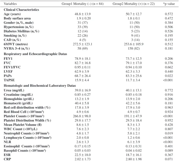
Table 1 Comparison of clinical characteristics and laboratory data in the groups
Variables Group1 Mortality (–) (n = 84) Group2 Mortality (+) (n = 22) *p value Clinical Characteristics
Age (years) 48.8 ± 13.9 50.7 ± 12.7 0.572
Body surface area 1.9 ± 0.20 1.8 ± 0.1 0.472
Gender (n,%, male) 31 (37) 11 (50) 0.384 Hypertension (n,%) 33 (39) 11 (50) 0.506 Diabetes Mellitus (n,%) 12 (14) 5 (23) 0.526 Smoking (n,%) 22 (26) 9 (41) 0.195 CAD (n,%) 4 (5) 3 (14) 0.155 6MWT (metres) 272.5 ± 123.1 253.6 ± 105.9 0.512 NYHA 3–4 (n,%) 58 (69) 158 (82) 0.181
Respiratory and Echocardiographic Datas
FEV1 78.9 ± 18.1 73.7 ± 12.5 0.206 FVC 82.7 ± 16.8 79.1 ± 17.0 0.376 FEV1/FVC 0.95 ± 0.11 0.94 ± 0.10 0.662 LVEF 62.9 ± 3.9 62.3 ± 3.3 0.440 PAPs 68.7 ± 26.4 83.3 ± 25.6 0.022 TAPSE 15.9 ± 4.4 11.7 ± 3.4 <0.001
Hematologic and Biochemical Laboratory Datas
Urea (mg/dL) 39.0 ± 16.9 40.1 ± 13.1 0.772
Creatinine (mg/dL) 0.85 ± 0.27 0.85 ± 0.18 0.916
Hemoglobin (gr/dL) 13.2 ± 1.9 13.9 ± 2.0 0.206
Hematocrit (gr/dL) 40.4 ± 5.8 42.2 ± 5.6 0.181
Red cell distribution width (%) 17.8 ± 3.9 17.9 ± 3.0 0.963 Red Blood Cell (×103/mm3) 4.9 ± 0.6 4.9 ± 0.7 0.924
Platelet Counts (×103/mm3) 266.8 ± 98.0 191.1 ± 47.9 <0.001
Platelet Distribution Width (%) 29.8 ± 17.7 29.5 ± 18.4 0.952
Mean Platelet Volume (fl) 8.6 ± 1.5 8.3 ± 1.3 0.428
WBC Count (×103/mL) 7.6 ± 2.3 7.7 ± 2.2 0.807 Neutrophil Counts (×103/mm3) 4.8 ± 1.7 5.8 ± 2.1 0.019 Lymphocyte Counts (×103/mm3) 2.0 ± 0.8 1.2 ± 0.6 <0.001 NLR 2.6 ± 1.3 6.1 ± 3.9 <0.001 Eosinophil Counts (×103/mm3) 0.17 ± 0.15 0.13 ± 0.31 0.401 Basophil Counts (×103/mm3) 0.05 ± 0.03 0.04 ± 0.02 0.089 ESR 22.5 ± 18.0 18.7 ± 16.1 0.367 CRP 2.02 ± 1.73 2.80 ± 1.98 0.071
Variables are expressed as mean ± standard deviation and percentage (%) for categorical variables. * Unpaired Student-t and Chi-square tests. CAD: coronary artery disease; 6MWT: 6 minutes walking test; NYHA: New York Heart Association; FEV: force expiratory volume; FVC: force vital capacity; LVEF: left ventricular ejection fraction; PAPs: systolic pulmonary arterial pressure; TAPSE: tricuspid annular plane systolic excursion; WBC: white blood cell; NLR: neutrophilto lymphocyte ratio; ESR: erythrocyte sedimentation rate; CRP: C-reactive protein
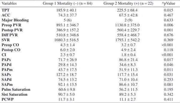
is presented in Table 2. There was no statistically signif-icant difference between the two groups with respect to age, gender, coronary artery disease risk factors. While white blood cell (WBC) and hemoglobin values were found to be insignificant between two groups, lymphocyte count was significantly lower and neutrophil count and NLR level was significantly higher in Group 2 patients (Table 1). Among operating times, while cross-clamp time was found to be insignificant, total perfusion time was found to be significantly longer in Group 2 than in Group 1 (Table 2).
Tricuspid Annular Plane Systolic Excursion (TAPSE), preoperative Cardiac output, Cardiac index, mean pul-monary arterial pressure, platelet level, neutrophil level, lymphocyte level, NLR, duration of totally perfusion time (minutes) (TPT), preoperative PVR and postopera-tive PVR, which were found significant as independent predictors of mortality in univariate analyses (Tables 1–2), were used to perform multivariate logistic regression analysis (Table 3). Despite systolic systemic arterial pres-sure (SAPs), systolic pulmonary arterial prespres-sure (PAPs) and diastolic pulmonary arterial pressure (PAPd) variables
are significant in univariate analysis, these are excluded in multivariate analysis firstly in order not to increase the number of independent variables unneccesarily, secondly due to the close relation between cardiac output (CO), car-diac index (CI) and SAPs and thirdly mean pulmonary arte-rial pressure (PAPm) value is calculated by using PAPs and PAPd values. According to this analysis; goodness of fit test, Nagelkerke R Square, Hosmer and Lemeshow
Test and Classification Table Percentage were found respectively as: p <0.0001, 55.5%, p >0.05 (p = 0,968) and 91%. Therefore, patients with higher NLR in admis-sion have a significantly higher mortality rate HR (hazard ratio): 2.767; 95% CI: 1.432–5.347; p = 0.002), TPT and postoperative PVR were found as second and third sta-tistically significant variables to predict the presence or absence of mortality (respectively, p = 0.0025, p = 0.007).
Table 2 Comparison of perioperative hemodynamic values and operating times in the groups Variables Group 1 Mortality (–) (n = 84) Group 2 Mortality (+) (n = 22) *pValue
TPT 185.9 ± 40.1 225.5 ± 68.4 0.015 ACC 74.3 ± 37.7 67.5 ± 42.8 0.467 Major Bleeding 5 (6) 2 (9) 0.633 Preop PVR 893.1 ± 346.7 1130.8 ± 375.0 0.006 Postop PVR 386.9 ± 157.2 560.4 ± 229.7 0.001 Diff PVR 510.8 ± 348.6 558.4 ± 468.7 0.676 SVR 1680.3 ± 516.5 1793.1 ± 542.2 0.369 Preop CO 4.3 ± 1.4 3.2 ± 0.7 <0.001 Postop CO 6.0 ± 2.0 4.9 ± 2.4 0.118 CI 2.3 ± 0.7 1.8 ± 0.4 <0.001 PAPs 71.7 ± 26.9 86.8 ± 21.4 0.017 PAPd 29.8 ± 14.3 34.6 ± 8.3 0.046 PAPm 43.7 ± 17.5 51.9 ± 11.3 0.011 SAPs 127.2 ± 18.7 117.7 ± 15.4 0.031 SAPd 74.5 ± 13.2 71.0 ± 10.4 0.253 SAPm 92.1 ± 13.5 86.6 ± 10.7 0.081 Pulm Saturation 60.6 ± 9.8 56.2 ± 11.5 0.195 Sist Saturation 90.7 ± 5.0 89.2 ± 5.3 0.342 PCWP 11.7 ± 3.1 11.1 ± 2.7 0.411
TPT: duration of totally perfusion time (minutes); ACC: aortic cross clamping time; PVR: pulmonary vas-cular resistance; Diff PVR: The gap between preoperative and postoperative PVR values; SVR: systemic vascular resistance; CO: cardiac output; CI: cardiac index; PAPs: systolic pulmonary arterial pressure; PAPd: diastolic pulmonary arterial pressure; PAPm: mean pulmonary arterial pressure; SAPs: systolic systemic arterial pressure; SAPd: diastolic systemic arterial pressure; SAPm: mean systemic arterial pressure; PCWP: pulmonary capillary wedge pressure
Table 3 Determination of significant independent predictors of mortality after performing multivariate logistic regression analyses
B S.E. Wald df Sig OR = Exp (B) 95 % CI. For EXP (B) Lower Upper NLR 1.018 0.336 9.177 1 0.002 2.767 1.432 5.347 Postop PVR 0.007 0.003 7.332 1 0.007 1.007 1.002 1.013 TPT 0.022 0.010 5.053 1 0.025 1.022 1.003 1.042 Constant –13.139 3.717 12.496 1 0.000 0.000
a. Variable(s) entered on step 1: NLR. (first important variable and statistically significant to predict the presence or absence of mortality). b. Variable(s) entered on step 3: TPT. (second important variable and statistically significant to predict the presence or absence of mortality). c. Variable(s) entered on step 2: POSTOP.PVR. (thirth important variable and statistically significant to predict the presence or absence of mortality). NLR: neutrophilto lympho-cyte ratio; PVR: pulmonary vascular resistance; TPT: duration of totally perfusion time (minutes)
ROC analysis revealed that using a cut-off point of 2.54, admission NLR predicts mortality with a sensitiv-ity of 86% and specificsensitiv-ity of 40% in CTEPH patients treated with PEA (Fig. 1). The area under the curve for this relationship is 0.825 and the 95% CI is 0.713–0.938. When patients divided with regard to cut off value, mean PVR is 872 ± 350 dyne ⋅ s ⋅ cm–5 in patients who have NLR lower than 2.54 and that is 1012 ± 367 dyne ⋅ s ⋅ cm–5 in patients who have NLR upper than 2.5 (p = 0.048).
Also, correlation analysis showed a significant cor-relation between preoperative PVR and NLR (r = 0.214, p = 0.027).
Discussion
This is the first study in CTEPH patients in which we demonstrated that clinical use of NLR may be a noninva-sive parameter for preoperative risk stratification of CTEPH patients. We showed that higher NLR level is associated with higher morbidity and mortality rate in patients with CTEPH who was operated PEA. Moreover we have shown that NLR value is an independent predic-tor of mortality in these patients. And also preoperative PVR is correlated with preoperative NLR.
One of the most important causes of PAH is CTEPH which is surgically curable.15) PEA is an effective and ideal therapeutic choice in CTEPH patients. It dramati-cally decreases PVR and leads to a significant healing of bad course. Nevertheless, the postoperative course is associated with significant morbidity and mortality. The postoperative mortality rate is remarkable and ranges from 1.3 to 24%.16) Previously researches have been demonstrated that severely affected patients, i.e. mPAP >50 mmHg, cardiac index <2.0 l/min m2, pulmonary vas-cular resistance >1000 dynes ⋅ s ⋅ cm–5 and/or New York Heart Association class IV disease, are at high risk for postoperative bad course and mortality after PEA.17,18) Among them, preoperative PVR is found the most important predictor of outcome after PEA.17,18) While mortality rate is 4% in patients who have preoperative PVR <900 dyne ⋅ s ⋅ cm–5, that is 10% in patients who have preoperative PVR 900–1200 dyne ⋅ s ⋅ cm–5 and that is 20% in patients who have preoperative PVR >1200 dyne ⋅ s ⋅ cm–5.17) Also, Jamiesson, et al. showed that postoperative PVR (more than 500 dyne ⋅ s ⋅ cm–5) is found as an independent predictor for mortality.18) In our study we showed that while preoperative and postopera-tive PVR is associated with mortality, only postoperapostopera-tive PVR is an independent predictor of mortality. Moreover, by using logistic regression analysis, we demonstrated that preoperative NLR and TPT are independent predic-tors of mortality too.
As is well known, the increased PVR in patients with CTEPH is mainly caused by endothelial dysfunction, vasoconstriction, vascular remodeling and proliferation, thrombosis and fibrosis of small and distal pulmonary arteries.8,19) There is increasing evidence of inflammation which plays an important role in the pathophysiology of PVR with elevated levels of many cytokines and chemo-kines in affected patients.20,21) Also, inflammatory cell infiltrates had been observed around the distal vascular area.8) It is now considered to be a distal vasculopathy, in which structural changes caused by vascular remodeling and inflammation play a major role.6–19) However, the pathophysiologic mechanism of increased PVR has not been well clarified so far.
White blood cell subtypes such as neutrophil and lym-phocyte have an important role in modulating the inflam-matory response in the PAH pathogenesis. Particularly, lymphocytopenia and neutrophilia are common findings in inflammatory states.19) As NLR reflects the balance between neutrophil and lymphocyte levels, it is an indi-cator of systemic inflammation.6,15) Increased NLR has
Fig. 1 ROC curve analysis of NLR predicts mortality. ROC: receiver operator characteristic; NLR: the neutrophil/lymphocyte ratio; AUC: area under curve.
been shown to be related to short and long term out-comes in various cardiac diseases.10–12) In particular, it is determined that NLR is a predictor of short term mortal-ity in acute pulmonary embolism.11)
Circulating monocyte chemoattractant protein-1 has been found to correlate with PVR in CTEPH patients.22) Our findings confirm and contribute previous studies, in which used different inflammatory biomarkers, on the usefulness of NLR to correlate preoperative PVR and also to predict mortality.9,23) In addition, NLR was positively correlated with PVR, supporting the hypothesis that inflammation is an important process on PVR in patients with CTEPH.
In our study we found that, the level of NLR ≥2.54 predicted the risk of mortality in CTEPH patients after PEA with 86% sensitivity and 40% specificity. More-over the defined cut-off level of NLR was reflected pre-operative PVR. As prepre-operative PVR was 872.6 ± 350 dyne ⋅ s ⋅ cm–5 in patients who have NLR <2.54 that was 1012.2 ± 367.7 dyne ⋅ s ⋅ cm–5 in patients who have NLR ≥2.54.
While NLR was significantly elevated in the mortality group, CRP levels did not statistically differ between the groups. Quarck, et al. stated that CRP could not be iden-tified as a prognostic factor to predict adverse outcome of PEA in CTEPH patients.9) Hence, we do not expect any relation between mortality and CRP.
There are some limitations to this study that need to be addressed. First is this study represents a small num-ber of patients. In this study, most of the patients were treated preoperatively with antiPAH drugs till surgery. The second point is the effects of medical treatment on NLR levels were not determined. The thirth one is inflammatory markers except CRP were not analyzed and compared with the NLR. Lastly, this is a retrospec-tive study. Therefore, a prospecretrospec-tively designed larger study should be performed to highlight the clinical importance and application of measurements of NLR in CTEPH patients.
Conclusion
We suggest that the NLR level may be a useful and noninvasive biomarker for operative risk stratifica-tion for mortality in early postoperative period after PEA. In addition to the preoperative NLR is correlated with preoperative PVR values, patients with higher NLR than 2.5 have higher preoperative PVR than 1000 dyne ⋅ s ⋅ cm–5.
Disclosure Statement
Mehmed Yanartas, Cengiz Koksal and Serpil Gezer Tas serve as a consultant to Marmara University School of Medicine, department of Cardiovascular surgery, Akin Arslan serve as a research assistant to Medipol Univer-sity Faculty of Medicine department of Cardiovascular Surgery, Nural Bekiroglu serve as a consultant to Mar-mara University School of Medicine Department of Bio-statistic, Bedrettin Yildizeli serve as a consultant to Marmara University School of Medicine, department of Thoracic Surgery. All authors declare that there is no any conflict of interest.
References
1) Thistlethwaite PA, Kaneko K, Madani MM, et al. Technique and outcomes of pulmonary endarterecto-my surgery. Ann Thorac Cardiovasc Surg. 2008; 14: 274-82
2) Mayer E. Surgical and post-operative treatment of chronic thromboembolic pulmonary hypertension. Eur Respir Rev 2010; 19: 64-7.
3) Kim NH, Lang IM. Risk factors for chronic thromboem-bolic pulmonary hypertension. Eur Respir Rev 2012;
21: 27–31.
4) Hoeper MM, Mayer E, Simonneau G, et al. Chronic thromboembolic pulmonary hypertension. Circulation 2006; 113: 2011-20.
5) Bonderman D, Wilkens H, Wakounig S, et al. Risk factors for chronic thromboembolic pulmonary hyper-tension. Eur Respir J 2009; 33: 325-31.
6) Fartoukh M, Emilie D, Le Gall C, et al. Chemokine macrophage inflammatory protein-1alpha mRNA expres-sion in lung biopsy specimens of primary pulmonary hypertension. Chest 1998; 114: 50S-51S.
7) Balabanian K, Foussat A, Dorfmüller P, et al. CX(3)C chemokine fractalkine in pulmonary arterial hyperten-sion. Am J Respir Crit Care Med 2002; 165: 1419-25. 8) Humbert M, Monti G, Brenot F, et al. Increased
interleukin-1 and interleukin-6 serum concentrations in severe primary pulmonary hypertension. Am J Respir Crit Care Med 1995; 151: 1628-31.
9) Quarck R, Nawrot T, Meyns B, et al. C-reactive protein: a new predictor of adverse outcome in pulmonary arterial hypertension. J Am Coll Cardiol 2009; 53: 1211-8.
10) Kaya MG, Akpek M, Lam YY, et al. Prognostic value of neutrophil/lymphocyte ratio in patients with ST-elevated myocardial infarction undergoing primary coronary intervention: a prospective, multicenter study. Int J Cardiol. 2013; 168: 1154-9.
11) Kayrak M, Erdo˘gan HI, Solak Y, et al. Prognostic value of neutrophil to lymphocyte ratio in patients with acute
pulmonary embolism: a retrospective study. Heart Lung Circ. 2014; 23: 56-62.
12) Kalkan ME, Açar G, Sahin M, et al. Mean platelet volume levels in patients with cardiac myxoma. Plate-lets. 2013. [Epub ahead of print]
13) Kim NH, Delcroix M, Jenkins DP, et al. Chronic thromboembolic pulmonary hypertension. J Am Coll Cardiol. 2013; 62 (25 Suppl): D92-9
14) Yıldızeli B, Ta¸s S, Yanarta¸s M, et al. Pulmonary end-arterectomy for chronic thromboembolic pulmonary hypertension: an institutional experience. Eur J Car-diothorac Surg. 2013; 44: e219-27
15) Mayer E, Jenkins D, Lindner J, et al. Surgical man-agement and outcome of patients with chronic throm-boembolic pulmonary hypertension: results from an international prospective registry. J Thorac Cardio-vasc Surg 2011; 141: 702-10.
16) Rahnavardi M, Yan TD, Cao C, et al. Pulmonary thromboendarterectomy for chronic thromboembolic pulmonary hypertension: a systematic review. Ann Thorac Cardiovasc Surg 2011; 17: 435-45.
17) Dartevelle P, Fadel E, Mussot S, et al. Chronic throm-boembolic pulmonary hypertension. Eur Respir J 2004;
23: 637-48.
18) Jamieson SW, Kapelanski DP, Sakakibara N, et al. Pul-monary endarterectomy: experience and lessons learned in 1,500 cases. AnnThorac Surg. 2003; 76: 1457-62. 19) Tuder RM, Groves B, Badesch DB, et al. Exuberant
endothelial cell growth and elements of inflammation are present in plexiform lessions of pulmonary hyper-tension. Am J Pathol 1994; 144: 275-85.
20) Bowers R, Cool C, Murphy RC, et al. Oxidative stress in severe pulmonary hypertension. Am J Respir Crit Care Med 2004; 169: 764-9.
21) DeMarco VG, Habibi J, Whaley-Connell AT, et al. Oxidative stress contributes to pulmonary hypertension in the transgenic (mRen2)27 rat. Am J Physiol Heart Circ Physiol 2008; 294: H2659-68.
22) Kimura H, Okada O, Tanabe N, et al. Plasma monocyte chemoattractant protein-1 and pulmonary vascular resis-tance in chronic thromboembolic pulmonary hyperten-sion. Am J Respir Crit Care Med 2001; 164: 319-24. 23) Surie S, Reesink HJ, Van der Plas MN, et al. Plasma
brain natriuretic peptide as a biomarker for haemody-namic outcome and mortality following pulmonary endarterectomy for chronic thromboembolic pulmo-nary hypertension. Interact Cardiovasc Thorac Surg. 2012; 15: 973-8.