3. McClelland JH, Wang X, Beckman KJ, Hazlitt HA, Prior MI, Nakagawa H, et al. Radiofrequency catheter ablation of right atriofascicular (Mahaim) accessory pathways guided by accessory pathway activation potentials. Circulation 1994; 89: 2655-66. [CrossRef]
4. Bohora S, Dora SK, Namboodiri N, Valaparambil A, Tharakan J. Electrophysiology study and radiofrequency catheter ablation of atriofas-cicular tracts with decremental properties (Mahaim fibre) at the tricuspid annulus: Europace 2008; 10: 1428-33. [CrossRef]
5. Parvez B, Pathak V, Schubert CM, Wood M. Comparison of lesion sizes produced by cryoablation and open irrigation radiofrequency ablation catheters: J Cardiovasc Electrophysiol 2008; 19: 528-34. [CrossRef] 6. Silver ES, Silva JN, Ceresnak SR, Chiesa NA, Rhee EK, Dubin AM, et al.
Cryoablation with an 8-mm tip catheter for pediatric atrioventricular nodal reentrant tachycardia is safe and efficacious with a low incidence of recurrence: Pacing Clin Electrophysiol 2010; 33: 681-6. [CrossRef] Address for Correspondence: Dr. Volkan Tuzcu,
İstanbul Medipol Üniversitesi Hastanesi, Pediyatrik Kardiyoloji Bölümü, Pediyatrik ve Genetik Aritmi Merkezi,
İstanbul-Türkiye Phone: +90 212 460 77 82 Fax: +90 212 471 94 94 E-mail: [email protected] Available Online Date: 25.06.2014
©Copyright 2014 by Turkish Society of Cardiology - Available online at www.anakarder.com DOI:10.5152/akd.2014.5291
Near zero fluoroscopy radiation exposure
during successful catheter ablation of
atrial tachycardia from the non-coronary
aortic cusp using 3-dimentional
electroanatomic mapping system
Ekrem Güler, Oğuz Karaca, Filiz Kızılırmak, Gamze Babür Güler, Fethi Kılıçaslan
Department of Cardiology, Faculty of Medicine, İstanbul Medipol University; İstanbul-Turkey
Introduction
Atrial tachycardia (AT) rarely originates from the paraHisian region (1). Catheter ablation of paraHisian AT carries a substantial risk of atrio-ventricular (AV) block. Cryoablation offers a safer ablation strategy for these patients (2). There is a small risk of AV block with cryoablation. Ablation of paraHisian AT from the non-coronary aortic cusp (NCC) is an option in these patients. We report a patient with paraHisian AT that was successfully ablated from the NCC by 3-Dimentional (3-D) mapping system with using near zero fluoroscopy (NZF).
Case Report
A 62-year-old woman, was referred to our institution due to symp-tomatic supraventricular tachycardia. She had undergone paraHisian AT ablation from right atrium at another center one year ago. She had recur-rence of AT after 3 months. At the time of her admission, electrocardio-gram (ECG) during tachycardia revealed AT. Echocardioelectrocardio-gram showed mild mitral regurgitation and normal left ventricular function. An
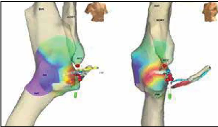
electro-physiological (EP) study was undertaken. Multi-electrode catheters were positioned into His bundle region and coronary sinus and an electro-anatomic mapping system was used (EnSite NavX® system, St. Jude Medical, Inc., St. Paul, MN, USA). His region was tagged on the 3-D anatomy of the right atrium (RA). Supraventricular tachycardia was induced by programmed atrial stimulation. AT was diagnosed by using the EP maneuvers. The tachycardia was mapped by using the En-site system and the earliest activation was found at the His region. Cryoablation cath-eter was advanced to the His region. Then, cryoablation could not be performed due to transient AV block during cryomapping of the tachycar-dia at the point of earliest activation. There was AV block even during manipulation of the cryoablation catheter. Then, 3-D anatomy of the aorta and aortic cusps were constructed (Fig. 1). The earliest local activation was detected within the NCC. Here, local atrial activity preceded onset of the p wave by 53 msec (Fig. 2). Location of the radiofrequency (RF) cath-eter was confirmed using fluoroscopy and electro mapping system. A single application of RF energy (power: 30 Watts, maximum temperature 55 degree) completely terminated the tachycardia. Total fluoroscopy time was 1 min 38 seconds. The patient was asymptomatic, ECG and rhythm Holter recordings showed no recurrence of AT.
Discussion
Focal ATs may originate in the right atrium around crista terminalis, coronary sinus, paraHisian region, tricuspid annulus and right atrial appendage. In left atrium, they primarily originate from pulmonary veins, mitral annulus, left atrial appendage and left septum (3). ATs originating Figure 1. 3-D geometry of aorta was constructed by En-Site system
HB - his bundle; IVC - inferior vena cava; NCC - non-coronary cusp; RA - right atrium; SVC - superior vena cava
Figure 2. Local atrial activity preceded onset of the P wave by 53 msec
Case Reports Anadolu Kardiyol Derg 2014; 14: 549-57
from paraHisian region are rare (1). Origin of AT is an important factor for the success and the complication of ablation procedures.
Ablation near His region has potential risk of damage of AV nodal conduction system. For this reason, cryoablation is preferred over RF abla-tion. Cryoablation has the advantage of cryomapping and if there is no AV block during cryomapping then it is generally safe to freeze to lower tem-peratures until permanent ablation occurs. In our case, we have seen AV block during cryomapping period. The superior part of the interatrial sep-tum has a close proximity to the NCC (4). Thus, ablation inside the NCC may terminate and cure ATs arising from this region. Actually, in certain patients, NCC offers the only available option for successful ablation.
Operator should be sure about the position of ablation catheter, because of the damage to aorta, aortic valve and coronary arteries dur-ing ablation in the NCC. Fluoroscopy and electro-anatomic mappdur-ing system may be used for this purpose. Also, 3-D mapping system facili-tates ablation, reduces radiation exposure and fluoroscopic time (5, 6). This system allows reconstruction of geometry and provides a high resolution map of the region with earliest activation (7, 8). Also, if avail-able intracardiac echocardiography may be helpful (9).
Ouyang et al. (10) performed RF ablation from NCC in 9 patients. Six of their patients had a history of failed ablation attempt. They reported that, all patients had successful ablation from the NCC. Our patient had RF abla-tion from the right atrium one year ago and had recurrence after 3 months.
Conclusion
We have described a successful catheter ablation procedure of focal AT originating from the NCC by using 3-D cardiac mapping system. Furthermore, very brief fluoroscopic use and low radiation exposure during the ablation procedure from the NCC is possible by using the electro-anatomic mapping system.
References
1. Poutiainen AM, Koistinen MJ, Airaksinen KE, Hartikainen EK, Kettunen RV, Karjalainen JE, et al. Prevalence and natural course of ectopic atrial tachycardia. Eur Heart J 1999; 20: 694-700. [CrossRef]
2. Pandozi C, Galeazzi M, Lavalle C, Ficili S, Russo M, Santini M. Navx-guided Cryoablation of Atrial Tachycardia Inside the Left Atrial Appendage. Indian Pacing Electrophysiol J 2011; 10: 556-61.
3. Roberts-Thomson KC, Kistler PM, Kalman JM, Atrial tachycardia: mecha-nisms, diagnosis, and management. Curr Probl Cardiol 2005; 30: 529-73. [CrossRef]
4. Raatikainen MJ, Huikuri HV. Successful catheter ablation of focal atrial tachycardia from the non-coronary aortic cusp. Europace 2007; 9: 216-9. [CrossRef]
5. Casella M, Pelargonio G, Dello Russo A, Riva S, Bartoletti S, Santangeli P, et al "Near-zero" fluoroscopic exposure in supraventricular arrhythmia ablation using the EnSite NavX™ mapping system: personal experience and review of the literature. J Interv Card Electrophysiol 2011; 31: 109-18. [CrossRef]
6. Jamil-Copley S, Linton N, Koa-Wing M, Kojodjojo P, Lim PB, Malcolme-Lawes L, et al. Application of ripple mapping with an electroanatomic mapping system for diagnosis of atrial tachycardias. J Cardiovasc Electrophysiol 2013; 24: 1361-9. [CrossRef]
7. Duru F. CARTO three-dimensional non-fluoroscopic electroanatomic mapping for catheter ablation of arrhythmias: a useful tool or an expen-sive toy for the electrophysiologist? Anadolu Kardiyol Derg 2002; 2: 330-7. 8. Casella M, Dello Russo A, Pelargonio G, Bongiorni MG, Del Greco M,
Piacenti M, et al. Rationale and design of the NO-PARTY trial: near-zero fluoroscopic exposure during catheter ablation of supraventricular arrhythmias in young patients. Cardiol Young 2012; 22: 539-46. [CrossRef] 9. Banchs JE, Patel P, Naccarelli GV, Gonzalez MD. Intracardiac echocar-diography in complex cardiac catheter ablation procedures. J Interv Card Electrophysiol 2010; 28: 167-84. [CrossRef]
10. Ouyang F, Ma J, Ho SY, Bänsch D, Schmidt B, Ernst S, et al. Focal atrial tachycardia originating from the non-coronary aortic sinus: electrophysi-ological characteristics and catheter ablation. J Am Coll Cardiol 2006; 48: 122-31. [CrossRef]
Address for Correspondence: Dr. Ekrem Güler, İstanbul Medipol Üniversitesi Tıp Fakültesi, Kardiyoloji Anabilim Dalı, İstanbul-Türkiye
Phone: +90 212 460 75 88 E-mail: [email protected] Available Online Date: 25.06.2014
©Copyright 2014 by Turkish Society of Cardiology - Available online at www.anakarder.com DOI:10.5152/akd.2014.5507
Case Reports