https://doi.org/10.1177/1120672120946287 European Journal of Ophthalmology 2021, Vol. 31(2) NP145 –NP150 © The Author(s) 2020 Article reuse guidelines: sagepub.com/journals-permissions DOI: 10.1177/1120672120946287 journals.sagepub.com/home/ejo
EJO
European Journal of OphthalmologyIntroduction
Coronavirus Disease 2019 (COVID-19) is a highly conta-gious newly recognized infection that has a significant worldwide impact on mortality and economic morbidity. Initial reports established from respiratory samples revealed severe acute respiratory syndrome coronavirus 2 (SARS-CoV-2) as the causative. Generally, patients infected with SARS-CoV-2 develop respiratory illness, with the first symptoms of fever, cough, and fatigue that quickly progress to pneumonia. Several patients were observed with atypical manifestations at the onset of the illness, such as conjunctivitis, or even presented with asymptomatic infection.
SARS-CoV-2 gains entry into host cells by binding to the angiotensin-converting enzyme 2 (ACE-2) receptor, which is distributed among various tissues, including the conjunctiva.1 During the SARS associated coronavirus
outbreak of 2003, a study indicated that healthcare
workers suffered from a higher risk of SARS infection when there was unprotected eye contact with secretions.2
There are increasing reports suggesting that a few COVID-19 pneumonia cases began with conjunctivitis as the initial symptom following contact with confirmed patients. Detection of viral RNA by reverse transcriptase polymer-ase chain reaction (RT-PCR) can be useful in early detec-tion of SARS-CoV-2 infecdetec-tion and taking appropriate quarantine measures. Therefore, determining whether SARS-CoV-2 is capable of transmitting through contact
Conjunctivitis as sole symptom of
COVID-19: A case report and review of
literature
Zeynep Kayaarasi Ozturker
Abstract
Introduction: Severe acute respiratory syndrome coronavirus 2 (SARS-CoV-2) is a novel virus causing an ongoing
pandemic in 2020. Although the symptomatic patients infected by SARS-CoV-2 generally show respiratory distress, atypical manifestations such as conjunctivitis are also observed. A series of cases are reported in which reverse transcriptase polymerase chain reaction (RT-PCR) testing on tears had demonstrated the presence of the virus. However, the transmission of the virus through ocular fluids remains unknown.
Case description: In this case report, the development of conjunctivitis is presented as the sole symptom of a new
coronavirus disease 2019 (COVID-19) in an emergency health care worker. The patient’s first application was to the ophthalmology clinic due to redness, stinging, tearing, and photophobia for one day in the right eye. The patient had no symptoms of fever, cough, shortness of breath, or fatigue. Two days later, the RT-PCR test, blood analysis, and chest computed tomography (CT) were applied to the patient for being in contact with a COVID positive patient. Conjunctival swabs did not identify SARS-CoV-2 by RT-PCR. However, nasopharyngeal swab and blood test confirmed the diagnosis of COVID-19. Chest CT did not show pneumonia.
Conclusion: This phenomenon shows that conjunctivitis may occur as a sole manifestation of COVID-19 which needs
to be carefully evaluated by health care workers and eye care professionals during the pandemic.
Keywords
Conjunctivitis, COVID-19, novel coronavirus, ocular infection, SARS-CoV-2
Department of Ophthalmology, Baskent University Istanbul Hospital, Istanbul, Turkey
Corresponding author:
Zeynep Kayaarasi Ozturker, Department of Ophthalmology, Baskent University Istanbul Hospital, Altunizade Mah. Kisikli Cad. Oymaci Sok. No: 7, Uskudar, Istanbul 34662, Turkey.
Email:[email protected]
with conjunctiva is an important consideration that war-rants for exploration.
Here, we present a case report of a health care worker diagnosed with COVID-19 who experienced conjunctivi-tis as the first and sole symptom of the disease. We believe it is essential for healthcare practitioners who play a sig-nificant role in the battle against the pandemic, to be knowledgeable about this problem and to take necessary steps to prevent the spread of the disease.
The study followed the tenets of the Declaration of Helsinki. The patient signed written informed consent for the research use of clinical records and data included in the study.
Case description
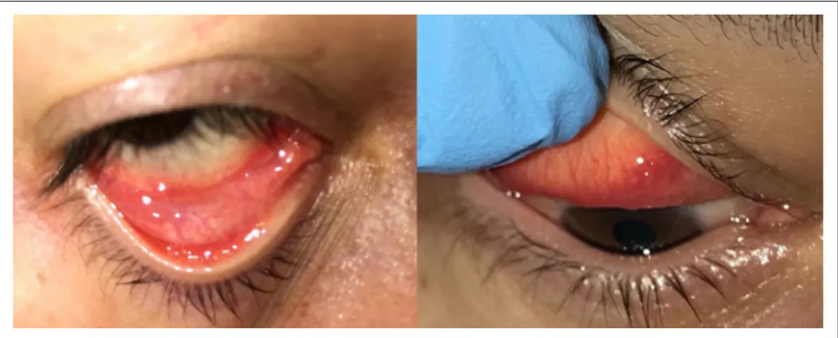
A 32-year-old otherwise healthy nurse working in the Emergency Department of Baskent University presented to the ophthalmology clinic on May 8, 2020, with one day history of redness, stinging, watery discharge, and photo-phobia in his right eye. The patient had no symptoms of fever, cough, shortness of breath, or general malaise. In his story, he did not declare any travel abroad in the last 14 days. The patient was diagnosed with idiopathic ante-rior uveitis in the right eye 2 years ago. Due to the regula-tions determined by the Turkish Republic Ministry of Health Department, oral and nasopharyngeal swab tests for SARS-CoV-2 were recommended for health care work-ers on demand. Upon the patient’s request, RT-PCR tests applied on April 14, 2020, and April 24, 2020, had nega-tive results consequently. In his ophthalmic examination, the visual acuity was 20/20 for both eyes without correc-tion. Intraocular pressure was 13 mmHg on the right and 14 mmHg on the left eye. Slit-lamp examination of the right eye revealed eyelid edema and serous secretion with 2+ conjunctival injection, mild chemosis, and follicular reaction in the upper and lower fornices (Figure 1). The
cornea was transparent, and no sign of inflammation was detected in the anterior chamber. Fundus examination revealed vital optic disc and macula. Anterior and poste-rior segment examination of the left eye was normal. Physical examination did not show any tenderness or enlargement of the submandibular, preauricular, or cervi-cal lymph nodes. The patient declared that he wore per-sonal protective equipment during close contact with suspected COVID-19 cases in the Emergency Department, but on some occasions, he had to remove his protective eyewear during the interventions.
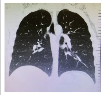
Considering acute conjunctivitis, moxifloxacin eye drop QID and artificial tears without preservative QID were prescribed for 7 days. On May 10, 2020, his mother, whom he lives together, developed a cough and fatigue, and RT-PCR test for nasopharyngeal swab was declared positive for COVID-19. Given his direct contact with his mother and occupational exposure, a nasopharyngeal RT-PCR test was applied to him on the same day, and a positive result was reported for SARS-CoV-2. Nasal swabs for Influenza a and b virus antigens were negative. His chest computed tomography and chest X-ray showed no signs of pneumonia (Figure 2). The routine blood examination showed high levels of glucose (114 mg/dL), C-reactive protein (13.00 mg/L), AST (44 U/L), ALT (108 U/L), LDH (251 U/L), and monocytes% (14.5%). He started taking systemic hydroxychloroquine and azithromycin for 5 days and instructed to self-quarantine until the complete resolution of the infection. On day 5 after diagnosis, the patient consulted his ophthalmologist by telemedicine service due to the worsening of his eye symptoms. Because of the infectious nature of COVID-19, quarantine protocols prevented access to the hospital during the active phase of the disease. Owing to the per-sistence of ocular complaints, the tear, and conjunctival swab samples were acquired by home visit. The samples were collected by conjunctival swab technique under the
Figure 1. A follicular conjunctival reaction in the upper and lower fornices, serous secretion, and mild chemosis in the right eye of the patient two days before the diagnosis of COVID-19.
patient’s approval. Upper and lower eyelids of both eyes were everted, and two separate samples were obtained by sweeping both fornices with sterile cotton swab without topical anesthesia. The swabs were inserted into a viral transport medium in ice before being tested for SARS-CoV-2. The patient had negative RT-PCR results for eye samples. Routine adenovirus tests also yielded a negative result.
Conclusion
Ocular diseases caused by coronaviruses are relatively rare compared to adenovirus and influenza viruses.3 Although
it is known that the main route of transmission of the SARS-CoV-2 is through the respiratory tract, several stud-ies have raised concerns due to infection in the unprotected eyes (Table 1). So far, it has not been clarified whether ocular secretions are contagious.
Based on current literature, SARS-CoV-2 can be detected in the swab samples taken from the conjunctiva by the RT-PCR method. In a study of 30 COVID positive patients, conjunctivitis developed in one patient (3.3%), which was the only symptom during the disease.4 Conjunctival
speci-men taken at days 3 and 5 gave positive results for viral RNA in the same patient, whereas all other patients had negative results. Conjunctival secretion of the patient was also tested for Herpes simplex virus, adenovirus, and other common viruses of conjunctivitis. However, the results were all negative indicating that the viral conjunctivitis of the patient might be related to SARS-CoV-2. Another study demonstrated conjunctivitis-like ocular findings in a much larger proportion of cases (31.6%).5 Conjunctival PCR was
found to be positive only in two cases (5.3%). Blood test
parameters, including white blood cell, neutrophil, procalci-tonin C, C-reactive protein, and lactate dehydrogenase lev-els, have also been reported to be higher in patients with conjunctivitis. In a similar study, the proportion of positive results for conjunctival SARS-CoV-2 was 2.5% which was significantly lower than the nasopharyngeal SARS-CoV-2 rate.6 However, it was suggested that a single time point
conjunctival sampling might lead to a low rate of virus detection in ocular samples.
The studies demonstrating serial conjunctival swab test-ing durtest-ing the disease have shown that SARS-CoV-2 RNA may persist for up to 21 days, even after the virus disappears in the nasal swabs.7 In addition, the RNA virus can be
re-detected in the conjunctiva after some time, indicating a per-sistent replication of the virus. The cytopathic effect of the RNA sample after inoculation in the Vero E-6 cell culture was also demonstrated. In contrast, another study suggested that the risk of SARS-CoV-2 transmission through tears was low. Among a total of 17 patients, only one patient had con-junctival injection and chemosis during the stay in the hos-pital. Conjunctival RT-PCR tests conducted on day three and day 20 were negative for all patients. The virus has been incubated in cell culture, and no cytopathic effect has been determined.8
In a separate case study of a 70- year-old man with symptoms of fever, cough, and fatigue, sequential con-junctival RT-PCR results remained positive until around two weeks after the nasopharyngeal RT-PCR results were negative.9 His left eye had previously been diagnosed with
nasolacrimal duct stenosis. In this clinically cured patient, the conjunctival swab of the left eye was still positive, sug-gesting that the obstruction of the lacrimal drainage system may reduce the virus clear through the eye and provide its shedding persistently.
Five studies have described biomicroscopic findings of COVID-19 patients with conjunctivitis.10–14 Ocular
symp-toms have emerged as the first symptom in all but one of these cases. Ocular complaints have been reported within a spectrum of red eye, watery discharge, photophobia, for-eign body sensation, and eyelid edema. Remarkable details in the slit-lamp examination were serous secretion, follicu-lar reaction in the upper and lower eyelid conjunctiva, che-mosis, keratoconjunctivitis, and pseudomembranous inflammation. In four of these cases, SARS-CoV-2 viral RNA was detected in the swab samples taken from the conjunctiva.10–13
Conjunctivitis remained the only sign and symptom of four active COVID-19 cases with a history of travel to the Lombardy region in Italy.15 None had a fever, general
malaise, or respiratory symptoms, consistent with our case. This report emphasizes the importance of ocular symptoms which can be overlooked if the patients do not show typical systemic symptoms of the disease.
Studies have shown that SARS-CoV-2 needs ACE-2 receptors for cell invasion.1 The ACE-2 receptors are
Figure 2. Computed tomography of the pulmonary parenchyma showing no sign of pneumonia.
found not only in human type 2 alveolar epithelial cells but also in the cornea and conjunctiva.16 This suggests that
ocular surface tissue may be a potential target tissue for SARS-CoV-2. Whether ocular contact with SARS-CoV-2 causes COVID-19 disease is unclear. However, according to one proposed theory, when the ocular surface comes into contact with SARS-CoV-2, virus particles can cause infection by draining the respiratory tract through the nasolacrimal canal.8
It is not yet known whether SARS-CoV-2 causes a more severe eye disease beyond keratoconjunctivitis. Robbins et al. demonstrated that a murine coronavirus was strongly tropic for retinal cells when introduced by cor-neal, anterior chamber, intravitreal, and intracerebral routes.17,18 Coronavirus was detected in the ganglion cell
layer ten days after inoculation, and long-term retinal pig-ment epithelium fibrosis and retinal atrophy have been detected after 14 weeks. Fibroblasts are suggested to play a significant role in the damaged tissue leading to gliosis of the retinal pigment epithelium.19
Serious eye problems may not be adequately assessed in patients with high SARS-CoV-2 virus loads who are
often treated in intensive care units for vital reasons. One report demonstrated severe ocular symptoms including tarsal hemorrhage, petechia and pseudomembranes in a 63-years-old patient who required intensive care surveil-lance and ventilatory support. Collected swabs did not identify any bacterial or viral etiology in conjunctival secretions and tears.20
While signs of conjunctivitis have usually been reported in adult patients, there has been one report on infected children.21 A 2-years-old asymptomatic boy was
detected through community screening with positive results of oropharyngeal SARS-CoV-2 RNA. He devel-oped conjunctivitis and eyelid dermatitis on day seven, which gradually disappeared in 5 days. As conjunctival swab was not taken, it is not clear whether a virus or sec-ondary bacterial infection caused ocular symptoms.
Telemedicine services also helped detect cases of con-junctivitis in the COVID-19 lockdown. One study reported a 27-years-old male patient who used telemedicine due to redness and foreign body sensation in his left eye.22
Examination revealed lid edema and conjunctival hypere-mia which required treatment. After 12 h, he admitted to
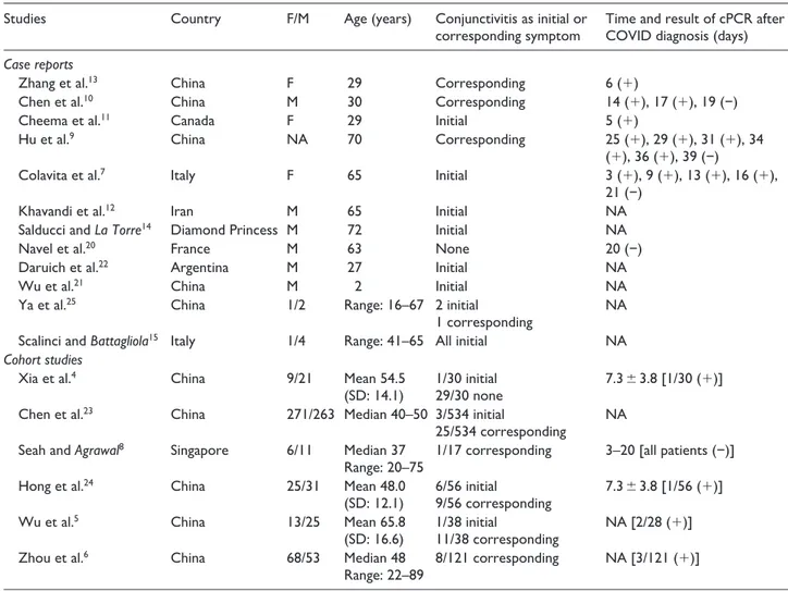
Table 1. Demographics and clinical data of COVID-19 patients with conjunctivitis.
Studies Country F/M Age (years) Conjunctivitis as initial or
corresponding symptom Time and result of cPCR after COVID diagnosis (days)
Case reports
Zhang et al.13 China F 29 Corresponding 6 (+)
Chen et al.10 China M 30 Corresponding 14 (+), 17 (+), 19 (−)
Cheema et al.11 Canada F 29 Initial 5 (+)
Hu et al.9 China NA 70 Corresponding 25 (+), 29 (+), 31 (+), 34
(+), 36 (+), 39 (−)
Colavita et al.7 Italy F 65 Initial 3 (+), 9 (+), 13 (+), 16 (+),
21 (−)
Khavandi et al.12 Iran M 65 Initial NA
Salducci and La Torre14 Diamond Princess M 72 Initial NA
Navel et al.20 France M 63 None 20 (−)
Daruich et al.22 Argentina M 27 Initial NA
Wu et al.21 China M 2 Initial NA
Ya et al.25 China 1/2 Range: 16–67 2 initial
1 corresponding NA
Scalinci and Battagliola15 Italy 1/4 Range: 41–65 All initial NA
Cohort studies
Xia et al.4 China 9/21 Mean 54.5
(SD: 14.1) 1/30 initial29/30 none 7.3 ± 3.8 [1/30 (+)]
Chen et al.23 China 271/263 Median 40–50 3/534 initial
25/534 corresponding NA
Seah and Agrawal8 Singapore 6/11 Median 37
Range: 20–75 1/17 corresponding 3–20 [all patients (−)]
Hong et al.24 China 25/31 Mean 48.0
(SD: 12.1) 6/56 initial9/56 corresponding 7.3 ± 3.8 [1/56 (+)]
Wu et al.5 China 13/25 Mean 65.8
(SD: 16.6) 1/38 initial11/38 corresponding NA [2/28 (+)]
Zhou et al.6 China 68/53 Median 48
Range: 22–89 8/121 corresponding NA [3/121 (+)]
the hospital because of added complaints of fever, cough, and dyspnea. In two other studies, ocular findings were evaluated retrospectively by making phone calls with the patients, and it was concluded that ocular symptoms were common in COVID-19 patients which might have clinical diagnostic significance.23,24
Recently, positive RT-PCR test findings in conjunctival swab samples of confirmed COVID-19 patients provide objective evidence for SARS-CoV-2 ocular surface infection. However, there are also patients with positive conjunctival RT-PCR results with no evidence of conjunctivitis.25 Therefore, the relationship between
ocu-lar surface infection and COVID-19 and whether the dis-ease can be transmitted through the ocular surface needs further research.
In conclusion conjunctivitis may appear to be the only sign and symptom of COVID-19, and these patients may not have fever, fatigue, or respiratory symptoms that may cause suspicion. The patients are generally those who report contact with COVID positive patients and there-fore undergo nasopharyngeal RT-PCR tests. According to a recent report, one-third of eye care professionals involved in the diagnosis and treatment of the patients during the pandemic accidentally acquired COVID-19 and demonstrated severe disease, including death.26
Thus, all physicians and ophthalmologists should be cau-tious when addressing a patient with conjunctivitis and adopt proper steps for the possible ocular transmission of SARS-CoV-2 until a vaccine is available. The RT-PCR test for nasopharyngeal or conjunctival swabs may help in early diagnosis of the disease if patients have no symp-toms other than conjunctivitis.
Acknowledgements
I thank Biologist Gulen Varan Ucak for her assistance in collect-ing nasopharyngeal and conjunctival swab samples for the RT-PCR analysis.
Declaration of conflicting interests
The author(s) declared no potential conflicts of interest with respect to the research, authorship, and/or publication of this article.
Funding
The author(s) received no financial support for the research, authorship, and/or publication of this article.
Informed consent statement
This case report was written using the clinical data of the patient in 2020. General consent was obtained in which the patient agrees with the research use of residual body parts and clinical records and data with anonymization.
ORCID iD
Zeynep Kayaarasi Ozturker https://orcid.org/0000-0002-
5513-9857
References
1. Wan Y, Shang J, Graham R, et al. Receptor Recognition by the Novel Coronavirus from Wuhan: An Analysis Based on Decade-Long Structural Studies of SARS Coronavirus. J
Virol. 2020; 94(7): e00127-20.
2. Raboud J, Shigayeva A, McGeer A, et al. Risk factors for SARS transmission from patients requiring intubation: A multicentre investigation 221 in Toronto, Canada. PLoS
ONE 2010; 5: e10717.
3. Belser JA, Rota PA and Tumpey TM. Ocular tropism of res-piratory viruses. Microbiol Mol Biol Rev. (2013) 77:144–56. 4. Xia J, Tong J, Liu M, Shen Y and Guo D. Evaluation of
coronavirus in tears and conjunctival secretions of patients with SARS-CoV-2 infection [published online ahead of print, 2020 Feb 26]. J Med Virol. 2020.
5. Wu P, Duan F, Luo C, et al. Characteristics of Ocular Findings of Patients With Coronavirus Disease 2019 (COVID-19) in Hubei Province, China. JAMA Ophthalmol. 2020;138(5): 575-578.
6. Zhou Y, Duan C, Zeng Y, et al. Ocular Findings and Proportion with Conjunctival SARS-COV-2 in COVID-19 Patients. Ophthalmology. 2020; S0161-6420 (20) 30405-X. 7. Colavita F, Lapa D and Carletti E, et al. SARS-CoV-2 isola-tion from ocular secreisola-tions of a patient with COVID-19 in Italy with prolonged viral RNA detection. Ann. Intern. Med. 2020.
8. Seah I and Agrawal R. Can the Coronavirus Disease 2019 (COVID-19) Affect the Eyes? A Review of Coronaviruses and Ocular Implications in Humans and Animals. Ocul
Immunol Inflamm. 2020; 28(3): 391-395.
9. Hu Y, Chen T, Liu M, et al. Positive detection of SARS-CoV-2 combined HSV1 and HHV6B virus nucleic acid in tear and conjunctival secretions of a non-conjunctivitis COVID-19 patient with obstruction of common lacrimal duct [published online ahead of print, 2020 May 14]. Acta Ophthalmol. 10. Chen L, Liu M and Zhang Z, , et al. Ocular manifestations
of a hospitalised patient with confirmed 2019 novel corona-virus disease. Br J Ophthalmol. 2020;104 (6): 748-751. 11. Cheema M, Aghazadeh H and Nazarali S, et al. Kerato-
conjunctivitis as the initial medical presentation of the novel coronavirus disease 2019 (COVID-19) [published online ahead of print, 2020 Apr 2]. Can J Ophthalmol. 2020; S0008-4182 (20) 30305-7.
12. Khavandi S, Tabibzadeh E, Naderan M and Shoar S. Corona virus disease-19 (COVID-19) presenting as conjunctivitis: atypically high-risk during a pandemic [published online ahead of print, 2020 Apr 27]. Cont Lens Anterior Eye. 2020; S1367-0484 (20) 30083-7.
13. Zhang X, Chen X, Chen L, et al. The evidence of SARS-CoV-2 infection on ocular surface. Ocul Surf. 2020; 18(3): 360-362.
14. Salducci M and La Torre G (2020). COVID-19 emergency in the cruise’s ship: A case report of conjunctivitis. La
Clinica Terapeutica. 2020; 171(3), e189-e191.
15. Scalinci SZ and Trovato Battagliola E. Conjunctivitis can be the only presenting sign and symptom of COVID-19.
IDCases. 2020; 20: e00774.
16. Zou X, Chen K and Zou J, , et al. Single-cell RNA-seq data analysis on the receptor ACE2 expression reveals the poten-tial risk of different human organs vulnerable to 2019-nCoV infection. Front Med. 2020;14(2):185-192.
17. Robbins SG, Detrick B and Hooks JJ. Retinopathy follow-ing intravitreal injection of mice with MHV strain JHM.
Adv Exp Med Biol. 1990; 276: 519–524.
18. Robbins SG, Detrick B and Hooks JJ. Ocular tropisms of murine coronavirus (strain JHM) after inoculation by various routes. Invest Ophthalmol Vis Sci. 1991; 32(6): 1883–1893. 19. Neri P and Pichi F. COVID-19 and the eye immunity:
Lesson learned from the past and possible new therapeutic insights. Int Ophthalmol. 2020; 40(5): 1057-1060.
20. Navel V, Chiambaretta F and Dutheil F. Haemorrhagic conjunctivitis with pseudomembranous related to SARS-CoV-2 [published online ahead of print, 2020 May 6]. Am J
Ophthalmol Case Rep. 2020; 100735.
21. Wu P, Liang L, Chen C, et al. A child confirmed COVID-19 with only symptoms of conjunctivitis and eyelid dermatitis [published online ahead of print, 2020 Apr 24]. Graefes Arch
Clin Exp Ophthalmol. 2020;1-2.
22. Daruich A, Martin D and Bremond-Gignac D. Ocular manifestation as first sign of Coronavirus Disease 2019
(COVID-19): Interest of telemedicine during the pandemic context. J Fr Ophtalmol. 2020; 43(5): 389-391.
23. Chen L, Deng C and Chen X, et al. Ocular manifestations and clinical characteristics of 534 cases of COVID-19 in China: A cross-sectional study. medRxiv [Preprint]. 2020 March 16. doi:10.1101/2020.03.12.20034678.
24. Hong N, Yu W and Xia J, et al. Evaluation of ocular symp-toms and tropism of SARS-CoV-2 in patients confirmed with COVID-19 [published online ahead of print, 2020 Apr 26]. Acta Ophthalmol. 2020;10.1111/aos.14445.
25. Ya Y, Yanping S and Ming Y, et al. Novel coronavirus pmneumonia combined with conjunctivitis: Three cases report. Zhonghua Shiyan Yanke Zazhi/Chinese Journal of Experimental Ophthalmology. 38. 242-244. 10.3760/cma.j .issn.2095-0160.2020.0006.
26. Qiao C, Zhang H, He M, et al. Symptomatic COVID-19 in Eye Professionals in Wuhan, China [published online ahead of print, 2020 Apr 18]. Ophthalmology. 2020; S0161-6420 (20) 30403-6.