Received Date: 01.10.2015 Accepted Date: 21.10.2015 © Copyright 2016 by Gaziosmanpaşa Taksim Training and Research Hospital. Available on-line at www.jarem.org DOI: 10.5152/jarem.2016.954 Address for Correspondence: Dr. Sevgi Şahin,
E-mail: [email protected]
Wegener Granulomatosis Complicated by Brain Abscess
Caused by Nocardia spp.
Sevgi Şahin
1, Baran Balcan
2, Şafak Kızıltaş
3, Mehtap Aydın
41Division of Nephrology, Acıbadem University School of Medicine, İstanbul, Turkey 2Department of Chest Diseases, Başkent University İstanbul Hospital, İstanbul, Turkey 3Department of Gastroenterology, Başkent University İstanbul Hospital, İstanbul, Turkey 4Department of Infectious Diseases, Başkent University İstanbul Hospital, İstanbul, Turkey
ABSTRACT
Wegener granulomatosis is a multisystemic disease associated with high mortality rate and characterized by necrotizing granulomatous vascu-litis predominantly in the respiratory tract and kidneys. Presence of kidney failure at the time of diagnosis describes a poor prognostic marker. We presented a Wegener granulomatosis case complicated with brain abscess caused by Nocardia. (JAREM 2016; 6: 126-8)
Keywords: Rapidly progressive glomerulonephritis, acute kidney failure, immunosuppression, infection INTRODUCTION
Wegener’s granulomatosis (WG) is a multisystemic disease that is associated with a high mortality rate and is characterized by nec-rotizing granulomatous vasculitis, predominantly in the respira-tory tract and kidneys. The presence of kidney failure at the time of diagnosis describes a poor diagnostic marker. We present a case of WG complicated with brain abscess caused by Nocardia during the treatment.
CASE PRESENTATION
A 66-year-old male patient with complaints of fatigue, elevated fever, weight loss, hemoptysis, and left ankle pain was examined over the course of 2 months in the infectious diseases clinic of a hospital. His PPD test revealed that he was anergic, and no infec-tion or malignancy focus was detected. The patient was admitted to the nephrology unit due to disruption observed in his kidney function tests. Verbal consent was obtained from the patient. In his laboratory findings, leucocytes were 10,200/mm3, Hb was
9.1 g/dL, Hct was 25.3%, thrombocytes were 287,000/mm3, urea
was 225 mg/dL, creatinine was 12.4 mg/dL, CRP was 52 mg/L, and sedimentation rate was 92 mm/h; proteinuria was observed in urinalysis, and plenty of erythrocytes were present in his urine sediment. Hemodialysis was started. He was positive (+) for anti-neutrophil cytoplasmic antibodies (c-ANCAs), while complement levels were within the normal range. On observing his kidney size to be within the normal range in his ultrasonography, kidney biop-sy was performed with the pre-diagnosis of “rapidly progressive glomerulonephritis”. Along with the elevation of sedimentation, the presence of c-ANCA positivity, lower respiratory tract involve-ment, and the compatibility of kidney biopsy findings with Pauci-immune Type III crescentic glomerulonephritis, the patient was diagnosed with WG. He received 500 mg methylprednisolone (Prednol; Mustafa Nevzat Pharmaceutical, İstanbul, Turkey) pulse treatment for three consecutive days, followed by 750 mg
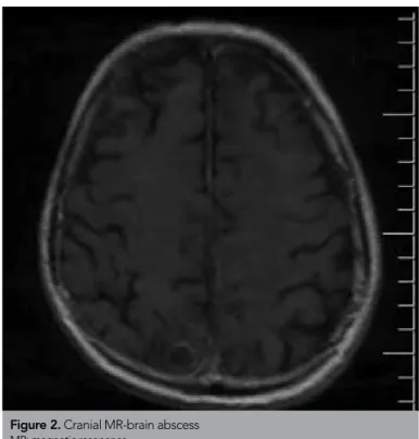
intra-venous (IV) cyclophosphamide (Endoxan; Eczacıbaşı-Baxter Hos-pital Products, İstanbul, Turkey) and five sessions of plasmapher-esis. In the first week of treatment, creatinine levels (creatinine: 1.2 mg/dL) dropped down. The recommendation of oral prednis-olone at a dose of monthly 750 mg IV cyclophosphamide+1 mg/ kg was registered in his polyclinic follow-up. In the third month of his treatment, the patient was admitted with complaints of coughing, elevated fever, and left side pain. Bronchopneumonia was detected in his chest computed tomography (CT) (Figure 1). IV 2×1000 mg/day ceftriaxone (Desefin; Deva Holding, İstanbul, Turkey) and 500 mg/day levofloxacin (Tavanic; Zentiva Medical Products, Lüleburgaz, Turkey) were started, and the steroid dose was lowered. On the second day of his treatment, the patient developed fatigue and loss of balance, and his cranial magnetic resonance (MR) imaging showed a brain abscess (Figure 2). Sur-gical treatment was not recommended because the abscess size was larger than 1.5 cm. Samples were obtained via lumbar puncture for culture. Antibiotic treatment was expanded to me-ropenem 3×1000 mg/day (Meronem; AstraZeneca, Macclesfield, England) + TMP/SMX (Bactrim; Deva Holding, Istanbul, Turkey). Because Nocardia spp. reproduced in his cerebrospinal fluid cul-ture, the treatment was planned as 8 weeks of parenteral 2×1000 mg ceftriaxone + 2×160/800 mg trimethoprim/sulfamethoxazole (TMP/SMX), followed by 6 months of oral TMP/SMX and monthly cranial MR controls. Immunosuppressive treatment directed at WG was limited to 16 mg/day methylprednisolone. At present, the controls are continued in the fourth month of the treatment, and the size of patient’s brain abscess is decreasing and his renal functions (serum creatinine level: 1.3 mg/dL) are stable.
DISCUSSION
WG is a necrotizing granulomatosis vasculitis with an unknown etiology and a prevalence of 3/100,000; it is observed in the fifth decade regardless of sex (1, 2). Its main symptoms are cough-ing, dyspnea, and hemoptysis. More than 90% of patients have
bilateral, multiple nodules and parenchymal infiltrations of vary-ing sizes, and lung imagvary-ing displays abnormalities (3). Pulmonary hemorrhage along with kidney function disorder increases the mortality rate.
Glomerulonephritis develops in approximately 75% of WG pa-tients. Histologically, focal necrotizing glomerulonephritis and crescentic glomerulonephritis are observed, and they can rapidly lead to kidney failure (4). Hematuria, proteinuria, and erythrocyte cylinders are observed as urinary findings. Two-thirds of patients
can have musculoskeletal complaints. Our patient had findings related to the kidneys, lungs, and skeletal system.
More than 90% of patients are c-ANCA positive (5, 6). In case of lung involvement accompanying hematuria/proteinuria, kidney or lung biopsy may need to be performed for a definitive diagno-sis. Despite the risk of hemorrhage in our uremic patient, kidney biopsy was performed in order to plan for the immunosuppres-sive treatment, given his age. Patients who require dialysis have been reported to benefit from an aggressive treatment (7). Fauci et al. (2) demonstrated in their study that a combination therapy of oral steroid + cyclophosphamide leads to 90% remis-sion. However, because of the adverse effects of medicines, cy-clophosphamide treatment is recommended to be administered parenterally (1, 3). We administered 0.5 g/m2 single-dose IV pulse
cyclophosphamide once a month for 3 days and 0.5 g/day IV methylprednisolone, followed by 1 mg/kg/day oral prednisolone follow-up treatment.
In a study comparing the effectiveness of plasmapheresis and pulse steroid in patients with kidney failure, the group that un-derwent plasmapheresis was reported to develop fewer late-stage kidney failures (8). Deaths are determined to occur within 3 months after the start of the treatment of this disease, which has a high mortality rate. In total, 54% of reported deaths are as-sociated with infections. While the treatment of our case was on-going with the diagnosis of lobar pneumonia that developed in the third month of immunosuppressive treatment, loss of balance occurred, which led to performing cranial MR that showed brain abscess in the parietal region. Reproduction of Nocardia spp. in the culture taken by lumber puncture established the diagnosis. Nocardia is a rare, mortal, and opportunistic gram-positive bac-terium that is particularly observed in cases with cellular immunity disorder (9, 10). Sole central nervous system involvement is very rare (9%) (9, 10). Slow growth in culture may result in mortality due to delayed diagnosis and treatment.
CONCLUSION
Patients who receive immunosuppressive treatments must be closely followed up as high-risk groups with regard to oppor-tunistic infections, and the treatment of infections must be per-formed energetically.
Informed Consent: Verbal informed consent was obtained from patient who participated in this case.
Peer-review: Externally peer-reviewed.
Author Contributions: Concept - S.Ş.; Supervision - S.Ş.; Resources - M.A., B.B.; Materials - Ş.K.; Analysis and/or Interpretation - S.Ş.; Literature Search - S.Ş.; Writing Manuscript - S.Ş.
Conflict of Interest: No conflict of interest was declared by the authors. Financial Disclosure: The authors declared that this study has received no financial support.
REFERENCES
1. Hoffman GS, Kerr GS, Leavitt RY, Hallahan CW, Lebovics RS, Travis WD, et al. Wegener granulomatosis: An analysis of 158 patients. Ann Intern Med 1992; 116: 488-98. [CrossRef]
127
Şahin et al.
Opportunistic Infection in Wegener Granulomatosis. JAREM 2016; 6: 126-8
Figure 1. Chest CT-bronchopneumonic infiltration
CT: computed tomography
Figure 2. Cranial MR-brain abscess
2. Fauci AS, Haynes BF, Katz P, Wolff SM. Wegener’s granulomatosis: prospective clinical and therapeutic experience with 85 patients for 21 years. Ann Intern Med 1983; 98: 76-85. [CrossRef]
3. Cordier JF, Valeyre D, Guillevin L, Loire R, Brechot JM. Pulmonary Wegener’s granulomatosis. A clinical and imaging study of 77 cases. Chest 1990; 97: 906-12. [CrossRef]
4. Jennette JC, Falk RJ, Andrassy K, Bacon PA, Churg J, Gross WL, et al. Nomenclature of systemic vasculitides. Proposal of an international consensus conference. Arthritis Rheum 1994; 37: 187-92. [CrossRef]
5. Niles JL, Pan GL, Collins AB, Shannon T, Skates S, Fienberg R, et al. Antigen-specific radioimmunoassays for anti-neutrophil cytoplasmic antibodies in the diagnosis of rapidly progressive glomerulonephri-tis. J Am Soc Nephrol 1991; 2: 27-36.
6. Kitching AR, Hutchinson P, Atkins RC, Holdsworth SR. The role of flow cytometric c ANCA detection in screening for acute pauci-immune
crescentic glomerulonephritis. Nephrol Dial Transplant 2004; 19: 365-70.
[CrossRef]
7. Lee T, Gasim A, Derebail VK, Chung Y, McGregor JG, Lionaki S, et al. Predictors of treatment outcomes in ANCA-associated vasculitis with severe kidney failure. Clin J Am Soc Nephrol 2014; 9: 905-13.
[CrossRef]
8. Jayne DRW, Gaskin G, Rasmussen N, Abramowicz D, Ferrario F, Gu-illevin L, et al. Randomized trial of plasma exchange or high-dosage methylprednisolone as adjunctive therapy for severe renal vasculitis. J Am Soc Nephrol 2007; 18: 2180-8. [CrossRef]
9. Corti ME, Villafañe Fioti ME. Nocardiosis: a review. Int J Infect Dis 2003; 7: 243-50. [CrossRef]
10. Beaman BL, Beaman L. Nocardia species, host-parasite relations-hips. Clin Microbiol Rev 1994; 7: 213-64. [CrossRef]