Evaluation of oxidative stress, hematological and biochemical
parameters during Toxoplasma gondii infection in gerbils
Nurgül ATMACA
1, Miyase ÇINAR
2, Bayram GÜNER
2, Ruhi KABAKÇI
1,
Aycan Nuriye GAZYAĞCI
3, Hasan Tarık ATMACA
4, Sıla CANPOLAT
4KırıkkaleUniversity, Faculty of Veterinary Medicine, 1
Department of Physiology; 2Department of Biochemistry; 3Department of Parasitology; 4Department of Pathology, Kırıkkale, Turkey.
Summary: The aim of the present study was to investigate the alterations of oxidative stress, hematological and biochemical
parameters in experimental infection caused by Toxoplasma gondii in gerbil. A total of 16 gerbil, 8 of which were control and 8 was infection group, were used in the study. The gerbils were infected by intraperitoneal inoculation of 5000 T. gondii RH strain tachyzoites. In group of, the gerbil were sacrificed at 7th day after inoculation. At the end of this period, blood samples collected and erythrocyte malondialdehyde (MDA) concentrations, superoxide dismutase (SOD), catalase (CAT) activities, plasma aspartat aminotransferase (AST) and alanine aminotranspherase (ALT) activities, total protein, albumin, globulin were determined. Besides, hematological parameters were analysed in whole blood. Aspartat aminotransferase and ALT activities and MDA concentrations and neutrophil percentage and total leukocyte counts increased significantly in infected group when compared to control. In infected group, SOD activities, albumin concentrations and lymhocyte percentage decreased when compared to control. The results of this study suggest that oxidative stress, hematological and biochemical alterations may contribute to the pathogenesis of toxoplasmosis in gerbils.
Key words: Blood parameters, gerbil, oxidative stress, Toxoplasma gondii.
Gerbillerde Toxoplasma gondii enfeksiyonu süresince oksidatif stres, hematolojik ve biyokimyasal
parametrelerin değerlendirilmesi
Özet: Bu çalışma Toxsoplasma gondii (T.gondii) ile deneysel olarak enfekte edilmiş gerbillerde oksidatif stres ile hematolojik
ve biyokimyasal parametrelerde meydana gelen değişikliklerin araştırılması amacıyla yapıldı. Çalışmada kontrol grubunda 8, enfeksiyon grubunda 8 olmak üzere toplam 16 tane gerbil kullanıldı. Gerbiller 5000 T. gondii RH takizoiti ile intraperitoneal yolla enfekte edildi. İnokülasyon sonrası 7. günde gerbiller sakrifiye edildi. Daha sonra kan örnekleri toplandı ve eritrosit malondialdehit (MDA) konsantrasyonu, süperoksit dismutaz (SOD) ve katalaz (CAT) aktiviteleri, plazma aspartat aminotransferaz (AST) ve alanin aminotransferaz (ALT) aktiviteleri, total protein, albumin ve globin düzeyleri belirlendi. Bunun yanısıra, tüm kanda hematolojik parametlerin analizi yapıldı. Kontrol ile karşılaştırıldığında enfekte grupta, AST ve ALT aktiviteleri ve MDA konsantrasyonu, nötrofil oranı ve toplam akyuvar sayısında anlamlı bir artış bulundu. Enfekte grupta, SOD aktivitesi, albumin konsantrasyonu ve lenfosit oranında kontrol ile karşılaştırıldığında düşme belirlendi. Bu çalışmanın sonuçlarına göre, gerbillerde toksoplazmozisin patogenezinde oksidatif stresin, hematolojik ve biyokimyasal değişikliklerin katkısının olduğu söylenebilir.
Anahtar sözcükler: Gerbil, kan parametreleri, oksidatif stres, Toxoplasma gondii.
Introduction
Toxoplasmosis is a parasitic disease caused by Toxoplasma gondii (30). Toxoplasma gondii is an obligate intracellular parasite that frequently infects warm-blooded animals, including humans (9, 11, 18). Toxoplasma gondii is one of the most common parasites throughout the world (18) which can cause either acute or chronic infection (36). Cats are the only known definitive hosts of T. gondii and excrete environmentally resistant oocysts in their feces (18, 35). Hosts may
become infected by ingesting food or drinks
contaminated with oocysts, or by ingesting undercooked
meat infected with T. gondii (18, 38). Toxoplasma gondii can be located in every vital organ, and especially in acute stage it can be seen in blood, cerebrospinal fluid, semen, tears, saliva and urine. It is a zoonotic disease that causes abort and fetal destruction due to placental transmission (30).
Infections can cause harmful effects on healthy hosts and stimulate the infected host’s immune system. In response, the immune system generates toxic oxidants known as free radical species (43). Reactive oxygen species degrade polyunsaturated lipids to form MDA (42), a reactive aldehyde that causes oxidative stress in
tissue and cells (26). Malondialdehyde damages proteins by generating covalent adducts, the accumulation of which likely participates in tissue damage (48). Malondialdehyde is used as a biomarker to measure lipid peroxidation in an organism (2, 39).
Free radicals are highly reactive chemical species, and antioxidant defense systems enable tissues to detoxify these toxic oxidants. Superoxide dismutase is an important antioxidant enzyme that is present in all aerobic cells. This enzyme catalyzes the dismutation of superoxide into oxygen and hydrogen peroxide, which, in turn, is reduced to water by the antioxidant enzymes catalase and glutathione peroxidase (2, 31). Catalase is a naturally occurring enzyme present in all living, and it protects cells from oxidative damage by preventing the formation of hydroxyl radicals by detoxifying hydrogen peroxide molecules (2).
There is some evidence that oxidative stress is involved in the pathogenesis of toxoplasmosis in infected animals (4, 5, 19, 23, 28) and humans (3, 10, 33). However, such evidence is sparse, and no studies on oxidative stress have been conducted in T. gondii-infected gerbils. Therefore, the main objective of this study was to determine the effects of toxoplasmosis on
lipid peroxidation, antioxidant defense systems,
hematological and biochemical parameters in gerbils. Results were compared to those obtained with uninfected animals for a better understanding of toxoplasmosis pathogenesis.
Materials and Methods
Acute infection was induced in sixteen 1-2 month-old Mongolian gerbil were used to study. Only 8 gerbils were used to acute Toxoplasma gondii infections. The virulent T. gondii RH strain, which is maintained in the Parasitology Laboratory of the Turkish Public Health Institution by continuous passage every 3–4 days in gerbils, was used to induce acute infection. Eight gerbils were infected by intraperitoneal inoculation of 5000 T. gondii RH tachyzoites. In group of, the gerbils were sacrificed at 7. day after inoculation. Another healthy 8 gerbils sacrificed at the beginning of study.
This study was conducted in University of Kirikkale, Faculty of Veterinary Medicine. The animal care and use protocol was reviewed and approved by the Ethics Committee of the Kirikkale University.
Collection of blood samples and preparing for analysis: At the end of the experiment, blood samples were collected into evacuated tubes containing EDTA solution as anticoagulant by the cardiac puncture to
determine some hematological and biochemical
parameters, MDA, indicator of lipid peroxidation, some enzymatic antioxidants such as SOD and CAT. Whole blood samples were centrifuged at 1600x g for 10 min at 4ºC to separate the plasma. Following separation of
plasma, the upper layer of the erythrocyte pellet that contains the buffy coat was removed. Erythrocytes hemolysates and plasma were stored at -80°C and -30οC respectively until analysis.
Hematological and biochemical analysis:
Anticoagulated blood samples were used to determine total leukocyte counts, erythrocyte counts, concentration of blood hemoglobin (Hb), packed cells volume (PCV), percentages of each of the five basic leukocytes were measured on hematology analyzer (Abacus Junior Vet 5, Austria). All samples were evaluated on the same day.
Plasma aspartate aminotransferase (AST) (EC 2.6.1.1), alanine aminotransferase (ALT) (EC 2.6.1.2) activities, total protein, albumin concentrations were determined using commercial test kits (Biolabo, France) by spectrophotometer (Shimadzu UV-1700, Japan). The plasma globulin concentrations were calculated by subtracting the albumin values from the total protein values.
Analysis of oxidative stress markers: The erythrocytes were washed three times with ice cold saline solution (0.9 % NaCl). The erythrocyte pellet hemolyzed with ice-cold bidistilled water. The hemolysates were used for the determination of the hemoglobin (Hb) and MDA concentrations, SOD and CAT activities.
Determination of hemoglobin in erythrocyte: The concentration of blood hemoglobin was determined by the cyanomethemoglobin method which is based on the measurement of cyanmethemoglobin at 540 nm in a spectrophotometer. Results were expressed as g/dl Hb (25).
Determination of lipid peroxidation: Lipid
peroxidation in erythrocytes was estimated by the thio-barbituric acid (TBA) method that determines aldehyde formed by degradation of hydroperoxide, including MDA. The absorbance of the reaction product between MDA and TBA was measured spectrophotometrically at 536 nm. The data were expressed as nmol/g Hb of erythrocyte hemolysate (15)
Determination of SOD activity: The activity of SOD (EC 1.15.1.1) was determined in erythrocyte hemolysates according to the method described by Sun et al. (46). This method provides that the rate of nitroblue tetrazolium (NBT) reduction to blue formazan by the superoxide anion generated by the xanthine oxidase (XOD) reaction is monitored spectrophotometrically at 560 nm. One unit of SOD was considered a 50% inhibition of reduction of NBT under the condition of the assay. The results were expressed as U/g Hb.
Determination of CAT activity: Catalase (EC 1.11.1.6) activity was assayed by the method of Aebi (1) in erythrocyte hemolysate. Decomposition of H2O2 was followed directly by monitoring the reduction of absorbance at 240 nm. Enzyme activity was calculated as catalytic content of a sample. The data were expressed as k/g Hb.
Statistical analysis: Statistical analysis was performed with SPSS software package (Version 15.0 for Windows). Data were expressed as mean±standard error (SE). For comparison of two groups of continuous variables, independent samples t-test was used. A probability value of P<0.05 were considered as significant difference for all statistical calculations.
Results
Evaluation of lipid peroxidation and antioxidant enzymes activity: Table 1 shows the erythrocyte MDA concentrations and some enzymatic antioxidants in gerbils infected toxoplasmosis. Gerbils infected with Toxoplasma gondii had significantly higher (P<0.05) erythrocyte MDA concentrations and lower SOD activity (P<0.01) in comparison to control gerbils. There was no significant difference in erythrocyte CAT activity between control and infected gerbils (P>0.05).
Table 1. Erythrocyte lipid peroxidation and some enzymatic antioxidant parameters in gerbils infected toxoplasmosis Tablo 1. Toksoplazma enfekte gerbillerde eritrosit lipit peroksi-dasyon ve bazı enzimatik antioksidant parametreler
Parameters Control (n=8) Infected (n=8) P value MDA (nmol/gHb) 119.76±17.09 198.89±20.30* P<0.05 SOD ( U /gHb) 1.46 ± 0.16 0.73 ± 0.14** P<0.01 CAT (k /gHb) 19.17 ± 1.57 16.82 ± 0.91 P>0.05
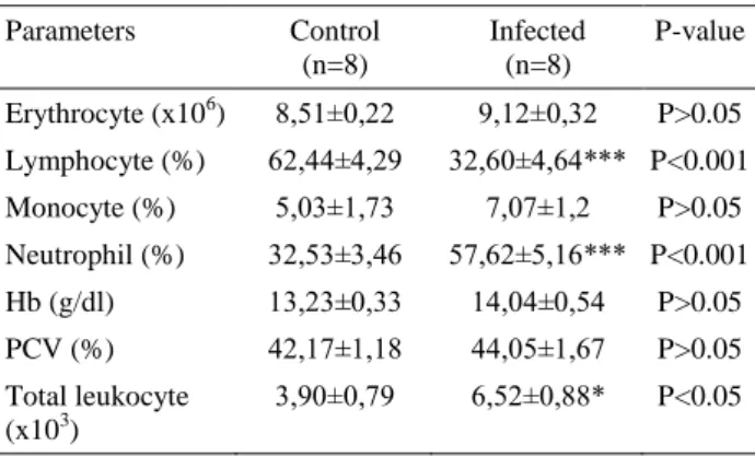
Table 2. Some hematological parameters in gerbils infected toxoplasmosis
Tablo 2. Toksoplazma enfekte gerbillerde bazı hematolojik parametreler Parameters Control (n=8) Infected (n=8) P-value Erythrocyte (x106) 8,51±0,22 9,12±0,32 P>0.05 Lymphocyte (%) 62,44±4,29 32,60±4,64*** P<0.001 Monocyte (%) 5,03±1,73 7,07±1,2 P>0.05 Neutrophil (%) 32,53±3,46 57,62±5,16*** P<0.001 Hb (g/dl) 13,23±0,33 14,04±0,54 P>0.05 PCV (%) 42,17±1,18 44,05±1,67 P>0.05 Total leukocyte (x103) 3,90±0,79 6,52±0,88* P<0.05
Hematological and biochemical analysis: The hematological findings in control and infected gerbils are presented in table 2. Neutrophils percentage (P≤0.001) and total leukocyte counts (P<0.05) gerbils infected with Toxoplasma gondii were increased, while lymphocytes percentage (P<0.001) were decreased significantly compared to control gerbils. There was no significant difference in RBC, monosit, Hb, Htc levels between control and infected gerbils (P>0.05).
Table 3 shows the plasma ALT and AST activities, total protein, albumin, globulin concentrations in gerbils infected toxoplasmosis. The activities of plasma AST (P<0.001), ALT (P≤0.01) were significantly increased, while the concentrations of plasma albumin (P< 0.001) were significantly decreased in infected gerbils when compared to control gerbils. No change was detected in the concentrations of plasma total protein and globulin between control and infected gerbils (P>0.05).
Table 3. Plasma liver enzyme activities and protein concentrations in gerbils infected toxoplasmosis
Tablo 3. Toksoplazma enfekte gerbillerde plazma karaciğer enzim aktiviteleri ve protein konsantrasyonları
Parameters Control (n=8) Infected (n=8) P-value Liver enzymes ALT (IU/L) 57.84 ± 7.95 207.75 ± 38.48** P<0.01 AST (IU/L) 140.85 ± 13.77 410.13 ± 33.36*** P<0.001 Protein concentrations Total protein (g/dl) 7.35 ± 0.79 6.19 ± 0.49 P>0.05 Albumin (g/dl) 3.31 ± 0.13 2.01 ± 0.21*** P<0.001 Globulin (g/dl) 4.03 ± 0.82 4.18 ± 0.62 P>0.05
Discussion and Conclusion
This study showed that T. gondii infection exert significant effects on erythrocyte MDA concentrations, SOD activities, plasma AST and ALT activities, albumin concentrations, total leukocyte counts, neutrophils and lymphocyte percentages
Lipid peroxidation, which generates free radical species, plays a role in the pathogenesis of many parasitic and protozoan infections (7, 34). Lipid peroxidation, as determined by erythrocyte MDA concentrations in the present study, was significantly increased due to toxoplasma infection. This may have resulted from the excess production of free radicals and oxidants following infection. Free radicals can react with DNA, resulting in genetic mutations or cytotoxicity. They also can bind to erythrocyte membranes, which are rich in polyunsaturated fatty acids (27), thereby causing membrane destruction and cellular damage (29). Karaman et al. (33) reported that serum MDA concentrations were significantly increased in T. gondii-infected humans, and Yang et al. (50) showed that serum concentrations of free oxygen radicals (NO, -OH, O2-) increased in T. gondii-infected mice. Our results are in accordance with Karaman et al. (33), which suggested that erythrocyte MDA concentrations were significantly increased by T. gondii infection. In contrast, Engin et al. (23) found no change in serum MDA concentrations of mice infected with T.gondii.
Preventing lipid peroxidation is essential in biological systems. To counteract the harmful effects of ROS, all
oxygen-metabolizing cells are equipped with cellular antioxidant defense systems (2). Superoxide dismutase is
an important physiological antioxidant defense
mechanism in aerobic organism. This enzyme prevents the formation of the hydroxyl radical by detoxifiying hydrogen peroxide (31). The decrease in SOD activity observed in our study may be related to the increasing the severity of parasitemia and oxidative stress (7). Moreover, erythrocytes infected with various protozoan parasites show decreased SOD activity (7, 22).
The assessment of the severity of disease and the prognosis of recovery of affected animals can only be made on the basis of clinical and laboratory data such as hematological and biochemical blood parameters (37). Polymorphonuclear leukocytes are one of the first cell types to arrive at a site of infection (32). Neutrophils are the first cells to be elicited during an inflammatory response and appear within minutes of chemokine release from the site of tachyzoite infection (17). Previous studies have suggested that neutrophils are important for controlling toxoplasmosis in mice (13, 16) and humans (12). With the T. gondii-infected group, our data showed that neutrophil percentage and the total leukocyte counts was increased compared to the control group. In contrast, the lymphocytes percentage was decreased in infected gerbils. Considering the lymphocyte cell types that are predominant in gerbils (45), the reason of the high percentage of neutrophil is probably an inflammatory response to the tachyzoite proliferation during infection. However, Tonin et al. (47) reported that the numbers of circulating lymphocytes were increased in T. gondii-infected rats. Tonin et al. (47) found that the number of lymphocytes was higher in toxoplasma-infected rats however in this study neutrophil percentage was found to be higher in toxoplasma-infected gerbils. This discrepancy can be related to the different sampling period of these studies.
Toxoplasmosis causes severe and progressive damage to the liver (8, 9, 40), resulting in alterations in liver metabolism (40). We found that plasma AST and ALT activities were increased in T. gondii-infected gerbils, consistent with previous studies conducted on rats (14, 41). Several studies have also demonstrated increased serum AST and ALT activities in T. gondii-infected mice, dogs and goats (6, 20, 51). Increased plasma AST and ALT activities reflects impairment of the liver. When the liver is impaired the liver cells release the enzymes into the blood raising the enzyme activities (44). However, these data conflict with the findings of studies by El-Shazly et al. (21) and Ustun et al. (49), who reported non-significant differences in the severity of liver damage between infected and non-infected rats. Boothroyd et al. (14) observed that toxoplasmosis led to an increase in serum protein and
globulin concentrations and a decrease in serum albumin concentrations during the acute stage. Yarim et al. (51) reported that T. gondii infection causes hypoalbuminemia in dogs. Our results are consistent with those of previous studies (6, 14, 51), except for the globulin and total protein concentrations measured. The reduction in plasma albumin concentrations in the present study may be explained by disturbances in liver function (24) or increase protein catabolism (14)
In conclusion, this study showed that toxoplasmosis
induces lipid peroxidation, decreases superoxide
dismutase activity and albumin concentrations, increases hepatic enzymes activities, such as AST and ALT. The neutrophil percentage was increased during immune responses in gerbils with acute toxoplasmosis. These findings showed that oxidative stress and blood parameters can be used to study the pathogenesis of toxoplasmosis in gerbils.
References
1. Aebi H (1983): Catalase. 273-286. In: Bergmeyer HU Ed: Methods of Enzymatic Analysis. Verlag Chemie, Weinhem. 2. Akkus I (1995): Serbest radikaller ve fizyopatolojik etkileri.
1-151. Mimoza Basım Yayım ve Dağıtım A.S. Konya. 3. Al-Azzauy AAM (2011): Evaluation of erytrocyte
malondialdehyde, glutathione concentration and serum nitric oxide levels in patients with Toxoplasma gondii. Ibn
Al-Haitham J For Pure&Appl Sci, 24, 1-6.
4. Al-Kennany ER (2007): Pathological study on the
capability of Toxoplasma gondii to induce oxidative stress and initation a primary lesion of atherosclerosis experimentally in broiler chickens. J Anim Vet Adv, 6,
938-942.
5. Al- Kennany ER (2009): Oxygen free radicals released in
placentae of ewes naturally infected with Toxoplasma gondii. Al-Anbar J Vet Sci, 2, 1-6.
6. Amany M, Al-Kaysi Eid RAA, Fahmy BGA (2010):
Biochemical studies on the effect of Toxoplasma infection on liver and kidney functions in mice. Egypt J Comp Path
& Clinic Path, 23, 174 – 185.
7. Asri-Rezaei S, Dalir-Naghadeh B (2006): Evaluation of
antioxidant status and oxidative stress in cattle naturally infected with Theileria annulata. Vet Parasitol, 142,
179-186.
8. Atmaca HT, Ocal N, Babur C, Kul O (2012):
Reactivated and clinical Toxoplasma gondii infection in young lambs: Clinical, serological and pathological evidences. Small Rum Res, 105, 335– 340.
9. Atmaca HT, Gazyagcı AN, Canpolat S, Kul O (2013):
Hepatic stellate cells increase in Toxoplasma gondii infection in mice. Parasite & Vectors, 6, 1-66.
10. Aziz BN, Umar FH, Ali WK (2006): Effect of
Toxoplasma gondii infestation on lipid peroxidation and certain antioxidants in pregnant women in Mosul City. Raf
Jour Sci, 17, 16-25.
11. Blader IJ, Saeij JP (2009): Communication between
Toxoplasma gondii and its host: impact on parasite growth, development, immune evasion, and virulence.
12. Bliss SK, Marshall AJ, Zhang Y, Denkers EY (1999):
Human polymorphonuclear leukocytes produce IL-12, TNF-α, and the chemokines macrophage-inflammatory protein-1α and −1β in response to Toxoplasma gondii antigens. J Immunol, 162, 7369–7375.
13. Bliss SK, Gavrilescu LC, Alcaraz A, Denkers EY (2001): Neutrophil depletion during Toxoplasma gondii
infection leads to impaired immunity and lethal systemic pathology. Infect Immun, 69, 4898–4905.
14. Boothroyd J, Black M, Bonnefoy S, Hehl A, Manger I, Tomavo S (1997): Genetic and biochemical analysis of
development in Toxoplasma gondii. Phil Trans R Scotland,
352, 1347-1354.
15. Buege JA, Aust SD (1978): Microsomal lipid
peroxidation. Methods Enzymol, 52, 302-310.
16. Del Rio L, Bennouna S, Salinas J, Denkers EY (2001):
CXCR2 deficiency confers impaired neutrophil recruitment and increased susceptibility during Toxoplasma gondii infection. J Immunol, 167, 6503–6509.
17. Denney CF, Eckmann L, Reed SL (1999): Chemokine
secretion of human cells in response to Toxoplasma gondii infection. Infect Immun, 67, 1547–1552.
18. Dubey JP, Lindsay DS (2004): Biology of Toxoplasma
gondii in cats and other animals. 1-21. In: Lindsay DS,
Weiss LM (ed) Opportunistic ınfections: Toxoplasma, Sarcocystis, and Microsporidia, Kluwer Academic Publishers, USA.
19. Elsheikha HM, El-Motayam MH, Abouel-Nour MF, Morsy AT (2009): Oxidative stress and
immune-suppression in Toxoplasma gondii positive blood donors: implications for safe blood transfusion. J Egypt Soc
Parasitol, 39, 421-428.
20. El-Manyawe SM, Abdel Rahman MAM, Abd El Aal AMI, Kamal AM, Snousi SA (2010): Prevalence of some
protozoa and its effects on biochemical changes in goats in Cairo, Marsa Matrouh, and El-Wadi El-Gadid provinces.
Egypt J Comp Path & Clinic Path, 23, 102-115.
21. Shazly AM, Soliman M, Kalla MR, Rezk H, El-Nemr H, Hgoussa AE, El-Aaty HE, Morsy TA (2001):
Evaluation of soluble adhesion molecules in the diagnosis of amoebiasis, giardiasis and toxoplasmosis. J Egypt Soc
Parasitol, 31, 691-700.
22. Erel O, Kocyigit A, Avcı S, Aktepe N, Bulut V (1997):
Oxidative stress and antioxidative status of plasma and erythrocytes in patients with vivax malaria. Clin Biochem,
30, 631-639.
23. Engin AB, Dogruman-Al F, Ercin U, Celebi B, Babur C, Bukan N (2012): Oxidative stress and trypthophan
degradation pattern of acute Toxoplasma gondii infection in mice. Parasitol Res, 111, 1725-1730.
24. Esmaeilnejad B, Tavassoli M, Asri-Rezaei S (2012):
Investigation of hematological and biochemical parameters in small ruminants naturally infected with Babesia ovis.
Vet Res Forum, 3, 31-36.
25. Fairbanks VF, Klee GG (1987): Biochemical aspect of
hematology. 803-806. In: Tietz NW (ed): Fundementals of
Clinical Chemistry, 3 rd edn. WB Saunders, Philadelphia. 26. Farmer EE, Davoine C ( 2007): Reactive electrop hile
species. Curr Opin Plant Biol, 10, 380-386.
27. Fisher CJ (2003): Lipid hydroperoxide (LOOH) of the
fatty acid (FA) nature. Free Rad Biol Med, 77, 1-11.
28. Garedaghi Y, Bahavarnia SR (2014): Repairing effect of
allium capa on testis degeneration caused by Toxoplasma gondii in the rat. Int J Women’s Hlth Reprod Sci, 2, 80-89.
29. Gilbert HS, Stump DD, Roth EF (1984): A method to
correct for errors caused by generation of interfering compounds during erythrocyte lipid peroxidation. Anal
Biochem, 137, 282- 286.
30. Guruz AY, Ozcel MA (2007): Toxoplasmosis. 141-189. In Özcel A (ed) Tıbbi Parazit Hastalıkları, Meta basım Matbaacılık Hizmetleri, İzmir.
31. Halliwell B, Chirico S (1993): Lipid peroxidation: its
mechanism, measurement, and significance. Am J Clin
Nutr, 57, 715-725.
32. Jackson SH, Gallin JI, Holland SM (1995): The p47phox
mouse knockout model of chronic granulomatous disease.
J Exp Med, 182, 751-758.
33. Karaman U, Celik T, Kıran TR, Colak C, Daldal NU (2008): Malondialdehyde, glutathione, and nitric oxide
levels in Toxoplasma gondii seropositive patients. Korean
J Parasitol, 46, 293-295.
34. Kiral F, Karagenc T, Pasa S, Yenisey C, Seyrek K (2005): Dogs with Hepatozoon canis respond to the
oxidative stress by increased production of glutathione and nitric oxide. Vet Parasitol, 131, 15-21.
35. Krugman S, Katz SL, Gershon AA, Wilfert CM (1985):
Infectious diseases of children. 388-397. 8th Ed CV Mosby
Company, USA.
36. Macquardt WC, Demaree R, Grieve RB (2000):
Toxoplasma and Toxoplasmosios. 165-168. In Parasitology
and vector biology, 2nd Ed, Academic Press, New York. 37. Matanovic K, Severin K, Martinkovic F, Simpraga M,
Janicki Z, Barisic J (2007): Hematological and biochemical
changes in organically farmed sheep naturally infected with Fasciola hepatica. Parasitol Res, 101, 1657-1661.
38. Montoya J, Liesenfeld O (2004): Toxoplasmosis, Lancet, 363, 1965- 1976.
39. Moore K, Roberts LJ (1998): Measurement of lipid
peroxidation. Free Radic Res, 28, 659-671.
40. Mordue DG, Monroy F, La Regina M,. Dinarello CA, Sibley LD (2001): Acute toxoplasmosis leads to lethal
overproduction of Th1 cytokines. J Immunol, 167,
4574-4584.
41. Portugal LR, Femanries LR, Cesar GE, Santiaçjjç, Oliveira DR, Silva NM, Silva AA, Gazzinelli RT, Alvarez-Leite JI (2004): Infection with Toxoplasma
gondii increases atherosclerotic lesion in Apo E-deficient mice. Infect Immun, 72, 3571-3576.
42. Pryor WA, Stanley JP (1975): Letter: A suggested
mechanism for the production of malonaldehyde during the antoxidation of polyunsaturated fatty acids. None enzymatic production of prostaglandin endoperoxides during antioxidation. J Org Chem, 40, 3615-3617.
43. Saleh MA, Mahran OM, Bassam Al-Salahy M (2011):
Circulating oxidative stress status in dromedary camels infested with sarcoptic mange. Vet Res Commun, 35,
35-45.
44. Sowjanya M, Kalyan Kumar K, Sunita K (2013):
Assesment of biochemical variotions and splenomegaly during Falciparum malaria in mice model. Bioscan Inter
45. Suckow MA, Stevens KA, Wilson RP (2012): The
Laboratory Rabbit, Guinea Pig, Hamster, and Other Rodents. 1131-1155. In: Gerbils, Academic Press, Elsevier,
London, UK.
46. Sun Y, Oberley LW, Li YAA (1988): A simple method
for clinical assay of superoxide dismutase. Clin Chem, 34,
497-500.
47. Tonin AA, Silva AS, Thorstenberg ML, Castilhos LG, França RT, Rosa Leal DB, Duarte MMMF, Vogel FSF, La RueML, Anjos Lopes ST (2013): Influence of
Toxoplasma gondii Acute Infection on Cholinesterase Activities of Wistar Rats. Korean J Parasitol, 51, 421-426.
48. Traverso N, Menini S, Maineri EP, Patriarca S, Odetti P, Cottalasso D, Marinari UM, Pronzato MA (2004):
Malondialdehyde, a lipoperoxidation-derived aldehyde, can bring about secondary oxidative damage to proteins. J
Gerontol A Biol Sci Med Sci, 59, 890–895.
49. Ustun S, Aksov U, Dagci F (2004): Incidence of
toxoplasmosis in patients with cirrhosis. World J
Gastroenterol, 10, 452-454.
50. Yang JF1, Yue HP, Hou YY, Liu ZS, Rao HX, He YX, Zhang TT, Huang CY, Guo LN (2010): Acute infection
of Toxoplasma gondii affects the level of oxygen free radicals in serum and testes of mice. Chinese J
Parasitol&Parasitic Dis, 28, 364-367.
51. Yarim GF, Nisbet C, Oncel T, Cenesiz S, Ciftci G (2007): Serum protein alterations in dogs naturally infected
with Toxoplasma gondii. Parasitol Res, 101, 1197-1202. Geliş tarihi: 16.07.2014/ Kabul tarihi: 24.10.2014
Address of correspondence:
Dr. Nurgül Atmaca KırıkkaleUniversity,
Faculty of Veterinary Medicine, Department of Physiology, 71450, Yahşihan/KIRIKKALE e-mail:[email protected]