Ankara Üniv Vet Fak Derg, 61, 309-311, 2014
Short Communication / Kısa Bilimsel Çalışma
Sweat gland carcinoma and concomitant plasmacytoma in a dog
Nihat YUMUŞAK1, Murat ÇALIŞKAN2, Osman KUTSAL3
1Harran University,Faculty of Veterinary Medicine, Department of Pathology, Şanlıurfa; Ankara University,Faculty of Veterinary
Medicine, 2Department of Surgery, 3Department of Pathology, Ankara, Turkey.
Summary: In this case study, sweat gland carcinoma associated with plasmacytoma was described in a 5.5-year-old male
mongrel dog suffering from a swelling in the pectoral region. Macroscopically, the biopsy material taken from the pectoral region was 3x5x3 cm in size, 12 g in weight, covered with skin and of elastic consistency, and presented with an ulcerous area nearly 2 cm in diameter on the upper surface. The cross-section had a multilobular appearance and contained cystic structures, which partly contained a serous fluid. Following routine tissue processing, the biopsy material was stained with haematoxylin-eosin (HE). Histologically, glandular structures composed of anaplastic sweat gland epithelial cells, which varied from round to oval in shape and contained hyperchromatic nuclei with evident nucleoli, were observed. In-between these glandular structures many anaplastic plasma cells existed.
Key words: Dog, plasmacytoma, sweat gland carcinoma.
Bir köpekte plazmasitomla seyreden ter bezi karsinomu
Özet: Bu olguda pektoral bölgesinde şişlik şikayeti bulunan 5.5 yaşlı, melez, erkek bir köpekte ter bezi karsinomu ile birlikte
seyreden plazmasitom olgusu tanımlandı. Pektoral bölgeden alınan biyopsi materyali makroskobik olarak, 3x5x3 cm boyutlarında, 12 gr ağırlığında, üzeri deriyle kaplı, elastik kıvamlı olup, üst yüzünde yaklaşık 2 cm çapında ülserli bir alan görüldü. Kesiti multilobuler görünümde, yer yer içlerinde seröz bir sıvının olduğu kistik yapılar bulunmaktaydı. Biyopsi materyali rutin doku takibinden sonra Hematoksilen eosin (HE) ile boyandı. Histolojik olarak, yuvarlaktan ovale kadar değişen şekillerde, hiperkromatik çekirdekli, belirgin çekirdekcikli anaplazik ter bezi epitel hücrelerinden oluşan bez yapıları görüldü. Bu bez yapılarının arasında ise çok sayıda anaplazik özellikler gösteren plazma hücrelerinin olduğu dikkati çekti.
Anahtar sözcükler: Köpek, plazmasitom, ter bezi karsinomu.
Sweat gland tumours constitute only 0.7-2.2% of canine skin-related tumours. These tumours are encountered more frequently in dogs aged 2-15 years (1, 4, 5, 10). It has been reported that female dogs are predisposed to these tumours. Generally, sweat gland tumours are located in the inguinal and axillary regions (4, 6, 8, 10). On the other hand, plasmacytomas are generally observed as solid and exuberant masses located in the neck, tail and head regions of 9 to 10-year-old dogs (2-4, 10). In this case, a plasmacytoma associated with a sweat gland carcinoma in a dog has been described.
Operative material belonging to a 5.5-year-old male mongrel dog, suffering from a swelling in the pectoral region, constituted the material of the study. Following macroscopic examination, the tissue sample was fixed in 10% buffered formaldehyde solution. Later, it was passed though graded alcohol and xylol series and embedded in paraffin. Five-micron-thick sections cut from the paraffin blocks were stained with haematoxylin-eosin (HxE) and examined under a light microscope. The
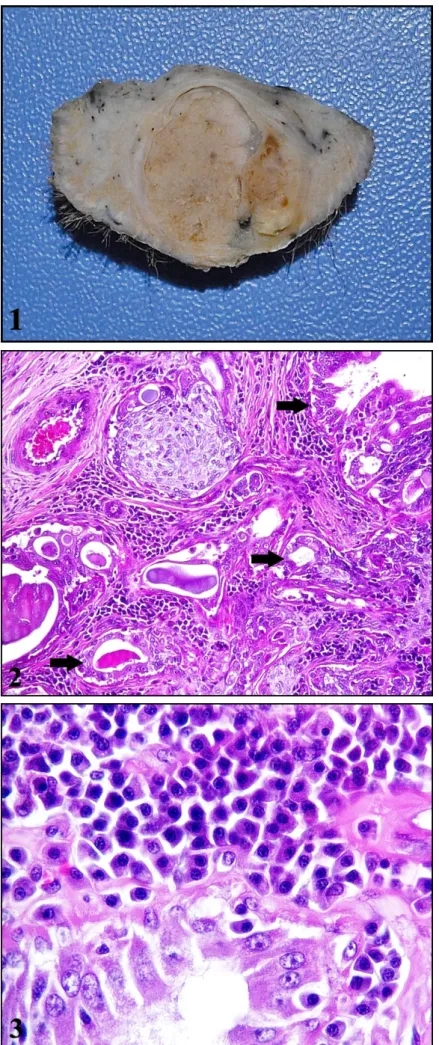
macroscopic examination of the operative material revealed a mass 3x5x3 cm in size and 12 g in weight, which was covered with skin and of elastic consistency and presented with an ulcerous area, 2 cm in diameter, on the upper surface. The cross-section of the mass had a multilobular appearance and contained cystic structures which were partly filled with a serous fluid (Figure 1). Microscopic examination demonstrated that the epidermis had lost its integrity in a certain region, where cell debris and many neutrophil leukocytes were observed. Glandular structures existed, which were composed of atypical sweat gland epithelial cells that varied from round to oval in shape and had hyperchromatic nuclei with evident nucleoli. Some of the glands were lined by a double layer of epithelium that formed papillary extensions towards the cystic lumen (Figure 2). The lumen of these structures contained an eosinophilic material. In-between the glandular structures were clusters comprised of multiple atypical plasma cells that displayed marked pleomorphism (Figure 3).
Nihat Yumuşak - Murat Çalışkan - Osman Kutsal 310
Figure 1. Macroscopical appearance of cut section of the mass.
Şekil 1. Tümöral kitlenin kesit yüzü.
Figure 2. Anaplastic sweat glands (arows), HxE, x40.
Şekil 2. Anaplaztik ter bezleri (oklar), HxE, x40.
Figure 3. Atypical plasma cells between anaplastic sweat glands, HxE, x100.
Şekil 3. Anaplaztik ter bezleri arasında atipik plazma hücreleri, HxE, x100.
Ankara Üniv Vet Fak Derg, 61, 2014 311
Both sweat gland tumours and plasmacytomas occur in the head, neck and extremities of senile cats and dogs (1, 3, 4, 10). In this case study, these tumours were identified in the left axillary region of a dog younger than that reported in available literature. While dogs of the Golden Retriever breed are predisposed to sweat gland tumours, plasmacytomas occur more frequently in Yorkshire terriers (2-4, 7). Furthermore, plasmacytomas are more common in male dogs, compared to female dogs. In compliance with literature reports, the material of this case study belonged to a male dog. While 70% of canine sweat gland tumours are of benign character, malignant tumours show a tendency of metastasis to regional lymph nodes and other organs and tissues by lymph vessels (1, 5, 8, 9). Sweat gland tumours, which are classified under tumours of the skin and its appendages by the World Health Organisation (WHO), histologically, are grouped under papillary syringadenoma, cystadenoma, spiradenoma and mixed tumours and carcinomas of the sweat glands (10). Mixed tumours of the sweat glands are uncommon and are of either benign or malignant character. Histologically, myoepithelial filaments may be associated with cartilaginous or osseous structures (1, 4, 5, 8). In this case study, the tumour tissue was characterized by the features of a plasmacytoma, rather than a mixed tumour, and presented with atypical plasma cells in-between the anaplastic sweat glands. Furthermore, nuclear atypia, necrosis and atypical mitosis may develop. Plasmacytomas are tumours composed of round shaped cells and although generally their diagnosis is not difficult they may be confused with mast cell tumours. In such cases, differential diagnosis can be made with toluidine blue staining (2, 3, 4, 7). When stained with toluidine blue, plasmacytomas do not display metachromasia. The histological findings obtained in this case study were in compliance with those indicated in literature reports.
In conclusion, in view of the scarcity of literature data available for sweat gland carcinoma and plasmacytoma, the diagnosis of these two different tumours in the same biopsy material was considered worth investigating.
References
1. Bahl A, Sharma D, Julka P, Das A, Rath G (2006): Sweat gland carcinoma with lung metastases. J Cancer Res Ther, 4, 209-211.
2. Canlas MS, Dillon ML, Loughrin JJ (1979): Primary cutaneous plasmacytoma. Arch Dermatol, 115, 722-724. 3. Gibson NR, Ness MG, McNeil PE (1997): Malignant
articular plasmacytoma in two dogs. Vet Rec, 141, 197-200.
4. Goldschmidt MH, Hendrick MJ (2002): Tumors of the Skin and Soft Tissues. 45-117. In: DJ Meuton, (ed), Tumors in Domestic Animals. 4 th ed. Iowa State Press, Iowa.
5. Kalaher KM, Anderson WI, Scott DW (1990): Neoplasms of the apocrine sweat glands in 44 dogs and 10 cats. Vet Rec, 127, 400-403.
6. Pagnoncelli M, Martins DB, França RT, Lopes STA, Zanette RA, Mazzanti CM (2012): Fine-needle aspiration cytology of the canine apocrine sweat gland carcinoma. Comp Clin Pathol, 21, 627-629.
7. Rusbridge C, Wheeler SJ, Lamb CR, Page RL, Carmichael S, Brearley MJ, Bjornson AP (1999): Vertebral plasma cell tumors in 8 dogs. J Vet Intern Med, 13, 126-133.
8. Simko E, Wilcock BP, Yager JA (2003): A retrospective study of 44 canine apocrine sweat gland adenocarcinomas. Can Vet J, 44, 38-42.
9. Souza CM, Damasceno KA, Gamba CO, Campos LC, Campos CB, Cassali GD (2011): Canine sweat gland mixed tumor. Acta Scientiae Veterinariae, 39, 1001. 10. Weiss E, Frese K (1974): Tumours of the skin. In:
BULLETIN of The World Health Organization, International Histological Classification of Tumours of Domestic Animals. WHO, Geneve, 50, 79-100.
Geliş tarihi: 21.08.2013 / Kabul tarihi: 31.10.2013 Address for correspondence:
Nihat Yumuşak
Harran University, Faculty of Veterinary Medicine Department of Pathology,
Şanlıurfa-TURKEY