Annals of Medical Research
DOI: 10.5455/annalsmedres.2019.11.780
Original Article
Investigation of the effects of tigecycline, a semi-synthetic
derivative of minocycline, on chondrocyte cultures
Yasin Emre Kaya1, Numan Karaarslan2, Ibrahim Yilmaz3, Duygu Yasar Sirin4, Onur Yaman5, Hanefi Ozbek6, Ozkan Ates7
1Abant Izzet Baysal University, Faculty of Medicine, Department of Orthopaedic and Traumatology, Bolu, Turkey 2Namik Kemal University, Faculty of Medicine, Department of Neurosurgery, Tekirdag, Turkey
3Istanbul Medipol University, Faculty of Medicine, Department of Medical Pharmacology, Istanbul, Turkey
4Namik Kemal University, Faculty of Arts and Sciences, Department of Molecular Biology and Genetics, Tekirdag, Turkey 5Memorial Bahcelievler Spine Center, Clinic of Neurosurgery, Istanbul, Turkey
6Istanbul Medipol University, Faculty of Medicine, Department of Medical Pharmacology, Istanbul, Turkey 7Istanbul Koc University Hospital, Clinic of Neurosurgery, Istanbul, Turkey
Copyright © 2020 by authors and Annals of Medical Research Publishing Inc.
Abstract
Aim: Although antibiotics are generally well-tolerated, they may have cytotoxic effects. The present randomized, double-blind, in
vitro study aimed to investigate the effects of tigecycline on cartilage tissue cells and the extracellular matrix.
Material and Methods: Cartilage tissues of patients (n = 8) were used for the preparation of primary cell cultures. Tigecycline-treated
cell cultures served as the study group. Non-treated cell cultures served as the control group. Analyses were performed at 0, 24, 48, and 72 h in both groups. The results obtained were statistically evaluated. The alpha significance value was determined to be 0.05.
Results: Proliferation remained unchanged in the tigecycline-treated cell cultures. The gene expressions of the markers involved in
anabolic pathways increased in the tigecycline-treated cell cultures. The results obtained were statistically significant (P < 0.05).
Conclusion: Although tigecycline had no toxic effect on the chondrocyte cell cultures and caused no damage to the extracellular
matrix, the present study was performed in an in vitro environment.
Keywords: Chondroadherin gene; Cartilage oligomeric protein; Interleukin-1beta; Matrix metalloproteinase; Primary chondrocyte culture; Tigecycline
Received: 29.11.2020 Accepted: 02.03.2020 Available online: 18.03.2020
Corresponding Author: Yasin Emre Kaya, Abant Izzet Baysal University School of Medicine, Department of Orthopaedic and Trauma,
14000, Bolu, Turkey E-mail: [email protected]
INTRODUCTION
Antibiotics are among the most misused drugs. Thus, inappropriate use of antibiotics may harm the liver and kidneys and lead to fungal infections, loss of probiotics in the intestine, and resistance development. Inappropriate antibiotic use also puts an unnecessary and excessive economic burden on healthcare systems (1-4).
The increase in the number of multi-drug resistant bacteria and potent resistance to antibiotics widely administered have become a major problem for many medical branches, particularly those that treat infectious diseases, in the last two decades (5).
The spread of extended-spectrum β-lactamases (ESBLs), which are resistant to antimicrobial treatment
of infections caused by Enterobacteriaceae, has become a global health problem. Carbapenems have thus been preferred for the treatment of serious infections caused by ESBL-producing organisms. Since the number of cases of resistance to carbapenem-group drugs has dramatically increased, older drugs, such as tigecycline, have gained popularity.
Tigecycline, a minocycline derivative, shows significant potency against Gram-positive and Gram-negative microorganisms, including methicillin-resistant Staphylococcus aureus (MRSA), vancomycin-resistant enterococci (VRE), ESBL-producing Escherichia coli, and Klebsiella pneumonia. It is also an effective pharmaceutical for atypical agents, including anaerobic bacteria and rapidly growing non-tuberculous mycobacteria. It is
widely prescribed for patients with renal insufficiency, as it is excreted through the bile tract, and dosage adjustments are not required (6).
Given the foregoing reasons, tigecycline can be used in clinics for complicated intraabdominal infections and skin and soft tissue infections. It is also commonly used for the perioperative washing of instruments and surgical site irrigation (1).
The most common adverse events related to tigecycline that have been reported, where tigecycline and other drugs used for the treatment of similar diseases were compared, are nausea and vomiting, pancreatitis, hepatic insufficiency, and increased levels of certain enzymes, such as alanine aminotransferase (7, 8). Some studies have also reported a variety of rare adverse events, including cholestasis, jaundice, and Stevens-Johnson syndrome (7, 8). However, no studies have investigated the effects of tigecycline on cartilage tissue elements, such as chondrocyte and extracellular matrix (ECM) structures. The present randomized, double-blind, in vitro study aimed to investigate the effects of tigecycline on human primary chondrocyte cultures.
MATERIAL and METHODS
The sensitivity of patients whose tissues were used for the preparation of primary cell cultures to tetracycline class antibiotics such as tigecycline, minocycline, and doxycycline was tested. Subsequently, the primary cell cultures were prepared using the tissues of patients who were not allergic to the aforementioned drugs.
The following patients were excluded from the study: a) patients who had used ketoconazole for the treatment of fungal diseases, as ketoconazole can reduce the activity of the P-gp enzyme; b) patients who had used cyclosporine to suppress their immune system; and c) patients who had used rifampicin for the treatment of tuberculosis, as rifampicin can also reduce the activity of the P-gp enzyme. The tissues of eight patients, four males and four females, were used for the preparation of the primary cell cultures. The age range of the patients (n = 8) varied from 22 to 44 years, and the mean age was 33 ± 10.69 years. The tissues were obtained from patients with gonarthrosis who had not responded to conservative and medical treatments (9). The tissues were placed in sterile containers containing penicillin-streptomycin and transferred to the laboratory at 4˚C where the primary cell cultures were prepared (9). Following the third passage, experiments were performed on primary chondrocyte cultures. The cells were stained with trypan blue, placed on a Thoma slide, and counted under an inverted light microscope (magnification × 10). The counted cells were plated at a density of 6 x 10⁴ cells per well in 96-well plates for 3-(4,5-dimethylthiazol-2-yl)-2,5-diphenyltetrazolium bromide (MTT) viability and proliferation and toxicity assays, inverted light microscopy and acridine orange (AO), and propidium iodide (PI) assays. The cells were also plated at a density of 6 x 10⁴
cells per dish in Petri dishes (100 mm) for chondrocyte and ECM analyses.
After incubating the plates and the Petri dishes overnight, tigecycline was added to perform experiments on the cell cultures that became confluent and adhered to the cell culture plates or dishes. The researchers were blinded to the dosages and drugs, namely the components in the culture medium, when performing the experiments and statistical analyses.
A 50-mg tigecycline lyophilized powder (TYGACIL®, Pfizer) was used to produce the main stock solution. A 10 mg/ml stock solution was prepared by dissolving 50 mg of tigecycline powder with a half-life of 36 hours (10) in Dulbecco’s Modified Eagle Medium (DMEM) containing 5% dimethyl sulfoxide (DMSO). This was then diluted to obtain a 20 µg/ml treatment solution. A 20 µg/ ml tigecycline solution was added to the study group samples. Untreated samples served as a control group. The untreated and treated samples were analyzed at 0, 24, 48, and 72 h on days 7 and 21. Using an inverted light microscope, cell surface morphologies and viability were examined under ×4, ×10, ×20, and ×40 magnifications. Membrane permeability assays were performed using fluorescence microscopy. The results of the assays were confirmed using the nucleic acid binding dyes AO and PI. AO generates green fluorescence and stains all nucleated cells, alive or dead. In contrast, PI stains only dead nucleated cells with poor membrane integrity and generates red fluorescence (11).
A commercial MTT kit (Vibrant MTT Cell Proliferation Assay, Cat #V13154, Thermo Fisher Scientific, USA), based on the principle that formazan crystals generate thiazole and blue coloration, was used to analyze the proliferation. This method is predicated on the measurement of color changes, which occur through purple formazan production as cells undergo proliferation using tetrazolium via increased dehydrogenase enzyme activity by spectrophotometry as absorbance. The initial untreated cell viability was taken as 100% for the MTT assays that were performed at a wavelength of 570 nm. The proliferation and the inhibition of the proliferation were calculated using “Test OD / Control ODX100” and “1- Test OD / Control OD,” respectively, and the data were recorded for statistical analysis (12).
Quantitative real-time polymerase chain reaction (qRT-PCR) was used to quantify both the DNA gene copies obtained from the primary cell cultures and the level of mRNA (Applied Biosystems 7300/7500 real-time PCR system, Thermo Fisher Scientific, USA; Thermocycling conditions: 2 min at 50˚C, 10 min at 95˚C, 15 sec at 95˚C, and 1 min at 60˚C for 40 cycles for each duration). Total RNA was extracted from the cultured primary chondrocytes using the PureLink™ RNA Mini Kit. A total of 50 ng of RNA were reverse-transcribed using a high-capacity cDNA RT kit to obtain cDNA. To determine the gene expression profiles, all genes were amplified using TaqMan Gene Expression Assays for the chondroadherin
gene (CHAD), β-actin (ACTβ), cartilage oligo matrix protein (COMP), matrix metalloproteinase (MMP)-7, MMP-19, and interleukine-1 beta (IL-1β.). As a result of the qPCR experiment, the relative quantity (RQ) values of each sample were obtained using the 7500 Fast SDS program V.2.3 (Thermo Fisher Scientific, USA). ACTβ was used as an endogenous control to normalize the targeted gene expressions. To obtain comparative results, a reference sample (Group 1, 0 h) was used, and RQ values were calculated using the 2-∆∆Cq method (13-18).
The statistical analyses were performed using Minitab (version 18.0) software, and the data were evaluated at a 95% confidence interval (CI). Descriptive statistics were presented as mean ± standard deviation (SD). Variance analysis (ANOVA) was used to analyze how many independent variables interacted with each other and the effects of those interactions on the dependent variable. As differences across groups were observed, Tukey’s honestly significant difference (HSD) post-hoc test was used for multiple pairwise comparisons. The alpha significance value was accepted as <0.05.
RESULTS
Cell viability and proliferation were time-dependently higher in the tigecycline-treated group compared to the control group from 0 h to 72 h. The AO/PI staining results confirmed the MTT ELISA results (Figure 1).
Figure 1. MTT-Cell viability, toxicity, and proliferation results. These
results were statistically significant (P < 0.05)
Microscopic images revealed that the ECM was preserved and had not disintegrated. The round-shaped cells that indicate the existence of cytotoxicity were not observed. Figure 2 first lane (a, b, c, and d) shows the inverted microscopy photographs of the tigecycline applied cartilage cultures at 0, 24, 48 and 72 hours, respectively. Figure 2 second lane is the inverted microscopy image of the 72th hour of control cartilage cultures (Figure 2, e) and fluorescence microscopy images of the same cultures after AO / PI staining (Figure 2, f).
Figure 2. Cellular organization and proliferation were very poor in tigecycline-treated cultures compared to the control group
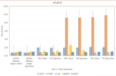
AO / PI staining was performed in all experimental groups and no apoptotic death was observed (Data not shown). Gene expressions changed time-dependently in the tigecycline-treated group from 0 h to 72 h (Figure 3). The expressions of COMP and CHAD, which are involved
in the anabolic pathways, increased in the tigecycline-treated group. IL-1β expression increased 0.9-fold in the tigecycline-treated group compared to the control group. The results obtained were statistically significant (P < 0.05).
Figure 3. Quantitative real-time PCR expression levels (shown as Fold Change, RQ) between sample groups
DISCUSSION
Tigecycline is the first type in the glycylcycline group of antibiotics. Although it is structurally similar to minocycline, it is a broad-spectrum antibiotic designed to evade common bacterial resistance mechanisms. The increase in resistant microorganisms has elevated the frequency of tigecycline use. Tigecycline, like minocycline, becomes effective by binding to the bacterial 30S ribosome. The binding point of tigecycline differs from that of tetracycline; therefore, it is not affected by Tet (M) protein and binds five times more effectively than tetracycline.
Tigecycline is not affected by common resistance mechanisms such as ribosome protection and efflux pumps, which makes it more potent than other antibiotics. Tigecycline shows strong activity against Gram-positive microorganisms, including S. aureus, S. epidermidis, Streptococcus agalactiae, Streptococcus pyogenes, E. faecium, and Listeria monocytogenes, as well as activity against gram-negative microorganisms, including Escherichia coli, Klebsiella pneumoniae, Enterobacter, and Serratia marcescens. It is also effective against resistant Gram-positive microorganisms, such as methicillin-resistant S. aureus (MRSA), methicillin-methicillin-resistant Staphylococcus epidermidis (MRSE), VRE, and resistant Gram-negative microorganisms, such as Acinetobacter baumannii and Stenotrophomonas maltophilia. Tigecycline has been approved for the treatment of adult patients with complicated intra-abdominal, skin, and soft-tissue infections. Some studies have recently suggested that tigecycline can be administered locally for the treatment of vascular graft infections caused by MRSE (19). It can also be administered perioperatively (1,20).
Commercial cell lines (21) or cell cultures prepared using animal tissues (22) are generally used to investigate the cytotoxicity of antibiotics. As is well-known, commercial cell lines contain only a single cell type, and the genetic
structure of the cells in the cell line has been modified; these cells do not carry the genotypic and/or phenotypic characteristics that they do in the human body. Hence, the results of experiments in which cell lines are used may be misleading (16-18). Many studies have also reported that the sensitivities of animal and human tissues are different; therefore, the results obtained through experiments using animal tissues differ from those using human tissues. This may cause misleading outcomes (16-18). In the present study, human cartilage tissues were used for the preparation of primary cell cultures. Hence, the results obtained from this study are believed to be reliable. No studies were found following a comprehensive and systematic search of electronic databases using the keywords “chondrocyte and tigecycline” and “cartilage tissue and tigecycline.” However, a study of the toxicity of tigecycline on human neuron cells was found when scanning the electronic databases using the keyword phrase “cytotoxicity of tigecycline.” In that study, the authors reported that tigecycline causes oxidative damage in neuron cells (21).
No studies have investigated the effect of tigecycline on chondrocyte and cartilage tissues in the literature. Therefore, the results obtained from the present study are believed to provide comprehensive insight into the potential effects of tigecycline.
CHAD is an NP-specific marker (23-25). To date, many studies have provided significant data on the enzymes that cause cartilage destruction. These enzymes include the members of the MMP family and the ADAMTS family. Studies on the regulation of this gene in cartilage tissue have become the new focus of research on the pathogenesis of OA. Previous studies have reported that inflammatory cytokines stimulate the ADAMTS-5 gene and increase cartilage damage (26,27). It should be noted that the loss of equilibrium between MMP-19, a member of the MMP family, and COMP, a member of the ADAMTS family, is responsible for ECM degradation, which may also cause the loss of proteoglycan. Proteolytic enzymes play a significant role in the pathophysiology of cartilage degeneration (28,29). Of these enzymes, those from the MMP family degrade the ECM by disintegrating its components. The increase in fibronectin and fibronectin fragments during degeneration stimulates MMP production. This stimulation has been reported to cause the suppression of proteoglycan production and the acceleration of cartilage-like tissue degeneration (30,31). Tigecycline was found to have no toxic effect when the control group and the tigecycline-treated group were compared. These results were statistically significant (P < 0.05).
The present study has a number of limitations. The cell cultures were obtained from a small number of patients who were from the same race. However, the cultures were established using human cartilage tissues, and all the experiments were repeated three times.
CONCLUSION
The present study was performed in an in vitro environment. Therefore, the results that were obtained may not be directly applicable to the clinical setting. Tigecycline was observed to have no toxic effect; however, it significantly changed gene expression levels. Further studies are needed to provide insight into the effects of tigecycline on cartilage tissue when used in clinics.
Competing interests: The authors declare that they have no competing interest.
Financial Disclosure: There were no financial supports.
Ethical approval: Istanbul Medpol University no. 29.11.2017- 10840098/604.01.01/E.44192).
Yasin Emre Kaya ORCID: 0000-0002-5412-8355 Numan Karaarslan ORCID: 0000-0001-5590-0637 Ibrahim Yilmaz ORCID: 0000-0003-2003-6337 Duygu Yasar Sirin ORCID: 0000-0002-1224-442X Onur Yaman ORCID: 0000-0002-2038-1643 Hanefi Ozbek ORCID: 0000-0002-8084-7855 Ozkan Ates ORCID: 0000-0002-3112-4839
REFERENCES
1. Karaarslan N, Yilmaz I, Ozbek H, et al. Is implant washing and wound irrigation with rifampicin effective for preventing surgical site infections in lumbar instrumentation? Turk Neurosurg 2018;28:904-909. 2. Dogan M, Isyar M, Yilmaz I, et al. Are the leading
drugs against Staphylococcus aureus really toxic to cartilage? J Infect Public Health 2016;9:251-258. 3. Bilir B, Isyar M, Yilmaz I, et al. Evaluation of
neutrophil-to-lymphocyte ratio as a marker of inflammatory response in septic arthritis. Eur J Inflam 2015;3:196-203.
4. Isyar M, Dogan M, Gumustas SA, et al. Evaluation of antibiotic alternatives used in orthopedic departments of 2 hospitals according to rational use of antibiotics. South Clin Ist Euras 2016;27:116-22.
5. Ranjbar R, Fatahian Kelishadrokhi A, Chehelgerdi M. Molecular characterization, serotypes and phenotypic and genotypic evaluation of antibiotic resistance of the Klebsiella pneumoniae strains isolated from different types of hospital-acquired infections. Infect Drug Resist 2019;20:603-11.
6. Srinivas NR. Tigecycline and cyclosporine interaction-an interesting case of biliary-excreted drug enhinteraction-ancing the oral bioavailability of cyclosporine. Eur J Clin Pharmacol 2009;65:543-4.
7. Chen Y, Zhu D, Zhang Y, et al. A multicenter, double-blind, randomized, comparison study of the efficacy and safety of tigecycline to imipenem/cilastatin to treat complicated intra-abdominal infections in hospitalized subjects in China. Ther Clin Risk Manag 2018;14:2327-39.
8. Kadoyama K, Sakaeda T, Tamon A, Okuno Y. Adverse event profile of tigecycline: data mining of the public version of the U.S. Food and Drug Administration
adverse event reporting system. Biol Pharm Bull 2012;35:967-70.
9. Oznam K, Sirin DY, Yilmaz I, et al. Iopromide- and gadopentetic acid-derived preparates used in MR arthrography may be harmful to chondrocytes. J Orthop Surg Res 2017;12:98.
10. Muralidharan G, Micalizzi M, Speth J, et al. Pharmacokinetics of tigecycline after single and multiple doses in healthy subjects, Antimicrob Agents Chemother 2005;49:220-9.
11. Kaplan N, Yilmaz I, Karaarslan N, et al. Evaluation of the effect of daptomycin, a glycopeptide agent, on intact intervertebral disc tissue. Turk Neurosurg 2019;29:522-9.
12. Kaplan N, Karaarslan N, Yilmaz I, et al. Are intervertebral disc tissue cells damaged when attempting to prevent thrombus formation using dabigatran, a new oral anticoagulant? Turk Neurosurg 2019;29:470-7.
13. Kaya YE, Karaarslan N, Sirin DY, et al. Investigation of the effects of methylphenidate, an amphetamine derivative, on intervertebral disc tissue cell cultures and matrix structures. Turk Neurosurg 2019;29:734-742.
14. Caliskan T, Sirin DY, Karaarslan N, et al. Effects of etanercept, a tumor necrosis factor receptor fusion protein, on primary cell cultures prepared from intact human intervertebral disc tissue. Exp Ther Med 2019;18:69-76.
15. Akgun FS, Sirin DY, Yilmaz I, et al. Investigation of the effect of dipyrone on cells isolated from intervertebral disc tissue. Exp Ther Med 2019;18:216-24.
16. Karaarslan N, Yilmaz I, Ozbek H, et al. Are radio-contrast agents commonly used in discography toxic to the intact intervertebral disc tissue cells? Basic Clin Pharmacol Toxicol 2019;124:181-9.
17. Karaarslan N, Yilmaz I, Sirin DY, et al. Pregabalin treatment for neuropathic pain may damage intervertebral disc tissue. Exp Ther Med 2018;16:1259-65.
18. Karaarslan N, Yilmaz I, Ozbek H, et al. Are specific gene expressions of extracellular matrix and nucleus pulposus affected by primary cell cultures prepared from intact or degenerative intervertebral disc tissues? Turk Neurosurg 2019;29:43-52.
19. Canbeyli ID, Kabalci M, Cirpar M, et al. Mesenchymal stem cells have significant anti-infective effect on methicillin-resistant Staphylococcus epidermidis vascular graft infections. Eklem Hastalik Cerrahisi 2019;30:201-11.
20. Polat M, Ozkaya-Parlakay A. Tigecycline salvage therapy for ventriculoperitoneal shunt meningitis due to extensively drug-resistant Acinetobacter baumannii. Eur J Pediatr 2019;178:117-8.
21. Xiao Y, Xiong T, Meng X, et al. Different influences on mitochondrial function, oxidative stress and cytotoxicity of antibiotics on primary human neuron and cell lines. J Biochem Mol Toxicol 2019;33:e22277. 22. Gao L, Schmitz HJ, Merz KH, et al. Characterization of
the cytotoxicity of selected Chelidonium alkaloids in rat hepatocytes. Toxicol Lett 2019;311:91-7.
23. Sirin DY, Karaarslan N. Evaluation of the effects of pregabalin on chondrocyte proliferation and CHAD, HIF-1α, and COL2A1 gene expression. Arch Med Sci 2018;14:1340-7.
24. Sirin DY, Kaplan N, Yilmaz I, et al. The association between different molecular weights of hyaluronic acid and CHAD, HIF-1α, COL2A1 expression in chondrocyte cultures. Exp Ther Med 2018;15:4205-12.
25. Kaplan N, Yilmaz I, Karaarslan N, et al. Does nimodipine, a selective calcium channel blocker, impair chondrocyte proliferation or damage extracellular matrix structures? Curr Pharm Biotechnol 2019;20:517-24.
26. 26. Liang Y, Fu Y, Qi R, et al. Cartilage oligomeric matrix protein is a natural inhibitor of thrombin. Blood 2015;126:905-14.
27. Yang CY, Chanalaris A, Troeberg L. ADAMTS and
ADAM metalloproteinases in osteoarthritis - looking beyond the ‘usual suspects’. Osteoarthr Cartilage 2017;25:1000-9.
28. Nagase H, Woessner JF Jr. Matrix metalloproteinases. J Biol Chem 1999;274:21491-4.
29. Wang G, Huang K, Dong Y, et al. Lycorine suppresses endplate-chondrocyte degeneration and prevents intervertebral disc degeneration by inhibiting NF-κB signalling pathway. Cell Physiol Biochem 2018;45:1252-69.
30. Akgun FS, Karaarslan N, Yilmaz I, et al. Evaluation of the effect of apixaban on the primary intact intervertebral disc cell cultures. Ann Med Res 2019;26:2414-22. 31. Karaarslan N, Yilmaz I, Sirin DY, et al. Does transcription
factor, induced by daptomycin and vancomycin, affect HIF-1α, chondroadherin, and COL2A1? Ann Med Res 2018;25:756-62.