Background: Antibiotic use in research volunteers partaking in dental studies varies widely, as inclusion and exclusion criteria used in participant selection lack strict rules. Aim: We aimed to investigate the most appropriate ending time of amoxicillin therapy in healthy subjects before their recruitment and participation in oral histology trials. A cross‑sectional design study was conducted in Istanbul, Turkey, between October 2018 and June 2019. Subjects and Methods: Study participants who had undergone a previous course of amoxicillin were divided into three groups according to the amount of time since their last antibiotic dose: group I, within 1 month; group II, within 4–6 months; and group III, within 7–12 months. Periodontal parameters were measured, and smear samples were obtained from buccal mucosa and keratinized gingiva. Gingival biopsies were taken from the upper premolars. Genetic damage and histopathological damage were analyzed with Feulgen reaction and with hematoxylin–eosin staining. Data were analyzed using the Mann–Whitney U, Kruskal–Wallis, and the Pearson Chi‑square tests. Results: There were no significant differences in the periodontal parameters and sociodemographic characteristics between the groups (P > 0.05). Higher genetic and histopathological damage scores were observed in group I, which were significantly different compared to the other groups (P < 0.05). No significant differences were observed between groups II and III (P > 0.05). Conclusions: When selecting healthy participants for oral histology trials, at least 3 months should pass after the final dose of amoxicillin is taken to avoid negative effects on gingival tissue and smear samples.
Keywords: Antibacterial agents, chromosome-defective micronuclei, cytology,
histopathology, periodontium
Effects of Amoxicillin on Gingival Biopsies and Oral Smears:
A Cross‑Sectional Study
B Alkan, P Koroglu1
Address for correspondence: Dr. B Alkan, Department of Periodontology, Faculty of Dentistry, Istanbul Medipol University, Birlik mah. Bahceler cad. No. 5, 34230, Esenler, Istanbul, Turkey. E‑mail: [email protected]
The World Health Organization[1] has reported that penicillin‑type antibiotics are the most frequently used globally, and within the European region, Turkey has been reported to have the highest rates of antibiotic consumption. Since 2014, Turkish health authorities have implemented an electronic prescription system that guides the appropriate use of antibiotics, monitors their medical use, and provides feedback to physicians and dentists.
I
ntroductIonD
ental studies use predetermined exclusion criteria that limit individual participation. While these criteria are essential for the design of high‑quality research protocols, setting rigorous exclusion criteria has disadvantages that include prolonging the study period and increasing the costs and challenges involved in finding appropriate participants. Moreover, the process of planning and conducting a study becomes more challenging for researchers if the majority of clinic patients possess characteristics that substantially differ from the eligibility criteria described in the literature. Department of Periodontology, Faculty of Dentistry, Istanbul Medipol University, Istanbul, 1Department of Histology and Embryology, Halic University, Faculty of Medicine, Istanbul, TurkeyA
bstr A ct Received: 05-Dec-2019; Revision: 20-Feb-2020; Accepted: 18-Jun-2020; Published: 19-Feb-2021This is an open access journal, and articles are distributed under the terms of the Creative Commons Attribution‑NonCommercial‑ShareAlike 4.0 License, which allows others to remix, tweak, and build upon the work non‑commercially, as long as appropriate credit is given and the new creations are licensed under the identical terms.
For reprints contact: [email protected]
How to cite this article: Alkan B, Koroglu P. Effects of amoxicillin on gingival biopsies and oral smears: A cross-sectional study. Niger J Clin Pract 2021;24:233-9.
Access this article online Quick Response Code:
Website: www.njcponline.com
DOI: 10.4103/njcp.njcp_660_19
A history of antibiotic use is a criterion that widely differs in clinical dental research. In some studies,[2‑4] antibiotic consumption within the previous 3 or 6 months is the basis for study exclusion, whereas in others,[5‑7] antibiotic use is not a criterion. Currently, there is no consensus regarding how long systemic antibiotics affect the oral mucosa and periodontal tissue, as the excretion time differs according to the type of antibiotic, its pharmacokinetics, the number of different antibiotics prescribed, and the method of delivery. Therefore, in this study, we assessed the length of the waiting period needed before systemic amoxicillin loses its effect on gingival tissue and oral mucosa cells.
We used commonly preferred exclusion criteria for amoxicillin exposure time and investigated the histopathological damage that occurs to gingival tissue and the genotoxic effects on oral mucosal cells. By comparing individuals last exposed to amoxicillin at different time points, we aimed to discern exclusion criteria that are most appropriate for oral histology trials.
s
ubjects AndM
ethodsThe institution’s ethics committee approved the study protocol (ethic number/date:
10840098 – [198.01.01 – E.47625] and
[604.01.01 – E.1460]/23 May 2018). The study followed the principles outlined in the Declaration of Helsinki of 1975, as revised in 2000.
Study design and population
This study used a single‑center, investigator‑blinded, cross‑sectional design that was carried out at the Faculty of Dentistry, Department of Periodontology in Istanbul, Turkey, between October 2018 and June 2019. Patients included those treated with 1 g of amoxicillin twice a day for 7 days for different durations. We compared participants based on the amount of time that had passed since their last antibiotic dose before conducting periodontal evaluations, oral smears, and biopsy sampling.
Systemically healthy participants between 18 and 60 years of age and without gingival inflammation were enrolled in the study. A diagnosis of a clinically healthy periodontium was assessed according to the 2017 World Workshop on the Classification of Periodontal Diseases and Conditions.[8] The evaluation of systemic status and antibiotic consumption was based on participant self‑reported medical histories and computerized medical records. During the study, no medication was given to any of the participants. After the study protocol was explained to eligible volunteers, those willing to enroll in the study signed an informed consent form. All participants answered a questionnaire that assessed
age, educational level, and frequency of dental visits. The identity of the subjects was kept confidential. The number of participants who attended the clinic during the research period determined the study size.
Inclusion criteria were as follows: participants requiring gingivectomy operations, crown lengthening procedures or tooth extractions, those with no clinical evidence of bleeding on probing, no gingival recession, no clinic attachment loss, or radiographic evidence of alveolar bone loss, and no periodontal treatment within the last 12 months. Participants should have undergone systemic amoxicillin therapy within the 12 months previous to the baseline examination. The exclusion criteria included systemic conditions that would potentially affect the periodontium (i.e., pregnancy, lactation, ovulation, and diabetes mellitus) and history of contagious disease. All of the participants were nonsmokers.
Periodontal evaluation and sample collection All clinical periodontal parameters including plaque index, gingival index, probing depth, bleeding on probing, gingival recession, and clinical attachment level were recorded from six sites (distobuccal, midbuccal, mesiobuccal, distopalatal, midpalatal, and mesiopalatal) immediately before taking smear samples and biopsies.[5] All periodontal assessments were performed using William’s periodontal probe (Hu‑Friedy, Chicago, IL, USA) by the same researcher. Clinical evidence of gingival recession was an exclusion criterion of this study; therefore, clinical attachment level was equal to the probing depth and was not further analyzed.
To process smear samples, participants were asked to rinse their mouths with water,[9‑14] then two separate (mascara‑type) sterile cytobrushes (Mediko Kimya, Istanbul, Turkey) were used to collect smear samples[12,15] from the keratinized gingiva of the upper premolar region and the buccal mucosa of the same cheek [Figure 1]. Samples were prepared on clean glass slides (Isolab, Istanbul, Turkey) and dried at room temperature (20–25°C and 85% relative humidity).[10,11,14] Within 2 h of collection, the samples of keratinized gingiva were fixed in a methanol–acetic acid (3:1) solution for 15 min,[15,16] and the samples of buccal mucosa were fixed in 80% methanol for 15 min.[9,12,13] Biopsy samples were obtained under local anesthesia from the vestibular region of the maxillary premolars during tooth extraction or crown lengthening procedures (Ultracain DS Forte, Sanofi‑Aventis Deutschland GmbH, Frankfurt, Germany) [Figure 2]. Internal bevel and intrasulcular incisions were subsequently performed around the tooth using a carbon steel surgical scalpel blade (No. 15, Braun, Aesculap,
Germany). The gingiva was then excised, including part of the pocket epithelium and the adjacent connective tissue. Following sample collection, tissues were immediately rinsed in sterile saline and fixed in 10% buffered formalin at room temperature until analysis. The surgical site was covered with periodontal dressing during the first week.
Histopathological evaluations and scoring criteria To assess cells containing micronuclei, the Feulgen reaction (Bio‑Optica, Milan, Italy) was applied to all smear samples according to the protocol described by Thomas et al.[17] Briefly, cells were placed in a fixative for nuclear staining and analysis using Schiff’s reagent (Bio‑Optica, Milan, Italy). Micronuclei in the buccal [Figure 3] and gingival [Figure 4] smear samples were analyzed by observing them in oil immersion at 1000 × magnification. Scoring was performed according to the criteria established by Tolbert et al.[18] Quantitative determination of the micronucleus index and analysis of cytogenetic damage was scored. The technique used to calculate the cells was a zigzag strategy. One thousand cells were screened, and the number of micronuclei present was identified. The following criteria were used for scoring micronuclei: (a) being round and smooth, (b) those with diameters <one‑third of the associated nucleus, (c) those that were dark and bright in the illumination field, (d) a similar staining intensity of the micronuclei and the nucleus, (e) a similar texture of the micronuclei and the nucleus, (f) micronuclei in the same focal plane as the nucleus, and (g) no overlap or bridge to the nucleus.
All microscopic sections were observed and photographed using a digital camera (OlympusCX41, Olympus, Tokyo, Japan) [Figure 5]. After fixation in 10% neutral buffered formalin, the tissue samples were embedded in paraffin wax, serially sectioned at a 4‑µm thickness, stained with hematoxylin and eosin for histopathological scoring, and examined at 400 × magnification under a light microscope. Five areas were randomly selected to avoid bias. All tissue and smear samples were analyzed at the same time and in the same laboratory by the same blinded investigator. A semiquantitative histological assessment of gingival damage was evaluated using modified histological criteria, as described by Jalayer‑Naderi et al.[6] Cellular components, vascularization, cell infiltration, necrosis, pyknotic nucleus, hyperemia, fibrosis, ulceration, atrophy, apoptosis, and rete peg configurations were scored using a scale ranging from zero to three (none: zero, mild: one, moderate: two, and severe: three) for each criterion.
Statistical methods
The NCSS2007 software program (NCSS LLC, Kaysville, UT, USA) was used for all statistical analyses. Descriptive statistical analyses (mean, standard deviation, median, frequency, ratio, minimums, and maximums) were performed. For the comparison of quantitative data, the Mann–Whitney U test was used to detect differences between two groups, and the Kruskal– Wallis test was used for intergroup comparisons in cases of more than two groups. The Pearson Chi‑square test was used for the analysis of qualitative data. For all analyses, P < 0.05 was considered statistically significant.
r
esultsStudy population
A total of 45 healthy participants with clinically healthy periodontium were included in the study. The mean age of the participants was 28.41 ± 8.06 years, ranging from 20 to 55 years. There were 36 women (80%)
Table 1: Patient sociodemographic characteristics by study group
Group I Group II Group III P
(n=13) (n=13) (n=19) Gender, n (%) Female 9 (69.2) 10 (76.9) 17 (89.5) 0.35* Male 4 (30.8) 3 (23.1) 2 (10.5) Age (years) Mean±SD 30±10.87 25.92±4.69 29.32±8.64 0.55† Min‑max (median) 21‑55 (24) 21‑35 (24) 20‑47 (26) Education, n (%) 1‑8 years 2 (15.4) 0 (0.0) 5 (26.4) 0.40* 9‑11 years 1 (7.7) 2 (15.4) 4 (21.2) 12 years or more 10 (76.9) 11 (84.6) 10 (52.6) Dental visit frequency, n (%) When there is a problem 5 (38.5) 8 (61.5) 12 (63.2) 0.34* Once a year 8 (61.5) 5 (38.5) 7 (36.8)
*The Pearson chi‑square test. †Kruskal–Wallis test. P<0.05, statistically significant. SD: Standard deviation
Figure 1: Smear samples collected with cytobrushes: (a) cytobrush
collection of exfoliated cells from buccal mucosa, and (b) cytobrush collection of exfoliated cells from keratinized gingival mucosa
b a
and 9 men (20%). Participants were divided into three groups based on a history of oral antibiotic therapy (1 g amoxicillin, twice a day, 7 days) for upper respiratory tract infections in the previous 12 months. In group Table 2: Clinical periodontal parameters by study group
Group I Group II Group III P†
(n=13) (n=13) (n=19) PI Mean±SD 0.32±0.28 0.55±0.38 0.61±0.33 0.06 Min‑max (median) 0‑0.75 (0.25) 0‑1 (0.5) 0‑1 (0.5) GI Mean±SD 0.05±0.15 0.03±0.14 0.05±0.13 0.82 Min‑max (median) 0‑0.5 (0) 0‑0.5 (0) 0‑0.5 (0) PD (mm) Mean±SD 2.15±0.33 2.21±0.25 2.21±0.35 0.87 Min‑Max (median) 1.75‑2.75 (2.25) 2‑2.75 (2.25) 1.75‑2.25 (2.25) BOP (%) Mean±SD 5.77±14.98 3.85±13.87 5.26±10.47 0.68 Min‑max (median) 0‑50 (0) 0‑50 (0) 0‑25 (0) †Kruskal–Wallis test. P<0.05, statistically significant. SD: Standard deviation; PI: plaque index; I: gingival index; PD: probing depth; BOP: bleeding on probing
Figure 5: Inner (right)–outer (left) epithelia of tissues belong to
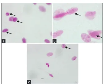
participants who used antibiotics within (a, b) 1 month, (c, d) 4–6 months, and (e, f) 7–12 months previous to sampling. Black arrows indicate pyknotic nuclei (hematoxylin and eosin staining, 40×) d c b f a e Figure 4: Gingival mucosa smear samples from all groups: the black arrow indicates the micronucleus. Photomicrographs of exfoliated epithelial cells belonging to participants who used antibiotics within (a) 1 month, (b) 4–6 months, and (c) 7–12 months previous to sampling (Feulgen staining, 1000×) c b a Figure 3: Buccal mucosa smear samples from all groups: the black arrow shows the micronucleus. Photomicrographs of exfoliated epithelial cells belong to participants who used antibiotics within (a) 1 month, (b) 4–6 months, and (c) 7–12 months previous to sampling (Feulgen staining, 1000×) c b a
Figure 2: Epithelial and connective tissue obtained by gingivectomy: (a)
preoperative facial view of a participant, (b) marking the depth of the suprabony pocket, (c) the bottom of pockets indicated by William’s periodontal probe, and (d) immediate postoperative view after a completed gingivectomy d c b a
I (n = 13; mean age = 30 years; range 21–55 years), participants had used amoxicillin within a 1‑month period before sampling. In group II (n = 13; mean age = 25.92 years; range 21–35 years), participants had used amoxicillin within 4–6 months before sampling. In group III (n = 19; mean age = 29.32 years; range 20–47 years), participants had used amoxicillin within 7–12 months before sampling and were used as the control group. An initial analysis of descriptive and sociodemographic characteristics including gender distribution, average age, educational level, and frequency of dental visits [Table 1], and clinical periodontal parameters including plaque index, gingival index, pocket depth, and bleeding on probing [Table 2] uncovered no significant (P > 0.05) differences between the groups. Gingival recession was not included in the analysis as all values were zero in the participants. Histopathological analysis
The values for atrophy, apoptosis, and rete peg configuration were zero for all participants and, therefore, not included in the analysis. No differences were found between the groups when the parameters of cellular components, vascularization, cell infiltration, necrosis, pyknotic nucleus, hyperemia, fibrosis, and tissue ulceration were analyzed separately (P > 0.05) (data not shown). However, the histopathological damage score of the periodontium and the micronuclei findings of both the gingival and buccal mucosa smear samples were higher in group I in comparison to the other two groups (P < 0.05) [Table 3]. There were no significant differences between groups II and III in any of the smear samples or histopathological damage data (P > 0.05).
d
IscussIonCurrent information in the literature about the effects of systemic antibiotic consumption on periodontal tissue and oral‑smear samples is insufficient. We hypothesized that study eligibility criteria regarding antibiotic
consumption in systemically and periodontally healthy participants participating in oral histology trials should be redefined. The results of our research confirmed this hypothesis. While no significant differences were found for the sociodemographic characteristics or clinical periodontal parameters between the groups, highly significant differences in histopathological damage and in the micronuclei findings were found between those who had taken antibiotics within the last month and those who had taken antibiotics within 4–12 months previously. We found that antibiotic consumption within the past 30 days resulted in chromosomal defects in the oral mucosa cells and histopathological damage in the periodontal tissues. These results indicate that potential volunteers should not consume antibiotics at least 3 months prior to participating in oral histology trials to allow for a sufficient period of time to pass and ensure the selection of healthy volunteers for control groups. The main limitation of this study was the lack of participants who had used antibiotics within 2 and 3 months before sampling. Nevertheless, we believe the current results are clinically useful as we took into account the most frequently predefined eligibility criteria for time intervals from the last antibiotic dose from the literature. A further study limitation concerns the possibility that the histopathological characteristics of the tissue samples may have varied because the density of the cell types differed from section to section. However, to avoid this, a blinded investigator randomly chose and examined five areas of each histologic section. One of the critical components of clinical research is selecting appropriate participants; thus, the determination of realistic eligibility criteria is the backbone of methodology. Taking the criteria of other published research as a guide is essential for predetermining eligibility criteria for new studies. However, understanding the biological reasons underlying the basis for these eligibility criteria is Table 3: Histopathological damage scores and smear examination findings by study group
Group I Group II Group III P (I-II)‡ P (I-III)‡ P (II-III)‡
(n=13) (n=13) (n=19) HDS Mean±SD 0.24±0.24 0.09±0.09 0.06±0.13 0.028 0.006 0.167 Min‑max (median) 0‑0.88 (0.13) 0‑0.25 (0.13) 0‑0.5 (0) Micronuclei findings Gingival mucosa Mean±SD 10.23±1.59 7.62±2.18 6.68±1.11 0.004 0.001 0.12 Min‑max (median) 8‑13 (10) 4‑11 (8) 5‑8 (7) Buccal mucosa Mean±SD 10.08±1.85 7.85±1.91 7.11±1.24 0.011 0.001 0.154 Min‑max (median) 7‑13 (10) 5‑11 (8) 4‑8 (8) ‡Mann–Whitney U test. P<0.05, statistically significant. SD: Standard deviation; HDS: Histopathological damage score
essential. Wells[19] has stated that exclusion criteria help researchers understand how a process works under ideal conditions. However, this may limit our understanding of the properties of a population in daily clinical practice. Antibiotics are essential for the treatment of bacterial infections, and amoxicillin is the most frequently prescribed drug by both dentists and physicians.[20,21] The time since the last administration of the final antibiotic dose is often considered as a predefined eligibility criterion in the study protocols in periodontology. Antibiotic consumption constitutes approximately 38% of the total pharmaceutical consumption in Turkey.[21] Therefore, if antibiotic consumption is considered a strict exclusion criterion for healthy controls, there is a danger of wasted time and labor during the search for potential research participants, as recruitment time needs to be extended until enough participants can be admitted to the study. Moreover, exposure to antibiotics via dietary intake of livestock products also occurs in populations; thus, exclusion criterion for antibiotic consumption should be further reviewed for clinical trials. While exclusion criteria are sometimes essential, we agree with Wells[19] in that some clinical trial exclusion criteria have created a gap between the ideals of research and the realities of clinical practice.
The applicability of our suggestions to other fields of study or different types of tissues is an important question, as the time since the final antibiotic dose also affects the eligibility criteria for nearly all clinical studies. The pharmacokinetic properties of amoxicillin tend to be different depending on the target tissue. Our research included only systemically and periodontally healthy participants treated with oral amoxicillin at different ending times, and we focused on the periodontium and oral‑smear samples from the gingiva and buccal mucosa.
c
onclusIonsThis study demonstrated that researchers should wait at least 3 months following the final administration of amoxicillin to avoid adverse effects of the antibiotic on gingival tissues and smear samples.
Declaration of patient consent
The authors certify that they have obtained all appropriate patient consent forms. In the form, the patient (s) has/have given his/her/their consent for his/ her/their images and other clinical information to be reported in the journal. The patients understand that their names and initials will not be published and due efforts will be made to conceal their identity, but anonymity cannot be guaranteed.
Financial support and sponsorship Nil.
Conflicts of interest
There are no conflicts of interest.
r
eferences1. World Health Organization. WHO report on surveillance of antibiotic consumption: 2016‑2018 early implementation. 2018. Available from: https://apps.who.int/iris/bitstream/handle/ 10665/277359/9789241514880‑eng.pdf.
2. Duarte P, Neto J, Casati M, Sallum E, Nociti F. Diabetes modulates gene expression in the gingival tissues of patients with chronic periodontitis. Oral Dis 2007;13:594‑9.
3. Kinney J, Morelli T, Oh M, Braun T, Ramseier C, Sugai J, et al. Crevicular fluid biomarkers and periodontal disease progression. J Clin Periodontol 2014;41:113‑20.
4. Hatipoglu H, Yamalik N, Berberoglu A, Eratalay K. Impact of the distinct sampling area on volumetric features of gingival crevicular fluid. J Periodontol 2007;78:705‑15.
5. Seymour G, Powell R, Aitken J. Experimental gingivitis in humans. A clinical and histologic investigation. J Periodontol 1983;54:522‑8.
6. Jalayer‑Naderi N, Semyari H, Elahinia Z. The impact of smoking on gingiva: A histopathological study. Iran J Pathol 2015;10:214‑20.
7. Egelberg J, Attstrom R. Comparison between orifice and intracrevicular methods of sampling gingival fluid. J Periodontal Res 1973;8:384‑8.
8. Caton J, Armitage G, Berglundh T, Chapple I, Jepsen S, Kornman K, et al. A new classification scheme for periodontal and peri‑implant diseases and conditions‑Introduction and key changes from the 1999 classification. J Periodontol 2018;89(Suppl 1):1‑8. 9. Zamora‑Perez A, Ortiz‑Garcia Y, Lazalde‑Ramos B,
Guerrero‑Velazquez C, Gomez‑Meda B, Ramirez‑Aguilar M,
et al. Increased micronuclei and nuclear abnormalities in buccal
mucosa and oxidative damage in saliva from patients with chronic and aggressive periodontal diseases. J Periodontal Res 2015;50:28‑36.
10. Srivastava P, Miles J, Lucas F. Y‑chromatin fluorescence in human buccal smear. Clin Genet 1974;6:201‑4.
11. Ravi K, Naithani M, Kaur S, Reddy K, Pasi R. Abnormal nuclear variations in response to radiotherapy‑ As a tool in treatment planning and assessment of prognosis. J Clin Diagn Res 2016;10:8‑12.
12. Tak A, Metgud R, Astekar M, Tak M. Micronuclei and other nuclear anomalies in normal human buccal mucosa cells of oral cancer patients undergoing radiotherapy: A field effect. Biotech Histochem 2014;89:464‑9.
13. Nersesyan A, Muradyan R, Kundi M, Knasmueller S. Impact of smoking on the frequencies of micronuclei and other nuclear abnormalities in exfoliated oral cells: A comparative study with different cigarette types. Mutagenesis 2011;26:295‑301.
14. Sarto F, Finotto S, Giacomelli L, Mazzotti D, Tomanin R, Levis A. The micronucleus assay in exfoliated cells of the human buccal mucosa. Mutagenesis 1987;2:11‑7.
15. Brandão P, Gomes‑Filho I, Cruz S, Passos‑Soares J, Trindade S, Souza L, et al. Can periodontal infection induce genotoxic effects? Acta Odontol Scand 2015;73:219‑25.
16. Cerqueira E, Meireles J, Lopes M, Junqueira V, Gomes‑Filho I, Trindade S, et al. Genotoxic effects of X‑rays on keratinized
mucosa cells during panoramic dental radiography. Dentomaxillofac Radiol 2008;37:398‑403.
17. Thomas P, Holland N, Bolognesi C, Kirsch‑Volders M, Bonassi S, Zeiger E, et al. Buccal micronucleus cytome assay. Nat Protoc 2009;4:825‑37.
18. Tolbert P, Shy C, Allen J. Micronuclei and other nuclear anomalies in buccal smears: Methods development. Mutat Res 1992;271:69‑77.
19. Wells K. Treatment research at the crossroads: The scientific interface of clinical trials and effectiveness research. Am J Psychiatry 1999;156:5‑10.
20. Kaptan R, Haznedaroglu F, Basturk F, Kayahan M. Treatment approaches and antibiotic use for emergency dental treatment in Turkey. Ther Clin Risk Manag 2013;9:443‑9.
21. Parameter on plaque‑induced gingivitis. American Academy of Periodontology. J Periodontol 2000;71 (5 Suppl):851‑2.