Primary Left Upper Quadrant (Palmer’s Point)
Access for Laparoscopic Radical Prostatectomy
İlter Tüfek, Haluk Akpınar, Cüneyd Sevinç, Ali Rıza Kural
Purpose: Although Palmer’s point approach is described for upper urinary
tract laparoscopy, we use this technique routinely for robotic and standard laparoscopic radical prostatectomy and we describe our experience with this approach.
Materials and Methods: Since 2004, Palmer’s point Veress entry has been
used to create pneumoperitoneum in 126 robotic and 21 standard laparoscopic radical prostatectomies. On the left side, a 2-mm transverse skin incision was made 3 cm below the left costal margin on the midclavicular line. Through this incision, a Veress needle was inserted to create pneumoperitoneum.
Results: The mean patients’ age and body mass index were 59.7 years
(range, 37 to 73 years) and 27.92 kg/m2 (range, 22 to 39 kg/m2), respectively.
Thirty-eight patients had prior abdominal operations. The mean number of punctures performed was 1.08 per case. In 93 % of the subjects, Veress needle was inserted during the first attempt. The mean time to establish pneumoperitoneum was 5.63 minutes (range, 4 to 8 minutes). No major entrance injuries occurred.
Conclusion: Palmer’s point upper quadrant Veress needle access may be
a safe and effective method of establishing pneumoperitoneum in patients subjected to robotic and standard laparoscopic radical prostatectomy.
Urol J. 2010;7:152-6. www.uj.unrc.ir Keywords: laparoscopy,
prostatectomy, prostatic neoplasms, robotics
Istanbul Bilim University, Istanbul, Turkey Corresponding Author: İlter Tüfek, MD Fulya Mah. Mehmetçik Cad. Hüseyin Cahit Yalcın Sok, No. 134394, Mecidiyeköy, Istanbul, Turkey Tel: +90 532 292 0069 Fax: +90 212 224 4982 E-mail: [email protected] Received September 2009 Accepted April 2010
INTRODUCTION
Laparoscopic surgery is animportant part of urologic practice. Establishing pneumoperitoneum is an important step in laparoscopic surgery; however, serious
complications may occur during this part of the procedure. In a recent review, establishing initial pneumoperitoneum accounted for 6% to 57% of injuries occurring during laparoscopy.(1)
Various techniques have been described to perform insufflation and place trocars. Classic closed pneumoperitoneum (Veress), open (Hasson) pneumoperitoneum,
direct trocar insertion through a small incision in the peritoneum, disposable shielded trocars, radially expanding trocars, and visual entry systems are used for laparoscopic entry.(2) The most widely utilized
techniques are the closed Veress technique, open Hasson technique, and direct trocar insertion without pre-existing pneumoperitoneum.(3)
To minimize entrance injuries, especially in those patients with prior abdominal operations and intraperitoneal adhesions, alternative entry sites to the closed insertion of a Veress needle at paraumbilical region
have been advocated. Palmer described the use of the left subcostal pararectus region as the primary puncture site, known as Palmer’s point (Figure 1). (4) This technique has mainly been
employed for procedures in the upper abdomen. In this study, we describe our experience with using Palmer’s point access for robotic and standard laparoscopic radical prostatectomy.
MATERIALS AND METHODS
Since 2004, Palmer’s point Veress entry has been used to create pneumoperitoneum in 126 robotic and 21 standard laparoscopic radical prostatectomies. All operations were performed by the same team. Surgical team consisted of 4 surgeons who had significant open radical prostatectomy experience. After completing the learning curve for the upper tract laparoscopy, 21 transperitoneal laparoscopic radical
prostatectomies were performed. The team completed 4-day da Vinci robotic laparoscopy training course in February, 2005. Our robot assisted radical prostatectomy program began in March, 2005. We used transperitoneal approach similar to Vattikuti Institute Prostatectomy technique with small modifications.
Under general anesthesia, the patients were prepped and draped from the nipples to the genitalia and the mid-thigh, including the perineum. In supine position, a nasogastric tube was placed. As described in the literature, on the left side, a 2-mm transverse skin incision was made 3 cm below the left costal margin on the
midclavicular line. Through this incision, a Veress needle was inserted to create pneumoperitoneum. Prior to insufflation, appropriate positioning of Veress needle in the peritoneal cavity was confirmed. Initial return of blood, urine, or intestinal content is the indicator of inappropriate positioning. A 10-mL syringe containing 2 mL of saline was attached to stopcock of Veress needle and was aspirated. Proper positioning should result in air return; not blood, urine, or intestinal content. Thereafter, the saline in the syringe was pushed through Veress needle and the syringe was removed leaving a small droplet on the stopcock. Proper positioning should result in uninhibited leakage of the saline into the abdomen.
Initial CO2 insufflation was done with low flow (1 L/min) and the intra-abdominal pressure was monitored. If the initial pressure did not exceed 8 mmHg, the flow rate was increased to 30 L/ min. Intra-abdominal pressure was set to 20 mmHg. The time between Veress needle incision and achieving the intra-abdominal pressure of 20 mmHg was defined as pneumoperitoneum time. Once pneumoperitoneum was established, a 12-mm optic trocar was inserted around the umbilicus and the remaining trocars were placed under direct laparoscopic vision. In the patients with prior abdominal operations, the initial trocar was most remote from the old incision. Depending on the size of this trocar, often 4 mm 30° cystoscope was used for initial exploration. After exploring intraperitoneal cavity, other ports were placed under direct vision following necessary adhesiolysis.
RESULTS
The mean patients’ age and body mass index (BMI) were 59.7 years (range, 37 to 73 years) and 27.92 kg/m2 (range, 22 to 39 kg/m2),
respectively. Thirty-eight patients had prior abdominal operations, including hernia repair, appendectomy, and cholecystectomy and 10 of them had more than 1 operation. Mean Veress puncture number was 1.08 (range, 1 to 3). In 93% of the patients, Veress needle was inserted successfully at the first attempt, while in 7%, more than 1 Veress needle insertion attempt was needed. This failure rate was not related with BMI Figure 1. Palmer’s point.
and prior abdominal operations.
Mean time to establish pneumoperitoneum was 5.63 minutes (range, 4 to 8 minutes). In 8 patients with prior abdominal operations and in 1 patient with virgin abdomen, there were significant intraperitoneal adhesions involving periumbilical Veress needle insertion site. During initial Palmer’s point Veress insertion, the omentum was traversed in 6 patients, but there were no adverse sequelae. No major entrance injury was encountered. In addition to patients’ demographics, prior abdominal operations, numbers of Veress puncture attempts, time to create pneumoperitoneum are shown in Table.
DISCUSSION
In recent years, laparoscopy has become a less invasive and acceptably safe surgical alternative to open surgery for treatment of urologic problems. Establishing pneumoperitoneum is the first and one of the most important steps for optimal surgical outcome. However, serious complications like visceral and vascular injuries may occur during this step. Veress needle insertion to
establish pneumoperitoneum is thought to be one of the most difficult parts of the laparoscopy. (5)
About 50% of all laparoscopic complications occur during laparoscopic entry and can be related to the entry technique.(6-8)
The closed Veress technique, the open Hasson, and direct trocar insertion through a small peritoneal incision without pre-existing pneumoperitoneum techniques are most commonly employed for laparoscopic entry. (3)
The Veress needle was developed in 1938 by
Veress, a Hungarian internist, to insufflate the pleural space to create a pneumothorax. It is the most commonly used, and also a quick method for establishing pneumoperitoneum.(9,10) The
classic open technique was first described in 1971 by Hasson.(11) This approach is associated with
minimal risks of gas embolism, pre-peritoneal insufflation, and bowel and vascular injury.(11-13)
It is favored by many laparoscopic surgeons, especially general surgeons. Direct trocar insertion without pneumoperitoneum was first described by Dingfelder in 1978 (14) and is the
least used entry technique. Potential benefits of the direct trocar insertion technique are shorter operation time, easy recognition of vascular/ visceral injuries, and low probability of entry failure.(15-19) This technique is claimed to reduce
the number of blind steps from three with Veress needle (insertion, insufflation, and trocar insertion) to just one (trocar insertion).(3)
The traditional site of initial access for closed Veress entry is the periumbilical area. Previous laparotomy, history of generalized peritonitis, inflammatory bowel disease, obesity or anorexia, pregnancy (>16 weeks gestation), or presence of large intra-abdominal mass are the risk factors for entrance injuries.(20) For patients with
increased risk of entrance injuries, open entry or an alternative site for closed entry has to be preferred. Open entry does not eliminate the risk of the bowel injury. It has been shown that up to 50% of patients with midline incision and 20% with low transverse incision have some degree of periumbilical bowel adhesions.(21) Also, good
vision of the peritoneal cavity through a 10-mm incision is not always possible with open entry. This is especially an issue with obese patients. Palmer, a gynecologist, described the left upper quadrant access as a safe alternative site for closed Veress entry, especially for patients with prior abdominal operations and intraperitoneal adhesions, to minimize entrance injuries.(4,22) As
Chung and colleagues suggested, we have been using Palmer’s point Veress needle insertion routinely for upper urinary tract laparoscopy since 2001.(23) Palmer’s point access is effective and
safe when splenomegaly and gastric distention are not present, especially in obese patients.(24-28)
Mean age (years) 59.7 (37 to 73)
Mean BMI (kg/m2) 27.92 (22 to 39)
History of prior abdominal operations 38†
Open appendectomy 18
Open cholecystectomy 9
Inguinal hernia repair 15‡ (13 open, 2 lap)
Lap cholecystectomy 4
Lap umbilical hernia repair 2
Mean Veress puncture number 1.08 (1 to 3) Mean pneumoperitoneum
establishment time (min)
5.63 (4 to 8) Demographic and operative characteristics.
†10 patients had more than 1 abdominal operation. ‡5 with polypropylene mesh.
In gynecologic literature, Palmer’s point Veress needle insertion has been restricted to small case series with no follow-up evaluations.(24,25) In a
relatively large series, left upper quadrant cannula insertion was performed in 60 patients for pelvic gynecologic surgery. In 51 (88%) of these patients, indications of left upper quadrant cannula
insertion were prior laparotomy or laparoscopy with suspected adhesions. In this series, in 21% of women, primary umbilical cannula insertion could have been unsafe due to adhesions. No intra-operative complications have been reported. Subcutaneous fat is less prominent in the
upper abdomen compared with the umbilical area. (28) The costal margin tents the peritoneum
anteriorly. Palmer’s point is also rarely affected by adhesions in part due to presence of the liver and the stomach. Furthermore, major vascular structures are distant from Palmer’s point with the interposition of other organs. In addition to these advantages, in our experience, Palmer’s point Veress insertion is easy to learn.
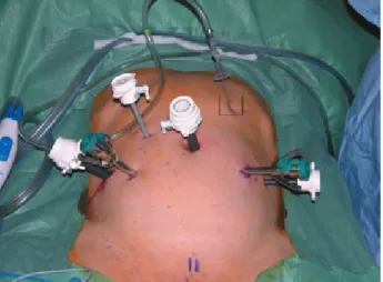
To the best of our knowledge, routine Veress needle insertion through Palmer’s point for robotic and standard laparoscopic radical prostatectomy has not been reported in the literature before. The extra 2-mm incision made for Palmer’s point Veress insertion is not used as a trocar site for pelvic laparoscopy. We have not found it to cause gas leakage, postoperative pain, or bleeding (Figure 2).
In low-risk patients, entry complications are rare in both open Hasson and closed Veress techniques. In a review article by Molloy and associates, entry related bowel and vascular complications occurred in approximately 0.1% of the patients.(3) There is no clear evidence for the
optimal form of laparoscopic entry in the low-risk patients. On the basis of evidence investigated in a recent review, there appears to be no verification of benefit in terms of safety of one technique over another.(29) While a randomized trial might define
which approach is the best, power calculations suggest that 800 000 laparoscopy procedures would be required for such a study.(20)
CONCLUSION
Instead of using different entry techniques
according to the patient’s characteristics, using the same method for every patient seems to be more feasible and easy to learn. Palmer’s point upper quadrant access can be routinely used for both upper and lower urinary tract laparoscopy in all patients.
ACKNOwLEDGEMENTS
The authors are thankful to Prof. Dean Assimos from Urology Department of Wake Forest University Medical School who assisted in preparing and reviewing this article.
CONFLICT OF INTEREST
None declared.REFERENCES
1. Chandler JG, Corson SL, way Lw. Three spectra of laparoscopic entry access injuries. J Am Coll Surg. 2001;192:478-90; discussion 90-1.
2. Vilos GA, Ternamian A, Dempster J, Laberge PY, The Society of Obstetricians and Gynaecologists of C. Laparoscopic entry: a review of techniques, technologies, and complications. J Obstet Gynaecol Can. 2007;29:433-65.
3. Molloy D, Kaloo PD, Cooper M, Nguyen TV. Laparoscopic entry: a literature review and analysis of techniques and complications of primary port entry. Aust N Z J Obstet Gynaecol. 2002;42:246-54. 4. Palmer R. Safety in laparoscopy. J Reprod Med.
1974;13:1-5.
5. See wA, Fisher RJ, winfield HN, Donovan JF. Laparoscopic surgical training: effectiveness and
Figure 2. Port placement for robotic radical prostatectomy with
a three arm da Vinci surgical system and Veress needle at Palmer’s point.
impact on urological surgical practice patterns. J Urol. 1993;149:1054-7.
6. Harkki-Siren P, Kurki T. A nationwide analysis of laparoscopic complications. Obstet Gynecol. 1997;89:108-12.
7. Jansen Fw, Kapiteyn K, Trimbos-Kemper T, Hermans J, Trimbos JB. Complications of laparoscopy: a prospective multicentre observational study. Br J Obstet Gynaecol. 1997;104:595-600.
8. Neudecker J, Sauerland S, Neugebauer E, et al. The European Association for Endoscopic Surgery clinical practice guideline on the pneumoperitoneum for laparoscopic surgery. Surg Endosc. 2002;16:1121-43. 9. wherry DC, Marohn MR, Malanoski MP, Hetz SP, Rich
NM. An external audit of laparoscopic cholecystectomy in the steady state performed in medical treatment facilities of the Department of Defense. Ann Surg. 1996;224:145-54.
10. Patel DN, Parikh MN, Nanavati MS, Jussawalla MJ. Complications of laparoscopy. Asia Oceania J Obstet Gynaecol. 1985;11:87-91.
11. Hasson HM. A modified instrument and method for laparoscopy. Am J Obstet Gynecol. 1971;110:886-7. 12. Ballem RV, Rudomanski J. Techniques of
pneumoperitoneum. Surg Laparosc Endosc. 1993;3:42-3.
13. Sigman HH, Fried GM, Garzon J, et al. Risks of blind versus open approach to celiotomy for laparoscopic surgery. Surg Laparosc Endosc. 1993;3:296-9. 14. Dingfelder JR. Direct laparoscope trocar insertion
without prior pneumoperitoneum. J Reprod Med. 1978;21:45-7.
15. Yerdel MA, Karayalcin K, Koyuncu A, et al. Direct trocar insertion versus Veress needle insertion in laparoscopic cholecystectomy. Am J Surg. 1999;177:247-9.
16. Borgatta L, Gruss L, Barad D, Kaali SG. Direct trocar insertion vs. Verres needle use for laparoscopic sterilization. J Reprod Med. 1990;35:891-4.
17. Byron Jw, Markenson G, Miyazawa K. A randomized comparison of Verres needle and direct trocar insertion for laparoscopy. Surg Gynecol Obstet. 1993;177:259-62.
18. Hasaniya Nw, Kosasa TS, Shieh T, Nakayama RT. Direct laparoscopic entry using a sharp and dull trocar
technique. Obstet Gynecol. 1996;88:620-1.
19. Copeland C, wing R, Hulka JF. Direct trocar insertion at laparoscopy: an evaluation. Obstet Gynecol. 1983;62:655-9.
20. Jansen Fw, Kolkman w, Bakkum EA, de Kroon CD, Trimbos-Kemper TC, Trimbos JB. Complications of laparoscopy: an inquiry about closed- versus open-entry technique. Am J Obstet Gynecol. 2004;190: 634-8.
21. Audebert AJ, Gomel V. Role of microlaparoscopy in the diagnosis of peritoneal and visceral adhesions and in the prevention of bowel injury associated with blind trocar insertion. Fertil Steril. 2000;73:631-5.
22. Mayol J, Garcia-Aguilar J, Ortiz-Oshiro E, De-Diego Carmona JA, Fernandez-Represa JA. Risks of the minimal access approach for laparoscopic surgery: multivariate analysis of morbidity related to umbilical trocar insertion. world J Surg. 1997;21:529-33. 23. Chung HJ, Meng MV, Abrahams HM, Stoller ML.
Upper quadrant access for urologic laparoscopy. Urology. 2003;62:1117-9.
24. Chang FH, Chou HH, Lee CL, Cheng PJ, wang Cw, Soong YK. Extraumbilical insertion of the operative laparoscope in patients with extensive intraabdominal adhesions. J Am Assoc Gynecol Laparosc.
1995;2:335-7.
25. Howard FM, El-Minawi AM, DeLoach VE. Direct laparoscopic cannula insertion at the left upper quadrant. J Am Assoc Gynecol Laparosc. 1997;4: 595-600.
26. Parker J, Reid G, wong F. Microlaparoscopic left upper quadrant entry in patients at high risk of periumbilical adhesions. Aust N Z J Obstet Gynaecol. 1999;39:88-92.
27. Patsner B. Laparoscopy using the left upper quadrant approach. J Am Assoc Gynecol Laparosc. 1999;6: 323-5.
28. Tulikangas PK, Nicklas A, Falcone T, Price LL. Anatomy of the left upper quadrant for cannula insertion. J Am Assoc Gynecol Laparosc. 2000;7: 211-4.
29. Ahmad G, Duffy JM, Phillips K, watson A.
Laparoscopic entry techniques. Cochrane Database Syst Rev. 2008CD006583.