ORIGINAL RESEARCH
Medicine Science 2019;8(4):774-81
Investigation of prognostic significance of CD44 expression in women with vulvar
squamous cell carcinoma
Yakup Yalcin1, Serenat Eris Yalcin2, Selda Uysal3, Burak Tatar4, And Yavuz5, Mehmet Ozgur Akkurt6, Seyran Yigit7 1 Istinye University School of Medicine, Department of Gynecologic Oncology, Istanbul, Turkey
2 Suleyman Demirel University School of Medicine, Department of Perinatology, Isparta, Turkey 3 Ataturk Training and Research Hospital, Department of Obstetrics and Gynecology, Izmır, Turkey
4 Samsun Training and Research Hospital, Department of Gynecologic Oncology, Samsun, Turkey 5 Zeynep Kamil Training and Research Hospital, Department of Perinatology, Istanbul, Turkey 6 Bursa Yuksek Ihtisas Training and Research Hospital, Department of Perinatology, Bursa, Turkey
7 Ataturk Training and Research Hospital, Department of Pathology, Izmır, Turkey Received 12 July 2019; Accepted 23 August 2019
Available online 27.09.2019 with doi:10.5455/medscience.2019.08.9085
Copyright © 2019 by authors and Medicine Science Publishing Inc.
Abstract
The aim of our study is to evaluate whether CD 44 isoform expression is a prognostic factor in vulvar carcinom and to correlate the expression with clinicopathological parameters. Methods: The study included 26 patients diagnosed with invasive squamous cell vulvar cancer. The data of patients were obtained from the oncology follow-up records and the hospital database. The haemotoxylene & eosin stained preparates of the cases were removed from the archives and re-evaluated by examination of the slides under a light microscope. The preparates of the patients were evaluated separately according to the extent and intensity of staining and the combined score applied with the immunohistochemical method in the anti-CD44 antibody in the epithelium and the stroma. Results: When the CD44 epithelial staining intensity and staining combined scores of the patients were examined according to other clinicopathological parameters, a statistically significant difference was found between the groups in terms of tumour grade, differentiation, age, lymph node positivity, and survival rates (p<0.05). As epithelial staining increased and the combined score increased, the tumour grade and lymph node positivity were observed to increase, differentiation worsened and survival reduced. Conclusions: The identification of additional new markers to present useful prognostic factors for vulvar carcinoma is critical. Aberrant expression of CD44 mediated adhesion is likely to constitute one of the main factors leading to the reduced cell-cell and cell-matrix adhesion characteristics of tumor cells and play a pivotal role in the acquisition of invasive and metastatic properties by neoplastic epithelial cells.
Keywords: CD44, expression, vulva, vulvar carcinom
Medicine Science International Medical Journal
Introduction
Vulvar cancer is rarely seen and constitutes 3-5% of all female genital system malignancies [1]. While squamous cell carcinoma comprises 90% of all primary vulva malignancies, malignant melanoma, adenocarcinoma, basal cell carcinoma and sarcoma are less frequently seen. There is no effective method for early diagnosis of vulva cancer showing the malignant transformation of pre-invasive lesions.
Cell adhesion molecules (CAM) function in the regulation of processes such as the orientation of cells to specific tissues, the recognition of each other, embryogenesis, cell growth, cell differentiation and inflammation. Proteases, which play a role in carcinogenesis, invasion and metastasis, may be an alternative *Coresponding Author: Yakup Yalcin, Istinye University School of Medicine, Department of Gynecologic Oncology,Istanbul, Turkey E-mail: [email protected]
in the determination of early stage disease and the evaluation of the prognosis of patients with cancer. The fragmentation of the extracellular matrix made by the proteases expressed from the tumour is the basis of the process required for tumour invasion of the cell. Changes in the structure of the extracellular matrix are important in the spread of the tumour. CD44 isoforms are cell transmembrane glycoproteins with various functions in the interaction between cell-cell or cell-matrix. They are molecules with important functions in the adhesion and migration of cells and which play a significant role in tumour spread and metastasis [2]. The aim of this study was to evaluate the correlation between clinicopathological parameters of CD44 expression in vulva carcinoma and the effect on prognosis. It was thought that CD44 may trigger the increase of some gene expressions or reduce the development of human tumours. A good understanding of these gene products may be helpful in the early diagnosis and treatment of vulvar cancers.
Material and Methods
The study included 26 patients diagnosed with invasive squamous cell vulvar cancer, who presented to the Gynaecology and Obstetrics Clinic of Izmir Katip Celebi University Training and Research Hospital between 1999-2010. The data of patient age, tumour diameter, localisation, histological type, grade, stage and treatment were obtained from the oncology follow-up records and the hospital database. The new FIGO 2009 grading system was used for surgical grading of the tumours.
The haemotoxylene & eosin stained preparates of the cases were removed from the archives and re-evaluated by examination of the slides under a light microscope. For each case, the most identifiable block was selected for immunohistochemical applications. From the selected paraffin blocks, new slices of 4-5 micron thickness were cut and placed on poly-L-lysine coated slides. The tissue samples were firstly stored for 16 hours in an incubator at 60ºC. The de-paraffinised slides were then kept in the Dako PT Link unit for 25 minutes and the initial procedures were applied. Staining procedures were applied with the Flex-envision system in the Dako Autostainer device. As primary antibody, anti-human CD44 antibody, Phagocytic Glycoprotein-1 (Mouse Monoclonal Antibody, Code no M7082 DakoCytomation Denmark) was diluted at a ratio of 1:25 and incubated for 30 mins. Then it was passed through increasing (80%, 96%, 99%) concentrations of alcohol. The slides clarified with xylol were closed with an adhesive medium. Ordinary tonsil tissue was used as a positive control [3].
Immunohistochemical Method
Evaluation of Immunohistochemical Staining
All the tumour areas were scanned by light microscope at low magnification (x10) and for each antibody in squamous areas, the staining density of tumour cells was defined semiquantitatively and subjectively at x20 magnification and at high magnification (x40) as the ratio of all the tumour cells (extent of staining). Cells other than tumour cells (lymphocytes, endothelial cells etc) showing staining were not included in the evaluation. Membranous and cytoplasmic staining was accepted as significant for CD44. To define the extent and intensity of the staining of CD44 in the epithelium, the study by Liang et al was taken as the basis and for extent and intensity of staining in the stroma, the study by Hamalainen et al [4, 5]. According to this, the percentage of cells stained positive for CD44 was defined by counting 10 consecutive high magnification (x40) areas.
The preparates of the patients were evaluated separately according to the extent and intensity of staining and the combined score applied with the immunohistochemical method in the anti-CD44 antibody in the epithelium and the stroma. According to this, positive stained tumour cells <5% were evaluated as 0 points, 5-25% as 1 point, 26-50% as 2 points, >50% as 3 points. Below 5% was accepted as negative and over 5% as positive. Staining intensity was scored as no staining =0, mild staining = 1, moderate staining = 2, severe staining =3. Finally evaluation was made with the combined score, which was the combination of the staining intensity and the staining percentage.
Combined score; 0 points: combined score 0 (negative) 1-2 points: combined score 1 (mild positive), 3-4 points: combined score 2 (moderate positive),
5-6 points: combined score 3 (strong positive)
In the current study, there were no cases of negative staining with a combined score of 0.
Statistical Analysis
All statistical analyses were performed with SPSS Statistical Package for Social Sciences) for Windows 15.0. When the data were evaluated they were summarised in table form. In the inter-group comparison of quantitative data, the Mann Whitney test was used and for qualitative data, the Chi Square test and Fisher’s Exact test. For correlations between the data, Spearman Correlation analysis was applied. Analysis of the survival of the 26 cases was applied with the Kaplan Meier method. The results were given at a 95% confidence interval and a value of p<0.05 was accepted as statistically significant.
Results
The study comprised 26 cases with vulva carcinoma with a mean age of 69.69 years (range 47-85 years). Mean tumour diameter was measured as 3.22 cm (range, 1.5-7cm). The tumour location was on the labium majus in 18 (69.25%) cases, the labium minus in 5 (19.25%) and on the clitoris in 3 (11.5%). When the patients were evaluated according to the tumour grade, 10 (38.5%) were well-differentiated, 7 (27%) were moderately differentiated and 9 (34.5%) were poorly differentiated. Radical vulvectomy + inguinofemoral lymph node dissection was applied in 21 cases and simple vulvectomy because of poor performance status associated with advanced age in 5 cases. The surgical borders were safe in all the vulvectomy material removed in the operations.
According to the evaluation using the FIGO surgical grading system, 1 case (3.8%) was Grade 1A, 15 (57.6%) were Grade 1B, 2 (7.6%) were Grade II, 3 (11.5%) were Grade IIIA and 5 (19.5%) weree Grade IIIB. No cases were in the Grade IV group. Of the 21 cases with vulva carcinoma to whom radical vulvectomy and lymph node dissection was applied, lymph node positivity was observed in 8 cases. In the follow-up of the surviving patients who were operated on, 15 died and 11 survived (Table 1).
No cases in the current study were CD44 negative stained. When the CD44 epithelial staining percentage and stromal staining percentage of the patients were examined according to tumour grade, differentiation, age, lymph node involvement, survival and radical surgery (Table 2), the CD44 epithelial staining percentage values were found to be statistically significantly high in those with moderate or poor differentiation, those with positive lymph node involvement, the exitus cases and those with advanced stage tumours (p<0.05). No statistically significant difference was determined between the groups in respect of CD44 stromal staining percentage values (p>0.05).
When the CD44 epithelial staining intensity and staining combined scores of the patients were examined according to other clinicopathological parameters, a statistically significant difference was found between the groups in terms of tumour grade, differentiation, age, lymph node positivity, and survival rates (p<0.05). As epithelial staining increased and the combined score increased, the tumour grade and lymph node positivity were observed to increase, differentiation worsened and survival reduced. No statistically significant difference was determined
between the groups in respect of age groups or radical operation rates (p>0.05) (Table 3). No statistically significant difference was determined between the groups in respect of CD44 stromal staining intensity and stromal staining combined score distributions with the other clinicopathological parameters (p>0.05).
When the invasion depth values were examined according to the evaluation of CD44 epithelial and stromal expression in the cancer patients (Table 4), a statistically significantly strong relationship was found between those with more than 50% epithelial staining and those with strong CD44 epithelial staining intensity and epithelial combined score and invasion depth values (p<0.05). It was seen that as CD44 epithelial expression increased, so invasion depth increased. No statistically significant difference was determined between the groups in terms of invasion depth values according to CD44 stromal staining percentage, CD44 stromal staining intensity and CD44 stromal staining combined score (p>0.05).
When survival was examined according to other clinicopathological
parameters (Table 5), survival was found to be statistically significantly low in those with moderate or poor histological differentiation, lymph node involvement, advanced tumour stage, CD44 epithelial staining of >50% and those with high CD44 epithelial staining intensity and epithelial staining combined scores (p<0.05). It was seen that general survival decreased with reduced differentiation, lymph node metastasis, advanced stage tumour grade and high CD44 expression. No statistically significant difference was determined between the groups in terms of survival according to radical surgery, age, stromal staining percentge, stromal staining intensity and stromal staining combined score (p>0.05).
A statistically significant difference was determined between general survival rates and tumour stage, lymph node positivity, differentiation and CD44 epithelial expression (p<0.05). A shorter survival period was observed in patients with advanced stage tumour, moderate or poor differentiation, lymph node positivity and positive CD44 epithelial expression.
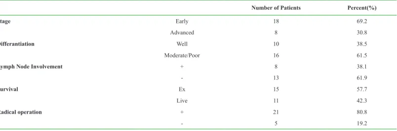
Table 1. The distribution of percentages of stage, differentiation, lymph node involvement, survival and radical operation
Number of Patients Percent(%)
Stage Early 18 69.2
Advanced 8 30.8
Differantiation Well 10 38.5
Moderate/Poor 16 61.5
Lymph Node Involvement + 8 38.1
- 13 61.9
Survival Ex 15 57.7
Live 11 42.3
Radical operation + 21 80.8
- 5 19.2
Table 2. The comparison of CD44 epithelial staining percentage and stromal staining percentage of the patients with tumour grade, differentiation, age, lymph node involvement, survival and radical surgery
CD44 epithelial staining percentage P Value CD44 stromal staining percentage P Value
Stage Well 23±18.14 0.001 49.5±34.6 0.363
Moderate/Poor 73.75±25.79 61.56±30.32
Differantiation <70 58.64±37.42 0.507 60.91±29.14 0.610
>70 51±32.08 54±34.5
Lymph Node Involvement + 86.25±15.06 0.001 46.88±37.32 0.644
- 40±30.05 61.39±29.25 Survival Ex 75.67±25.97 0.001 56.67±34.1 0.799 Live 25±17.46 57.27±30.28 Radical operation + 54.52±33.01 0.801 52.86±33.26 0.178 - 53±41.77 74±19.49 Early 40±30.05 0.001 61.39±29.25 0.644 Advanced 86.25±15.06 46.88±37.32
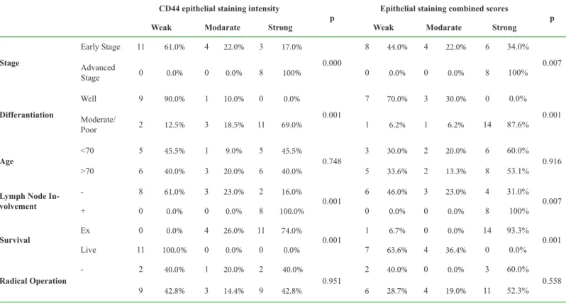
Table 3. The comparison of the CD44 epithelial staining intensity and staining combined scores of the patients with other clinicopathological parameters CD44 epithelial staining intensity
p Epithelial staining combined scores p
Weak Modarate Strong Weak Modarate Strong
Stage Early Stage 11 61.0% 4 22.0% 3 17.0% 0.000 8 44.0% 4 22.0% 6 34.0% 0.007 Advanced Stage 0 0.0% 0 0.0% 8 100% 0 0.0% 0 0.0% 8 100% Differantiation Well 9 90.0% 1 10.0% 0 0.0% 0.001 7 70.0% 3 30.0% 0 0.0% 0.001 Moderate/ Poor 2 12.5% 3 18.5% 11 69.0% 1 6.2% 1 6.2% 14 87.6% Age <70 5 45.5% 1 9.0% 5 45.5% 0.748 3 30.0% 2 20.0% 6 60.0% 0.916 >70 6 40.0% 3 20.0% 6 40.0% 5 33.6% 2 13.3% 8 53.1%
Lymph Node In-volvement - 8 61.0% 3 23.0% 2 16.0% 0.001 6 46.0% 3 23.0% 4 31.0% 0.007 + 0 0.0% 0 0.0% 8 100.0% 0 0.0% 0 0.0% 8 100% Survival Ex 0 0.0% 4 26.0% 11 74.0% 0.001 1 6.7% 0 0.0% 14 93.3% 0.001 Live 11 100.0% 0 0.0% 0 0.0% 7 63.6% 4 36.4% 0 0.0% Radical Operation - 2 40.0% 1 20.0% 2 40.0% 0.951 2 40.0% 0 0.0% 3 60.0% 0.558 9 42.8% 3 14.4% 9 42.8% 6 28.7% 4 19.0% 11 52.3%
Table 4. The examination of the invasion depth values according to the evaluation of CD44 epithelial and stromal expression in the cancer patients
Invasion Depth p
CD44 epithelial staining percentage
%0-5 4.63±2.3
0.024
%6-25 2.75±0.71
%26-50 5.81±3.25
>%50 5.89±4.82
CD44 epithelial staining intensity
Weak 2.7±0.88
0.001
Moderate 5.96±1.33
Strong 7.08±3.01
CD44 epithelial staining combined score
Weak 3.22±1.37
0.001
Moderate 2.56±1.3
Strong 6.82±2.76
CD44 stromal staining percentage
%0-5 4.5±1.49
0.563
%6-25 6.13±0.18
%26-50 3.5±1.32
>%50 5.34±3.45
CD44 stromal staining intensity
Weak 4.19±1.72
0.535
Moderate 6.29±4.37
Strong 4.97±2.52
CD44 stromal staining combined score
Weak 5.04±1.43
0.218
Moderate 3.13±1.32
Table 5. The comparison of survival with other clinicopathological parameters
Overall survival (month) P
Differantiation Moderate/PoorWell 24.38±19.3647.1±19.9 0.007
Age <70 32.91±26.85 0.507
>70 33.27±19.18
Lymph Node Involvement - 40.56±22.37 0.004
+ 16.38±9.36
Radical Operation - 23±5.92 0.486
+ 35.52±24.07
Stage Early 40.56±22.37 0.004
Advanced 16.38±9.36
CD44 epithelial staining percentage
%0-5 36.5±13.44
0.013
%6-25 51±20.59
%26-50 47±26.76
>%50 21±15.56
CD44 epithelial staining intensity
Weak 47±22.17
0.007
Moderate 37.25±24.97
Strong 17.73±8.84
CD44 epithelial staining combined score
Weak 47.38±19.33
0.014
Moderate 46±27.66
Strong 21.29±15.69
CD44 stromal staining percentage
%0-5 42.25±16.58
0.202
%6-25 11.5±2.12
%26-50 37.67±23.67
>%50 32.71±23.82
CD44 stromal staining intensity
Weak 39.22±23.38
0.542
Moderate 30.43±23.93
Strong 29.5±21.18
CD44 stromal staining combined score
Weak 32±20.45
0.447
Moderate 46.25±25.85
Strong 30.25±22.27
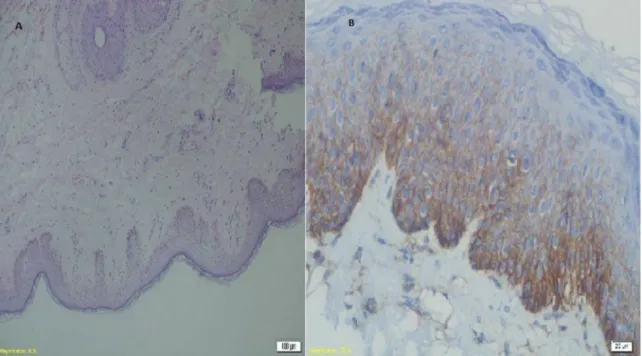
Figure 1. A: Normal vulvar tissue stained with hematoxylin and eosin (HE ×20 magnification) and B: Staining of CD 44 antibody in normal vulvar tissue (at ×40 magnification)
Discussion
Various genetic factors play a role in the development of cancer. Changes in some gene expressions cause the formation and development of tumours. CD44 and isoforms, which have been recently investigated in various cancer types, play a significant role in carcinogenesis. Increased expression of CD44 isoforms is seen to be a potential prognostic marker, showing both the early phase and the metastatic phase of carcinogenesis in most epithelial and non-epithelial malignancies [6, 7].
CD44 is an integral membrane glycoprotein and functions as a hyaluronic acid receptor. It is known as extracellular matrix receptor III. CD44 is expressed in epithelial, mesenchymal, lymphoid and glial cells. It is responsible for cell and cell-extracellular matrix adhesion. It is a mediator in the rolling of lymphocytes over endothelial cells, cell migration and in the stimulation of hematopoietic cell differentiation [6].
It has been shown that CD44 expression may be a determining factor of the metastatic capacity and progression of tumours in gastro-intestinal cancers, renal cell cancer, lung cancer, breast cancer and non-Hodgkin lymphomas [8-14]. There are also studies showing a relationship of low expression CD44 in transitional cell carcinoma and neuroblastoma and poor prognostic factors [15, 16]. In a study by Hong et al on the importance of CD44 expression in various gynaecological malignancies, a relationship was reported between increased CD44 expression in early stage cervix cancers and stromal invasion. However, in more than 5mm stromal invasion, expression was observed to decrease. It has been
shown that CD44 expression in patients with endometrium cancer could be related to cell differentiation and in contrast to Grade 1 tumours, expression has been shown to be reduced in Grade 2 and 3 tumours. In addition, it was also reported that it could be used as a prognostic marker in the early diagnosis of endometrium cancer patients. However, no relationship was determined between CD44 expression and grade, myometrial invasion, tumour size and isthmus-cervix involvement. When ovarian tumours were examined, there was no expression in benign tumours and increased expression was noticeable in borderline and malignant tumours. A relationship has been found between CD44 expression and the development of borderline or malignant ovarian tumours [17].
In a study by Hamalainen et al, CD44 epithelial and stromal expressions were examined in vulva carcinoma, vulvar intraepithelial neoplasia (VIN), lichen sclerosus (LS) and normal vulvar tissue. In malignant cases, the epithelial CD44 expression was found to be statistically significantly reduced compared to VIN, LS and normal vulvar tissue. In the evaluation in terms of stromal staining intensity, no statistically significant relationship was found between cases [5]. In the current study, similar findings were found. However, the CD44 epithelial expression was seen to be increased in malignant cases compared to other cases in this study.
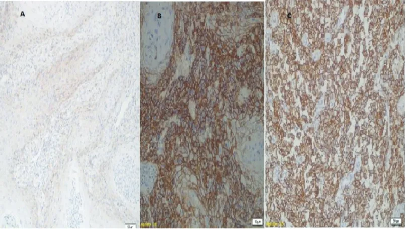
In a study by Tempfer et al of patients with vulva cancer, a comparison was made of cases with positive CD44 expression with those with negative CD44 expression and it was reported that the positive cases had shorter and worse survival rates. However, no relationship was determined between CD44 expression and Figure 2. A: CD44 expression in well-differentiated tumor, B: in moderately differentiated tumor and 2C: in poor differentiated tumor (at ×20 magnification).
tumour stage, histological grade and type of treatment [18]. In another study by Tempfer et al, the effect of CD44 expression on prognosis was investigated in early stage vulva cancers. It was concluded that CD44 expression affected prognosis negatively and reduced disease-free survival in the general survival period. The recurrence rates of patients with CD44 expression were determined to be greater. However, no significant difference was found when the relationship of CD44 expression and tumour differentiation was examined [19].
Hefler et al determined a relationship between the reduction of disease-free survival and the general survival rate and over-expression of CD44 in a multi-centre study of patients with vulva cancer. In previous studies, the relationship between CD44 expression and lymph node metastasis had not been investigated because of the low number of patients and no dissection of lymph nodes. In that study there were sufficient patients and lymph node dissection but no statistically significant correlation was determined between CD44 expression and tumour stage, lymph node involvement or tumour differentiation. It was shown that CD44 expression could give important information about survival, independently of the other clinicopathological parameters. In addition, in that study no correlation was found between the CD44 expression values examined immunohistochemically in the tumour tissue and the serum CD44 values which were examined at the time of treatment [20]. In the current study, a statistically significant relationship was determined between CD44 expression and tumour stage, lymph node involvement, invasion and tumour differentiation. This showed that it could not have affected survival independently of other clinicopathological parameters.
The determination of new markers is of critical importance in addition to the existing prognostic factors of stage, differentiation and lymph node involvement which may be useful for patients with vulvar carcinoma. Although there are ongoing studies on this subject, the ideal prognostic factors for this disease have not yet been identified. The most important parameters currently for the determination of prognosis are stage and lymph node metastasis. However, even these parameters are insufficient to determine prognosis in some patients. Therefore, many studies are being conducted on this subject to identify new prognostic factors. The determination in recent years of new immunohistochemical markers with tumour cytogenitics, the presence of some markers in the serum with the immunoblotting method and the effect of these on prognosis has resulted in studies related to the use of new agents in treatment continuing at a rapid rate. In this study an investigation was made into the effect on prognosis and the relationship with clinicopathological parameters of markers which play a significant role in the development of the invasive and metastatic properties of neoplastic cells, such as CD44 which has a function in cell-cell and cell matrix adhesion. Some of the results of this study were found to be consistent with literature and some were different. However, there are many different results in studies in literature, which can be considered to be due to differences in the technique used, the use of different dilutions of different antibody clones and different methods used in the evaluations. In the light of these data, to be able to arrive at a conclusion, there is a need for further studies of larger numbers of cases with a standard immunohistochemical technique and standard evaluations.
Conflict of interest
The authors declare that there are no conflicts of interest.
Financial Disclosure
All authors declare no financial support.
Ethical approval
There is ethics commıtee approval Yakup Yalcin ORCID:0000000288266481 Serenat Eris Yalcin ORCID:000000026465325X Selda Uysal ORCID:0000000307686159 Burak Tatar ORCID:0000000264950174 And Yavuz ORCID:0000000259694444
Mehmet Ozgur Akkurt ORCID:0000000176878977 Seyran Yigit ORCID:000000023530988X
References
1. Jemal A, Siegel R, Ward E, et al. Cancer Statistics. CA Cancer J Clin. 2006;56:106-30.
2. Leblanc M, Poncelet C, Soriano D, et al. Alteration of CD44 and cadherins expression: possible association with augmented aggressiveness and invasiveness of endometrial carcinoma. Virchows Arch. 2001;438:78–85. 3. Ingvarsson S, Dahlenborg K, Carlsson R, et al. Co-ligation of CD44 on
naive human tonsillar B cells induces progression towards a germinal center phenotype. Int Immunol. 1999;11:739-44.
4. Liang Y, Fang T, Xu H, et al. Expression of CD44v6 and Livin in gastric cancer tissue. Chin Med J. 2012;125:3161-5.
5. Hämäläinen K, Kosma VM, Eloranta ML, et al. Downregulated CD44 and hyaluronan expression in vulvar intraepithelial neoplasia and squamous cell carcinomas. Acta Obstet Gynecol Scand. 2010;89:108-19.
6. Kaufmann M, Heider KH, Sinn HP, et al. CD44 variant exon epotopes in primary breast cancer and length of survival. Lancet. 1995;345:615-9. 7. Woodman AC, Sugiyama M, Yoshida K, et al. Analysis of anomalous CD44
gene expression in human breast, bladder and colon cancer and correlation of observed mRNA and protein isoforms. Am J Pathol. 1996;149:1519-30. 8. Galluzzo E, Albi N, Fiorucci S. Involvement of CD44 variant isoform
in hyaluronate adhesion by human activated T cells. Eur J Immunol. 1995;25:2932-9.
9. Xin Y, Grace A, Gallagher MM, et al. CD44v6 in gastric carcinoma: A marker of tumor progression. Appl Immunohistochem Mol Morph. 2001;9:138-42. 10. Yamaguchi A, Goi T, Yu J, et al. Expression of CD44v6 in advenced gastric
cancer and its relationship to hematogenous metastasis and long-term prognosis. J Surg Oncol. 2002;79:230-5.
11. Shimbori M, Kijima H, Sato S. Expression of CD44 in primary lung carcinomas using histological and cytological analyses. Anticancer Res. 2003;23:115-21.
12. Lucin K, Matusan K, Dordevic G, et al. Prognostic significance of CD44 molecule in renal cell carcinoma. Croat Med J. 2004;45:703-8.
13. Ma W, Deng Y, Zhou L. The prognostic value of adhesion molecule in women with pimary breast carcinoma: A clinicopathologic study. Clin Oncol. 2005;17:258-63.
14. Lockhart MS, Waldner C, Mongini C, et al. Evaluation of soluble CD44 in patients with breast and colorectal carcinomas and non-Hodgkin’s lymphoma. Oncol Rep. 1999;6:1129-33.
15. Toma V, Hauri D, Schmid U, et al. Focal loss of CD44 variant protein expression is related to recurrence in superficial bladder carsinoma. Am J
Pathol. 1999;155:1427-32.
16. Comito MA, Savell VH, Cohen MB. CD44 expression in neuroblastoma and related tumors. J Pediatr Hematol Oncol. 1997;19:292-6.
17. Hong SC, Song JY, Lee JK, et al. Significance of CD44v6 expression in gynecologic malignancies. J Obstet Gynaecol Res. 2006;32:379-86. 18. Tempfer C, Gitsch G, Haeusler G, et al. Prognostic value of
immunohistochemically detected CD44 expression in patients with carcinoma
of the vulva. Cancer. 1996;78:273-7.
19. Tempfer C, Sliutz G, Haeusler G, et al. CD44v3 and v6 variant isoform expression correlates with poor prognosis in early-stage vulvar cancer. Br J Cancer. 1998;78:1091-4.
20. Hefler L, Concin N, Mincham D, et al. The Prognostic Value of Immunohistochemically Detected CD44v3 and CD44v6 Expression in Patients with Surgically Staged Vulvar Carcinoma. A Multicenter Study American Cancer Society. Cancer. 2002;94:125–30..