Comparative Study of Inhibitory Potential of Dietary
Phytochemicals Against Quorum Sensing Activity of and Biofilm
Formation by Chromobacterium violaceum 12472, and Swimming
and Swarming Behaviour of Pseudomonas aeruginosa PAO1
https://doi.org/10.17113/ftb.57.02.19.5823Elif Burcu Bali
1* ,
Kübra Erkan Türkmen
2,3,
Demet Erdönmez
4and
Necdet Sağlam
51 Gazi University, Vocational School
of Health Services, Department of Medical Services and Techniques, Programme of Medical Laboratory Techniques, 06830 Gölbaşı, Ankara, Turkey
2 Karamanoğlu Mehmetbey University,
Faculty of Science Department of Biology, 70100 Karaman, Turkey
3 Hacettepe University, Faculty of
Science Department of Biology and Biotechnology, 06800 Beytepe, Ankara, Turkey
4 Aksaray University, Faculty of Science
and Letters, Department of Biology, 68100 Aksaray, Turkey
5 Hacettepe University, Graduate
School of Science and Engineering, Nanotechnology and Nanomedicine Division, 06800 Beytepe, Ankara, Turkey Received: 27 April 2018 Accepted: 8 February 2019 *Corresponding author: Phone: +903124845635175 E-mail: [email protected], [email protected]
SUMMARY
Quorum sensing (QS) and biofilm formation are important mechanisms related to antibiotic resistance of many pathogens. Alternative treatments are needed to prevent recurrent or chronic infections caused by multi-resistant pathogens. Therefore, the aim of this study is to investigate and compare the inhibitory potential of the dietary phyto-chemicals: curcumin, quercetin, apigenin, pyrogallol, gallic acid and luteolin against QS of and biofilm formation by Chromobacterium violaceum ATCC 12472 and the swimming and swarming abilities of Pseudomonas aeruginosa PAO1. Anti-QS potential of the phyto-chemicals was evaluated qualitatively and quantitatively using C. violaceum via the disk diffusion assay based on violacein pigment inhibition at the subminimal inhibitory con-centrations ranging from 46.87 to 750 µg/mL. The results of anti-QS and antibiofilm ac-tivities on C. violaceum demonstrated that all the phytochemicals except pyrogallol and gallic acid inhibited violacein production (from (11.0±0.1) to (88.2±0.1) %) in a concentra-tion-dependent manner. In addition, the biofilm formation was also significantly inhib-ited (p<0.05) in the presence of all the phytochemicals ((1.38±0.08)–(84.2±0.2) %). In the present study, the results revealed that quercetin, curcumin, apigenin and luteolin could be promising QS and biofilm inhibitory agents against the C. violaceum 12472 biosensor system. Our findings also suggest that all the phytochemicals, especially curcumin, querce-tin and pyrogallol, might be anti-pathogenic agents against P. aeruginosa PAO1 infections due to the ability to control QS. However, more comprehensive studies at the molecular level, explaining their anti-QS mechanisms, need to be conducted to confirm these results and identify the genes involved.
Key words: phytochemicals, anti-quorum sensing, Chromobacterium violaceum ATCC
12472, Pseudomonas aeruginosa PAO1, antibiofilm activity
INTRODUCTION
Chromobacterium violaceum obtained from water, soil, and human skin is a facultative
anaerobic, Gram-negative opportunist, which often produces violacein, a characteristic purple, water-insoluble pigment with antibacterial activity. It causes severe morbidity and mortality associated with infections such as bacteremia and abscesses. It is also resistant to multiple antimicrobials, thereby causing recurrent infections (1–3). The production of the violacein pigment in C. violaceum is controlled by quorum sensing (QS), a process of bacterial cell-cell communication in which cells regulate the transcription of the specific genes responsible for the production of antibiotics, biofilm differentiation, cell division, bioluminescence and other processes (4,5).
The QS systems of Gram-negative bacteria are composed of LuxI-type autoinducer syn-thases that synthesize specific acylated homoserine lactone (AHL) autoinducers and the AHL binds specifically to LuxR receptor protein to trigger specific gene expressions (6). For ex-ample, C. violaceum ATCC 12472 produces and responds to cognate autoinducer molecules
C6-AHL and C4-AHL to induce the production of the violacein
pigment(7). The compounds preventing the violacein
produc-tion by this pathogen, without any bacterial inhibiproduc-tion, could be promising QS inhibitors, capable of attenuating bacterial pathogenicity with a lower risk of resistance development than in the case of antibiotics (8). On the other hand, Pseudomonas
aeruginosa is the most prevalent and important opportunistic
human pathogen, causing for example fatal lung disease in pa-tients with cystic fibrosis. It can display virulence partially ow-ing to its motility, which plays a significant role in its coloniza-tion in various environments, the attachment of the bacteria to surfaces, and biofilm formation. Moreover, the bacterium uses different types of QS signal molecules to synchronize partic-ular gene expressions, including those involved in its biofilm formation and virulence (9–11).
The overuse of conventional antibiotics has resulted in the emergence of diseases caused by multi-resistant bacte-ria. The disadvantages of conventional antimicrobials include their natural selective pressure and failure to treat infections caused by bacterial biofilms (12). Compared to conventional antimicrobials, plant-derived compounds, especially bioac-tive phytochemicals, are not generally associated with many side effects, and they have a significant anti-infective poten-tial against infectious diseases (13). Therefore, many bioactive compounds in the form of plant phenols have been used ex-tensively as antimicrobials for decades in traditional medi-cine (14). In addition, the inhibition of bacterial QS system is being evaluated as a new target for developing anti-infective therapies since blocking of QS would weaken virulence of the pathogens, making them more susceptible to treatment and facilitating easy clearance by host defence mechanisms (15). Therefore, research focused on the discovery of novel phytochemicals and plant extracts specifically targeting QS signalling systems and their biofilm inhibition has increased recently (14–17). Apart from the development of potent ther-apeutics, the detection of anti-pathogenic phytochemicals interfering with QS and virulence factor production may re-veal promising anti-infective compounds. Hence, the aim of the present study is to comparatively investigate anti-QS and antibiofilm potential of the dietary phytochemicals querce-tin, curcumin, apigenin, pyrogallol, gallic acid and luteolin, against C. violaceum ATCC 12472 as well as their inhibitory activities against swimming and swarming abilities of P.
aeru-ginosa PAO1. A few of the phytochemicals, particularly
quer-cetin and curcumin, have already been reported to act as an-ti-QS and antibiofilm agents (6,18–21); however, this study is the first one to compare their inhibitory effects on QS and biofilm formation by C. violaceum 12472, and swimming and swarming motility of P. aeruginosa PAO1.
MATERIALS AND METHODS
Bacterial strains and culture conditions
In this study, the wild-type strain Chromobacterium
violace-um ATCC 12472 and Pseudomonas aeruginosa PAO1 were used
as biosensor strains for quorum sensing (QS) and motility as-says, respectively. C. violaceum 12472 was a kind gift from Prof. Dr Robert J.C. McLean (University of Texas, TX, USA), and P.
ae-ruginosa PAO1 was also a kind gift from Daniel Lopez, PhD
(Na-tional Centre for Biotechnology (CNB), Autonomous University of Madrid, Madrid, Spain). The strains were routinely grown in Luria-Bertani (LB) broth (1 % tryptone, 0.5 % yeast extract and 1 % NaCl (Sigma-Aldrich, Merck, St. Louis, MO, USA) medium; pH=7.0). Flasks with C. violaceum and P. aeruginosa PAO1 were incubated at 30 and 37 °C for 24 h, respectively.
Preparation of dietary phytochemicals
The dietary phytochemicals (quercetin, curcumin, api-genin, pyrogallol and luteolin) were purchased from Sig-ma-Aldrich, Merck. Gallic acid and all solvents were from Merck (Darmstadt, Germany). The stock solutions of api-genin and luteolin were prepared in 10 % dymethylsulphox-ide (DMSO), those of curcumin and quercetin in 50 % metha-nol, and pyrogallol and gallic acid were dissolved in distilled water. The final volume fraction of DMSO or methanol was adjusted to ≤0.5 %, which did not have any discernible ef-fect on the growth of C. violaceum 12472. The stock solutions were prepared daily and diluted to the desired volume frac-tion immediately prior to use. The control groups were 0.5 % DMSO, 0.5 % methanol and the bacteria cultured in LB broth.
Determination of minimum inhibitory concentrations
Minimum inhibitory concentration (MIC) values of the di-etary phytochemicals were determined by the broth microdi-lution test in 96-well plates as previously described (22) with some modifications. A volume of 100 μL of the stock solutions of the phytochemicals (50 % V/V) was added to an equal volume of LB broth, and twofold serial dilutions (1500, 750, 375, 187.5, 93.75 and 46.87 µg/mL) were prepared in the microtiter plate. Overnight cultures of C. violaceum 12472 and P. aeruginosa PAO1
were adjusted to the absorbance value A600 nm=0.4 using
ste-rile broth, and 100 μL of each culture were added to the plates, after which the plates were incubated at 30 and 37 °C for 24 h, respectively. Inhibition of the bacterial growth in the wells con-taining the phytochemicals was assessed by a comparison with the growth in blank control wells. Every experiment included negative (medium, 0.5 % DMSO, and 0.5 % methanol) and posi-tive control (medium including the inoculum). The MIC was re-corded as the lowest concentration at which there was no visi-ble growth of the bacteria. The MIC assay was repeated at least twice. The subminimal inhibitory concentrations (sub-MICs) of each phytochemical (46.87–750 µg/mL) were tested for the assessment of anti-QS and antibiofilm activity.
Quorum sensing inhibition
Qualitative anti-QS activity: Disc diffusion method
The standard disc diffusion assay, with few modifications, was used for the detection of the anti-QS potential of the
dietary phytochemicals using the wild-type pigmented bio-sensor strain C. violaceum 12472 (23). It was grown in LB broth or on the LB agar (1.2 % m/V). A volume of 5 mL of molten LB agar (0.3 % m/V) was inoculated with 50 μL of the C. violaceum 12472 culture grown overnight in LB broth. The agar solution with the culture was immediately poured over the surface of LB agar plates. A volume of 20 μL of each phytochemical solution (46.87, 93.75, 187.5, 375 and 750 µg/mL) was pipetted
on sterile paper disks (6 mm diameter; Bioanalyse
®
, Ankara,Turkey), which was placed on the solidified agar. The plates were incubated overnight at 30 °C and examined for violace-in pigment production. QS violace-inhibition, violace-in sub-MIC values, was detected by a ring of colourless but viable cells around the disks. The measurements were made from the outer edge of the disks to the edge of the zones suggesting anti-QS inhi-bition. Controls were 0.5 % DMSO and 0.5 % methanol. This experiment was carried out at least three times.
Quantitative anti-QS activity: Violacein inhibition
Inhibitory potential of the dietary phytochemicals on the violacein pigment production was also measured spectro-photometrically using the method of Blosser and Gray (24), with a few modifications. Quantitative evaluation of anti-QS activity of the phytochemicals was carried out based on their ability to inhibit the production of the purple pigment vi-olacein by C. violaceum 12472. Briefly, the phytochemicals were added to 200 µL of bacterial culture (in LB broth) at the sub-MIC concentrations (46.87, 93.75, 187.5, 375 and 750 µg/ mL) and incubated at 30 °C until complete pigmentation was achieved in the blank, i.e. untreated culture. First, 200 µL of treated and untreated cultures were placed in an Eppendorf tube and lysed by addition of 200 µL of 10 % SDS, vortexed for 5 s and incubated at room temperature for 5 min. Sub-sequently, 900 µL of water-saturated butanol (50 mL n-bu-tanol mixed with 10 mL distilled water) were added to the cell lysate, followed by vortexing for 5 s and centrifugation at 13 000×g for 5 min. The upper (butanol) phase containing the violacein was collected and the absorbance was read at 585 nm in UV-Vis spectrophotometer (UV-1800; Shimadzu, Kyoto, Japan). The percentage of violacein inhibition was calculated using the following formula:
Inhibition=(A585 nm(control)–A585 nm(test)/A585 nm(control))·100 /1/ where A is the absorbance, controls were 0.5 % DMSO, 0.5 % methanol and the untreated bacteria, and the test was the culture treated with the phytochemicals. The experiments were performed at least in triplicate.
Antibiofilm activity
Biofilm formation in 96-well U-bottom polystyrene micro-titer plates (Nunc™, Thermo Fisher Scientific, Waltham, MA, USA) was assayed via a previously described, slightly modified method (25). An overnight culture of C. violaceum 12472 was di-luted 1:100 with LB broth and grown for another hour. After the
addition of sub-MICs of the phytochemicals (46.87, 93.75, 187.5, 375 and 750 µg/mL), 100 µL of the culture were pipetted into the wells of the microtiter plates and the plates were incubated for 24 h at 30 °C. Then, the medium was removed and washed with 1×PBS buffer three times. The plates were dried at 65 °C
in a universal oven (UNB 100; Memmert
®
, Schwabach,Germa-ny) and then 100 µL of a 1 % m/V aqueous solution of crystal violet were added. The stain was allowed to fix at room tem-perature for 20 min, after which the dye was removed from the wells by washing thoroughly with sterile water. For the quantification of the attached biomass, the bound dye was dissolved with 30 % acetic acid solution, and the absorbance was determined at 595 nm. Inhibitor-mediated reduction of biofilm formation was assessed by comparing it to the posi-tive control without phytochemicals. The biofilm assay was performed three times, and six wells per treatment were used each time.
Swimming and swarming assays
The swimming and swarming motility assays were per-formed using a previously described, slightly modified meth-od (26). In swimming assay, 5 µL of overnight culture of the
Pseudomonas aeruginosa PAO1 (A600 nm=0.4) were point in-oculated at the centre of an agar medium consisting of 1 % tryptone, 0.5 % NaCl and 0.3 % agar with the sub-MIC con-centration of the phytochemicals (93.75 µg/mL). For swarm-ing assays, the agar medium comprised 1 % peptone, 0.5 % NaCl, 0.5 % agar and 0.5 % filter-sterilized d-glucose with the
same sub-MIC concentration of phytochemicals (93.75 µg/mL). The plates were then incubated at 37 °C in an upright position for 16 h. The reduction in swimming and swarming migra-tion was recorded by measuring the swimming and swarm-ing zones of the bacterial cells after 16 h compared to the negative controls.
Growth curve assay
To confirm the anti-QS activity of the dietary phytochemi-cals, the growth curve assay of C. violaceum 12472 cultivated in the presence and absence of phytochemicals was
perfor-med. Overnight culture of the bacteria (1 %; A600 nm=0.4) was
inoculated in a 250-mL Erlenmeyer flask containing 50 mL of LB broth supplemented with the sub-MIC concentrations (375 and 750 µg/mL) of each phytochemical. The flasks were incubated at 30 °C and 180 rpm in a rotary shaker (KS300; IKA
®
-Werke GmbH & Co. KG, Staufen, Germany). The cell den-sity was measured by UV-Vis spectrophotometer (UV-1800; Shimadzu) at 1-hour intervals up to 20 h. The control was the bacteria without the treatment with phytochemicals (19).Statistical analysis
The results were analysed using SPSS (Statistical Package for the Social Sciences) v. 20.0 (27) and expressed as mean val-ue±standard deviation (S.D.). The differences between the
control and test samples were analysed using t-test and one--way ANOVA. Differences at p<0.05 were considered statis-tically significant.
RESULTS AND DISCUSSION
Minimum inhibitory concentration of dietary phytochemicals against test strains
MIC was determined for each of the dietary phytochemi-cals at the concentrations ranging from 1500 to 46.87 µg/mL against Chromobacterium violaceum 12472. The MIC value of gallic acid was found to be ≥1500 µg/mL. This result corrobo-rates well the findings of Borges et al. (12), who reported the MIC value of gallic acid of >1000 μg/mL for the same strain. The effect of phenolic acids on the physicochemical proper-ties of bacterial cell surface has shown that these compounds, in particular gallic acid, change bacterial hydrophobicity. As known, phenolic acids alter the polar, nonpolar and electron acceptor components of bacterial cells (28).
In addition to these results, the MIC values of curcumin and pyrogallol were found to be 1500 µg/mL and the MIC values of quercetin, luteolin and apigenin were found to be 750 µg/mL. Gopu et al. (6) found that the MIC value of quer-cetin was 120 μg/mL against C. violaceum CV026. This result differs from our data, which may be due to the use of differ-ent methods employed to detect the MIC values, variations in the preparation of the solutions of phytochemicals and/or due to the use of different biosensor strains. In our study, we believe that dietary phytochemicals affect bacterial bioche-mical activities responsible for bacterial growth.
Phytochemicals are known to have strong antimicrobi-al effects, which mainly cause structurantimicrobi-al or functionantimicrobi-al da-mage to the bacterial cell membrane (29). They could also
show different target mechanisms of antimicrobial activity on bacterial cells, such as the degradation of the cell wall, the leakage of the cell contents, the depletion of the proton motive force, or cytoplasmic protein coagulation or inhibiti-on (30). Ohemeng et al. (31) and Mirzoeva et al. (32) reported that quercetin inhibited DNA gyrase and disrupted the bac-terial membrane potential. Sorrentino et al. (33) also repor-ted that gallic acid caused irreversible changes in permeabi-lity profile, rupture and pore formation of the bacterial cell membranes. In our study, the dietary phytochemicals may use one of the target mechanisms to inhibit the growth of C.
violaceum 12472.
Determination of anti-QS activity by disc diffusion method
The production of the purple pigment violacein in C.
vi-olaceum is controlled by QS (4). Loss of the violacein is a
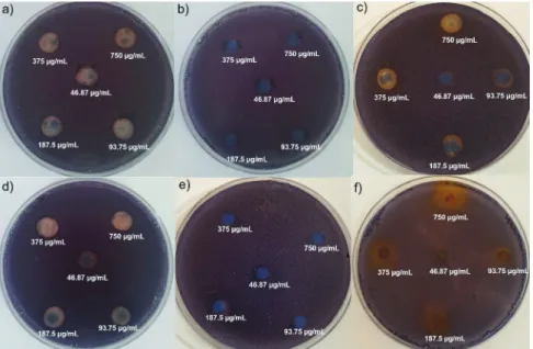
hall-mark of QS inhibition in C. violaceum 12472 by the dietary phytochemicals. The phytochemicals showed promising an-ti-QS activity, and a white opaque zone of inhibition was ob-served in the biosensor plate containing the reference strain
C. violaceum 12472 (Fig. 1). The sub-MICs of quercetin, lute-olin and apigenin were found to be <750 μg/mL, and of cur-cumin, pyrogallol and gallic acid <1500 µg/mL. The sub-MICs of each tested phytochemical ranged from 46.87 to 750 µg/ mL. As shown in Table 1, the anti-QS activity was concen-tration-dependent, showing an increase in the diameter of the QS inhibition zones with increasing concentrations of the phytochemicals (p<0.05). Among all the phytochemi-cals screened for the QS inhibition, curcumin, quercetin, lu-teolin and apigenin exhibited the anti-QS activity, but
py-rogallol and gallic acid did not.Al-Hussaini and Mahasneh
(34) reported the QS inhibition zones for different herbal ex-tracts with diameters 8–10.5, 13 and 18 mm, corresponding
Fig. 1.The anti-quorum sensing activity of the dietary phytochemicals at concentrations from 46.87 to 750 μg/mL against biosensor strain
to weak, moderate and strong anti-QS activity, respectively. In our study, the QS inhibition zones (Table 1) were found for curcumin ((7.0±0.3)–(13.3±0.1) mm), quercetin ((7.5±0.1)– (13.3±0.5) mm), luteolin ((9.1±0.1)–(14.2±0.1) mm) and api-genin ((9.3±0.7)–(14.3±0.0) mm) at the concentration range of 46.87–750 µg/mL. At low concentrations (46.87 and 93.75 µg/ mL), these phytochemicals had weak anti-QS activities. In ad-dition, curcumin ((9.7±0.1) mm) and quercetin ((9.5±0.7) mm) exhibited weaker anti-QS activities than luteolin ((12.3±0.6) mm) and apigenin ((11.5±0.5) mm) at 187.5 µg/mL. However, curcumin ((12.5±0.3) mm), luteolin ((13.6±0.5) mm) and api-genin ((13.0±0.1) mm) showed stronger anti-QS activities than quercetin ((12.0±0.2) mm) at 375 µg/mL (p<0.05). At the est sub-MIC (750 µg/mL), curcumin demonstrated the high-est QS inhibition (the diameter of zone of inhibition being (13.3±0.1) mm; Table 1). The concentration-dependent an-ti-QS activity of the phytochemicals observed in this study is in agreement with some of the findings reported by Borges
et al. (12), Husain et al. (15), and Vasavi et al. (20).
Determination of quantitative anti-QS activity by violacein inhibition
To confirm the QS inhibitory activity of the dietary phyto-chemicals, the extraction and the quantification of the viol-acein from C. violaceum 12472 culture was also performed in the presence and absence of the phytochemicals (curcumin, quercetin, pyrogallol, luteolin, gallic acid and apigenin) at the same sub-MIC levels (46.87–750 µg/mL). The results showed a concentration-dependent inhibition of the violacein pro-duction by curcumin, quercetin, luteolin and apigenin. Com-pared to the control, all concentration ranges of curcumin, quercetin, luteolin and apigenin (46.87–750 µg/mL) demon-strated a significant drop in the violacein content of C.
violace-um 12472, without the inhibition of bacterial growth (p<0.05).
As shown in Fig. 2, curcumin, quercetin, luteolin and apigenin inhibited the violacein production ((14.0±0.3)–(88.2±0.1) %, (11.1±0.1)–(55.03±0.07) %, (37.49±0.08)–(59.48±0.08) % and (36.3±0.2)–(58.95±0.08) %) in a concentration-dependent manner. Among all the phytochemicals, curcumin exhibited the most powerful QS inhibitory effect ((54.7±0.3), (72.2±0.2) and (88.2±0.1) %) at 187.5, 375 and 750 µg/mL. At all concen-trations, luteolin and apigenin demonstrated stronger viol-acein inhibition than quercetin (Fig. 2). The results revealed
that the percentage of inhibition of QS by apigenin and lu-teolin was similar at the tested concentrations. This may be because apigenin and luteolin are the flavonoid compounds with similar structures. Thus, the QS inhibitory effect of the phytochemicals was found to be in the following order: cur-cumin>apigenin>luteolin>quercetin.
Table 1.Zone of violacein inhibition obtained with the subminimal inhibitory concentrations (46.87–750 μg/mL) of the dietary phytochemicals
γ(phytochemical)/(µg/mL) d(inhibition zone)/mm
CR QT PG GA LT AP 46.87 (7.0±0.3)a (7.5±0.1)a NA NA (9.1±0.1)a (9.3±0.7)a 93.75 (8.5±0.0)b (8.0±0.1)b NA NA (10.1±0.4)b (10.3±0.7)b 187.5 (9.7±0.1)c (9.5±0.7)c NA NA (12.3±0.6)c (11.5±0.5)c 375 (12.5±0.3)d (12.0±0.2)d NA NA (13.6±0.5)d (13.0±0.1)d 750 (13.3±0.1)e (13.3±0.5)e NA NA (14.2±0.1)e (14.3±0.0)e
CR=curcumin, QT=quercetin, PG=pyrogallol, GA=gallic acid, LT=luteolin, AP=apigenin; NA=no activity. The values in the same column with different letters in superscript are significantly different (p<0.05)
0 10 20 30 40 50 60 70 80 90 100
Curcumin Quercetin Luteolin Apigenin
Vi ol ac ei n i nhi bi tion/ % γ/(µg/mL) 46.87 93.75 187.5 375 750 γ/(µg/mL) * * * * * * * * * * * * * * * * * * * *
Fig. 2. Quantitative analysis of violacein inhibition in
Chromobacte-rium violaceum ATCC 12472 by the dietary phytochemicals at the
subminimal inhibitory concentrations (46.87–750 μg/mL). Data are presented as a percentage of violacein inhibition. Mean values of triplicate independent experiments and S.D. are shown. The con-trol groups were: 0.5 % DMSO, 0.5 % methanol and the bacteria cul-tured in Luria-Bertani broth. *Statistically different from the control (p<0.05)
Brackman et al. (35) found that curcumin inhibited viola-cein production by (18±12) % in C. violaceum CV026 at a con-centration of 184 µg/mL. In our findings, curcumin inhibited violacein production by (54.7±0.3) % at 187.5 µg/mL. In addition, in the present study, the violacein inhibition by 750 µg/mL cur-cumin ((88.2±0.1) %) is comparable with that of Packiavathy et
al. (19), who reported 89 % violacein inhibition in C. violaceum
CV026 when treated with 100 µg/mL curcumin. Moreover, the violacein inhibition by quercetin at the maximum sub-MIC of 375 µg/mL ((39.9±0.1) %) is comparable with that of Gopu et
al. (6), who reported that quercetin exhibited maximum
viol-acein inhibition of 83.23 % in C. violaceum CV026 at a concen-tration of 80 μg/mL. Similarly, 50 % inhibition of the violace-in production violace-in C. violaceum 12472 by quercetviolace-in (50 µg/mL) was reported by Vasavi et al. (20) using a different method.
Apigenin and luteolin inhibited violacein ((58.4±0.2)and (55.13±0.04) %) at 375 µg/mL. Vandeputte et al. (36) reported that apigenin and luteolin at the concentration of 4 mM (ap-prox. 1.2 mg/mL) exhibit no QS inhibiton, but a bactericidal or bacteriostatic activity on C. violaceum CV026. Our study showed that apigenin and luteolin at the sub-MICs of 375, 187.5, 93.75 and 46.87 µg/mL showed anti-QS activity on C.
violaceum 12472 without inhibition of bacterial growth (Fig. S1, Fig. 1a, Fig. 1d and Fig. 2).
Pyrogallol and gallic acid did not exhibit the anti-QS ac-tivity when evaluated by the disk diffusion method. There-fore, the percentage of the violacein inhibition by these phy-tochemicals was evaluated only at the highest concentration (1500 µg/mL), and the obtained results were (76.95±0.02) and (58.76±0.05) % for pyrogallol and gallic acid respectively (data not shown). Considering that no QS inhibition zones were de-tected on the agar, these results were observed solely due to the inhibition of bacterial growth since gallic acid and py-rogallol only inhibited microbial growth and not the violace-in synthesis. The obtaviolace-ined results are also violace-in agreement with those of Borges et al. (12), who reported that gallic acid exhib-ited no anti-QS activity on C. violaceum biosensor systems.
Previous research of Borges et al. (12,37) also revealed that as a sustainable source of new broad-spectrum antimicrobial products, gallic acid can cause irreversible changes in some Gram-negative and Gram-positive bacteria. In addition, Tinh
et al. (38) reported that pyrogallol also exhibited the
antibac-terial activity against Vibrio parahaemolyticus. Although py-rogallol is known as the phytochemical with anti-QS activity and the ability to inhibit autoinducer-2 (AI-2)-mediated QS in
Vibrio harveyi (39,40), it has been revealed in further
investi-gations that this AI-2-mediated QS inhibition is a side effect of the peroxide-producing activity of this compound rather than true QS inhibition (41). Our findings also show that py-rogallol does not inhibit QS system of the C. violaceum 12472 biosensor strain.
Determination of antibiofilm activity of dietary phytochemicals
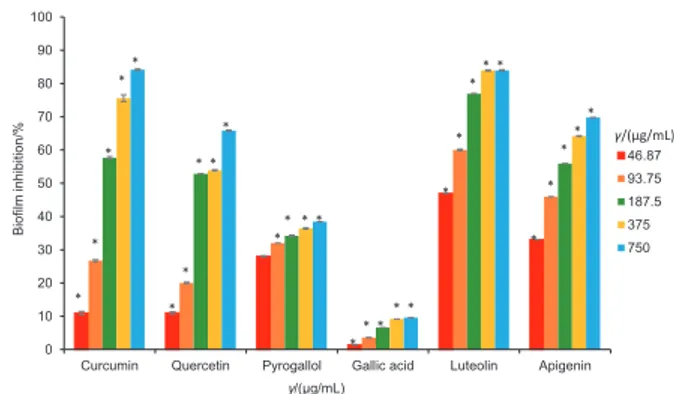
In the present study, the biofilm inhibition potential of all the phytochemicals (curcumin, quercetin, pyrogallol, luteo-lin, gallic acid and apigenin) was evaluated at the same sub- -MIC range (46.87–750 µg/mL) (Fig. 3). All the phytochemicals exhibited a significant concentration-dependent antibiofilm activity (p<0.05). Gallic acid showed the weakest biofilm in-hibition (from (1.38±0.08) to (9.57±0.06) %) at 46.87–750 µg/ mL, although it did not possess antimicrobial activity against
C. violaceum 12472 at <1500 µg/mL. Compared to the
oth-er compounds (pyrogallol, apigenin, quoth-ercetin and luteolin), curcumin showed the weakest biofilm inhibition ((11.0±0.5) and (26.7±0.3) % at 46.87 and 93.75 µg/mL, respectively). Our results are comparable with the results of Packiavathy et al. (18), who found that curcumin (>100 μg/mL) inhibited the bio-film formation of Vibrio harveyi (69 %), Vibrio
parahaemolyti-cus (56 %) and Vibrio vulnifiparahaemolyti-cus (79 %), without affecting their
planktonic growth. Packiavathy et al. (19) also reported that curcumin (100 µg/mL) efficiently inhibited the biofilm bio-mass growth of some uropathogens, including Escherichia coli (52 %), Pseudomonas aeruginosa PAO1 (89 %), Proteus mirabilis (52 %) and Serratia marcescens (76 %). In addition, the com-bined effects of curcumin and honey, as a traditional medi-cine, and epigallocatechin gallate, as a green tea polyphe-nol, were reported to enhance significantly the inhibition of biofilm formation in wastewater bacteria (52 to 99 %) and P.
aeruginosa PAO1 (20 to 94.6 %) (21,42). Therefore, the
anti-biofilm activity of curcumin may vary depending on the type of strain, the method used and/or its combination with some other constituent. Our results (Fig. 3) showed that although curcumin exhibited a weak biofilm inhibition potential at concentrations of 46.87 and 93.75 µg/mL, at higher sub-MICs of 187.5, 375 and 750 µg/mL, it had a strong concentration-dependent activity ((57.7±0.4), (75.6±1.0) and (84.2±0.2) %; p<0.05). Fig. 3 shows that the biofilm inhibitory potentials of the phytochemicals at concentrations of 46.87 and 93.75 µg/ mL increased in the following order: gallic acid<curcumin< pyrogallol<quercetin<apigenin<luteolin. At these concentra-tions, luteolin had the maximum biofilm inhibition values of (46.94±0.05) and (60.0±0.2) %, respectively. Quercetin, api-genin and pyrogallol also significantly reduced (p<0.05) the biofilm formation of C. violaceum 12472 (Fig. 3).
0 10 20 30 40 50 60 70 80 90 100
Curcumin Quercetin Pyrogallol Gallic acid Luteolin Apigenin
Bio film in hib itio n/ % γ/(µg/mL) 46.87 93.75 187.5 375 750 * * * * * * * * * * * * * * * * * * * * * * * * * * * * * γ/(µg/mL)
Fig. 3. The percentage of biofilm inhibition in Chromobacterium
viol-aceum ATCC 12472 by the dietary phytochemicals at the subminimal
inhibitory concentrations (46.87–750 μg/mL). Data are presented as a percentage of violacein inhibition. Mean values of triplicate inde-pendent experiments and S.D. are shown. The control groups were: 0.5 % DMSO, 0.5 % methanol and the bacteria cultured in Luria-Ber-tani broth. *Statistically different from the control (p<0.05)
Pyrogallol showed weak biofilm inhibition with (34.2±0.2), (36.4±0.2) and (38.5±0.0) % at higher sub-MICs of 187.5, 375 and 750 µg/mL, respectively (Fig. 3). Thus, the biofilm in-hibitory potentials of all the phytochemicals at higher sub-MICs of 187.5 and 375 µg/mL increased in the following order: gallic acid<pyrogallol<quercetin<apigenin<curcu-min<luteolin. Luteolin had the maximum biofilm inhibition ((46.94±0.05)–(83.9±0.2) %) at the sub-MIC values of 46.87 to 375 µg/mL. Our results are in accordance with the find-ings of Vikram et al. (43), who reported that quercetin and apigenin reduced the biofilm formation in V. harveyi and E.
coli O157:H7. In addition, Shehzad et al. (44) and Lee et al. (45)
also reported that curcumin, pyrogallol and apigenin were effective polyphenols against the biofilm formation of
Can-dida albicans.
Swimming and swarming motility
In this study, the motility (swimming and swarming be-haviour) of P. aeruginosa PAO1 in the presence and absence of the dietary phytochemicals at the sub-MIC of 93.75 µg/ mL was evaluated using agar plates. Our results demonstrate that all the phytochemicals significantly (p<0.05) blocked the swimming and swarming motility of P. aeruginosa PAO1 with-out impairing its growth capacity. Its swimming and swarm-ing motilities after the treatment with the phytochemicals were observed to be significantly poor (p<0.05; Table 2). Compared to the control, curcumin exhibited the maximum reduction in the swimming and swarming motility assays. These results are in accordance with the previous reports of Packiavathy et al. (19) and Jadaun et al. (42), who observed a remarkable decrease in the swimming and swarming motil-ity of P. aeruginosa PAO1 when treated with curcumin. Table 2 also indicates the inhibition of swimming and swarming behaviour of P. aeruginosa PAO1 by quercetin.
motility. The results also suggest a correlation between the inhibition of the swimming and swarming motility for each phytochemical.
Bacterial growth curve
The bacterial growth curve revealed that the sub-MIC concentrations of dietary phytochemicals used in this study (375 and 750 µg/mL) did not have a growth inhibitory effect on C. violaceum 12472 (Fig. S1).
CONCLUSIONS
This study investigated the inhibitory potentials of six di-etary phytochemicals (curcumin, quercetin, apigenin, luteo-lin, gallic acid and pyrogallol) against the violacein pigment production that is controlled by quorum sensing (QS) and biofilm formation in Chromobacterium violaceum ATCC 12472 biosensor system, and their impact on swimming and swarm-ing behaviour of Pseudomonas aeruginosa PAO1. Our results also demonstrated that all the phytochemicals, except py-rogallol and gallic acid, inhibited the violacein production. Moreover, the biofilm formation was significantly hindered by all the phytochemicals at each of the sub-MICs in the range of 46.87–750 µg/mL (p<0.05). Although some of the phyto-chemicals, especially curcumin and quercetin, possess QS and biofilm inhibitory potentials, this study reveals that apigenin and luteolin could also inhibit QS and biofilm formation. Our results also show that all the phytochemicals, especially cur-cumin, quercetin and pyrogallol, may be used as anti-patho-genic agents, particularly against P. aeruginosa PAO1, owing to QS control; however, more detailed experiments should be performed to find out their anti-QS mechanisms. Since these phytochemicals are the active compounds in foods such as onion, broccoli, apples, avocado, mango, banana, parsley, cab-bage, carrots, mustard, pepper and radish, the consumption of these foods might be beneficial in the treatment of bacte-rial infections. In addition, the studies of QS inhibitory poten-tials of the phytochemicals, especially apigenin and luteolin, against other biosensor strains, and the antibiofilm effect of the phytochemicals against different pathogens are scarce in the literature. Therefore, we suggest that more detailed ex-periments be conducted to reveal the anti-QS mechanisms and the pharmaceutical potential of the phytochemicals used in this study to confirm these results at the molecular level.
FUNDING
This work was supported by the Scientific Research Projects Unit of Gazi University of Turkey (grant number 21/2015-01).
CONFLICT OF INTEREST
The authors declare that there are no conflicts of interest.
SUPPLEMENTARY MATERIAL
All supplementary material is available at www.ftb.com.hr. Table 2. Effect of the dietary phytochemicals at the subminimal
in-hibitory concentration (93.75 µg/mL) on swimming and swarming motility of Pseudomonas aeruginosa PAO1
Phytochemical l(migration)/mm
Swimming motility Swarming motility
Control (44.0±3.5)a (32.0±1.7)a CR (11.3±0.4)b* (12.5±0.7)b* QT (15.7±0.6)c* (14.5±0.7)c* PG (19.3±1.2)d* (16.0±1.4)d* GA (33.3±0.6)e* (25.5±2.1)e* LT (28.6±0.4)f* (23.5±1.3)f* AP (26.4±0.5)g* (17.5±0.4)g*
CR=curcumin, QT=quercetin, PG=pyrogallol, GA=gallic acid, LT=luteolin, AP=apigenin. The data represent the mean values of three independent experiments. The values in the same column with different letters in superscript are significantly different (p<0.05). *Statistically different from the control (p<0.05)
Our findings are consistent with the reports of Vasavi et al. (20), who found that the flavonoid fraction of Psidium
guaja-va leaves included an active compound,
quercetin-3-O-arab-inoside, inhibits the swarming motility of P. aeruginosa PAO1. In addition, Gopu et al. (6) reported that quercetin signifi-cantly reduced the swimming and swarming behaviour of foodborne isolates of Pseudomonas aeruginosa PUFSTb04 at the concentration of 80 μg/mL. Table 2 shows that the in-hibition potential of the swimming and swarming motilities of the bacterial strain by the investigated phytochemicals decreased in the following order: curcumin>quercetin>py-rogallol>apigenin>luteolin>gallic acid.
The obtained results clearly indicate that curcumin and quercetin exhibited the most powerful inhibition of the
ORCID IDs
E.B. Bali https://orcid.org/0000-0002-8797-0573
K. Erkan Türkmen https://orcid.org/0000-0002-4813-7941
D. Erdönmez https://orcid.org/0000-0003-3955-1734
N. Sağlam https://orcid.org/0000-0002-5463-8355
REFERENCES
1. Meher-Homji Z, Mangalore RP, Johnson PDR, Chua KY. Chro-mobacterium violaceum infection in chronic granulomatous
disease: A case report and review of the literature. JMM Case Rep. 2017;4(1).
https://doi.org/10.1099/jmmcr.0.005084
2. Richard KR, Lovvorn JJ, Oliver SE, Ross SA, Benner KW, Kong
MYF. Chromobacterium violaceum sepsis: Rethinking con-ventional therapy to improve outcome. Am J Case Rep. 2015;16:740–4.
https://doi.org/10.12659/AJCR.894509
3. Morohoshi T, Kato M, Fukamachi K, Kato N, Ikeda T.
N-acyl-homoserine lactone regulates violacein production in
Chro-mobacterium violaceum type strain ATCC 12472. FEMS
Mi-crobiol Lett. 2008;279(1):124–30.
https://doi.org/10.1111/j.1574-6968.2007.01016.x
4. Stauff DL, Bassler BL. Quorum sensing in Chromobacteri-um violaceChromobacteri-um: DNA recognition and gene regulation by the
CviR receptor. J Bacteriol. 2011;193(15):3871–8.
https://doi.org/10.1128/JB.05125-11
5. Zhu H, He CC, Chu QH. Inhibition of quorum sensing in Chro-mobacterium violaceum by pigments extracted from Auricu-laria auricular. Lett Appl Microbiol. 2011;52(3):269–74.
https://doi.org/10.1111/j.1472-765X.2010.02993.x
6. Gopu V, Meena CK, Shetty PH. Quercetin influences quorum
sensing in food borne bacteria: in-vitro and in-silico evidence. PLoS One. 2015;10(8):e0134684.
https://doi.org/10.1371/journal.pone.0134684
7. Bai AJ, Vittal RR. Quorum sensing inhibitory and anti-biofilm
activity of essential oils and their in vivo efficacy in food sys-tems. Food Biotechnol. 2014;28(3):269–92.
https://doi.org/10.1080/08905436.2014.932287
8. Husain FM, Ahmad I, Khan MS, Ahmad E, Tahseen Q, Khan
MS, Alshabib NA. Sub-MICs of Mentha piperita essential oil and menthol inhibits AHL mediated quorum sensing and biofilm of Gram-negative bacteria. Front Microbiol. 2015;6: Article No.420.
https://doi.org/10.3389/fmicb.2015.00420
9. Overhage J, Lewenza S, Marr AK, Hancock REW.
Identifica-tion of genes involved in swarming motility using a
Pseu-domonas aeruginosa PAO1 mini-Tn5-lux mutant library.
J Bacteriol. 2007;189(5):2164–9.
https://doi.org/10.1128/JB.01623-06
10. Chow S, Gu K, Jiang L, Nassour A. Salicylic acid affects
swim-ming, twitching and swarming motility in Pseudomonas
aeruginosa, resulting in decreased biofilm formation. J
Ex-perim Microbiol Immunol. 2011;15:22–9.
11. Alayande AB, Aung MM, Kim IS. Correlation between
quo-rum sensing signal molecules and Pseudomonas
aerugi-nosa’s biofilm development and virulency. Curr Microbiol.
2018;75(7):787–93.
https://doi.org/10.1007/s00284-018-1449-5
12. Borges A, Serra S, Abreu AC, Saavedra MJ, Salgado A, Simões
M. Evaluation of the effects of selected phytochemicals on quorum sensing inhibition and in vitro cytotoxicity. Biofoul-ing. 2014;30(2):183–95.
https://doi.org/10.1080/08927014.2013.852542
13. Akkoç N, Akçelik M, Haznedaroglu IC, Goker H, Turgut M,
Aksu S, et al. In vitro anti-bacterial activities of Ankaferd me-dicinal plant extract. Turkiye Klinikleri J Med Sci. 2009; 29(2): 410–5.
14. Reen FJ, Gutiérrez-Barranquero JA, Parages ML, O'Gara F.
Coumarin: A novel player in microbial quorum sensing and biofilm formation inhibition. Appl Microbiol Biotechnol. 2018;102(5):2063–73.
https://doi.org/10.1007/s00253-018-8787-x
15. Husain FM, Ahmad I, Al-thubiani AS, Abulreesh HH, AlHazza
IM, Aqil F. Leaf extracts of Mangifera indica L. inhibit quorum sensing–regulated production of virulence factors and bio-film in test bacteria. Front Microbiol. 2017;8:Article no. 727.
https://doi.org/10.3389/fmicb.2017.00727
16. Rajkumari J, Borkotoky S, Murali A, Suchiang K, Mohanty SK,
Busi S. Attenuation of quorum sensing controlled virulence factors and biofilm formation in Pseudomonas aeruginosa by pentacyclic triterpenes, betulin and betulinic acid. Mi-crob Pathog. 2018;118:48–60.
https://doi.org/10.1016/j.micpath.2018.03.012
17. Zhou JW, Luo HZ, Jiang H, Jian TK, Chen ZQ, Jia AQ.
Horde-nine, a novel quorum sensing inhibitor and antibiofilm agent against Pseudomonas aeruginosa. J Agric Food Chem. 2018;66(7):1620–8.
https://doi.org/10.1021/acs.jafc.7b05035
18. Packiavathy IASV, Sasikumar P, Pandian SK, Ravi AV.
Preven-tion of quorum-sensing-mediated biofilm development and virulence factors production in Vibrio spp. by curcum-in. Appl Microbiol Biotechnol. 2013;97(23):10177–87.
https://doi.org/10.1007/s00253-013-4704-5
19. Packiavathy IASV, Priya S, Pandian SK, Ravi AV. Inhibition
of biofilm development of uropathogens by curcumin – An anti-quorum sensing agent from Curcuma longa. Food Chem. 2014;148:453–60.
https://doi.org/10.1016/j.foodchem.2012.08.002
20. Vasavi HS, Arun AB, Rekha PD. Anti-quorum sensing activity
of Psidium guajava L. flavonoids against Chromobacterium
violaceum and Pseudomonas aeruginosa PAO1. Microbiol
Immunol. 2014;58(5):286–93.
21. Lade H, Paul D, Kweon JH. Combined effects of curcumin
and (-)-epigallocatechin gallate on inhibition of N-acylho-moserine lactone-mediated biofilm formation in wastewa-ter bacwastewa-teria from membrane bioreactor. J Microbiol Biotech-nol. 2015;25(11):1908–19.
https://doi.org/10.4014/jmb.1506.06010
22. Zgoda J, Porter J. A convenient microdilution method for
screening natural products against bacteria and fungi. Pharm Biol. 2001;39(3):221–5.
https://doi.org/10.1076/phbi.39.3.221.5934
23. Bauer AW, Kirby WMM, Sherris JC, Turck M. Antibiotic
sus-ceptibility testing by a standardized single disk method. Am J Clin Pathol. 1966;45(4):493–6.
https://doi.org/10.1093/ajcp/45.4_ts.493
24. Blosser RS, Gray KM. Extraction of violacein from Chromo-bacterium violaceum provides a new quantitative bioassay
for N-acyl homoserine lactone autoinducers. J Microbiol Methods. 2000;40(1):47–55.
https://doi.org/10.1016/S0167-7012(99)00136-0
25. O'Toole GA, Kolter R. Initiation of biofilm formation in Pseu-domonas fluorescens WCS365 proceeds via multiple,
con-vergent signalling pathways: A genetic analysis. Mol Micro-biol. 1998;28(3):449–61.
https://doi.org/10.1046/j.1365-2958.1998.00797.x
26. Packiavathy IASV, Agilandeswari P, Musthafa KS, Pandian SK,
Ravi AV. Antibiofilm and quorum sensing inhibitory poten-tial of Cuminum cyminum and its secondary metabolite me-thyl eugenol against Gram negative bacterial pathogens. Food Res Int. 2012;45(1):85–92.
https://doi.org/10.1016/j.foodres.2011.10.022
27. SPSS software, v. 20, IBM Corporation, Armonk, NY, USA;
2011. Available from: https://www.ibm.com/analytics/
spss-statistics-software.
28. Borges A, Ferreira C, Saavedra MJ, Simões M. Antibacterial
activity and mode of action of ferulic and gallic acids against pathogenic bacteria. Microb Drug Resist. 2013;19(4):256–65.
https://doi.org/10.1089/mdr.2012.0244
29. Silva V, Igrejas G, Falco V, Santos TP, Torres C, Oliveira AMP, et al. Chemical composition, antioxidant and antimicrobial
activity of phenolic compounds extracted from wine indus-try by-products. Food Control. 2018;92:516–22.
https://doi.org/10.1016/j.foodcont.2018.05.031
30. Gutiérrez-del-Río I, Fernández J, Lombó F. Plant
nutraceu-ticals as antimicrobial agents in food preservation: Terpe-noids, polyphenols and thiols. Int J Antimicrob Agents. 2018;52(3):309–15.
https://doi.org/10.1016/j.ijantimicag.2018.04.024
31. Ohemeng KA, Schwender CF, Fu KP, Barrett JF. DNA gyrase
inhibitory and antibacterial activity of some flavones (1). Bioorg Med Chem Lett. 1993;3(2):225–30.
https://doi.org/10.1016/S0960-894X(01)80881-7
32. Mirzoeva OK, Grishanin RN, Calder PC. Antimicrobial
ac-tion of propolis and some of its components: The effects
on growth, membrane potential and motility of bacteria. Microbiol Res. 1997;152(3):239–46.
https://doi.org/10.1016/S0944-5013(97)80034-1
33. Sorrentino E, Succi M, Tipaldi L, Pannella G, Maiuro L,
Stur-chio M, et al. Antimicrobial activity of gallic acid against food-related Pseudomonas strains and its use as biocontrol tool to improve the shelf life of fresh black truffles. Int J Food Microbiol. 2018;266:183–9.
https://doi.org/10.1016/j.ijfoodmicro.2017.11.026
34. Al-Hussaini R, Mahasneh AM. Microbial growth and quorum
sensing antagonist activities of herbal plants extracts. Mol-ecules. 2009;14(9):3425–35.
https://doi.org/10.3390/molecules14093425
35. Brackman G, Hillaert U, Van Calenbergh S, Nelis HJ, Coenye
T. Use of quorum sensing inhibitors to interfere with biofilm formation and development in Burkholderia multivorans and Burkholderia cenocepacia. Res Microbiol. 2009;160(2): 144–51.
https://doi.org/10.1016/j.resmic.2008.12.003
36. Vandeputte OM, Kiendrebeogo M, Rasamiravaka T,
Stévi-gny C, Duez P, Rajaonson S, et al. The flavanone naringenin reduces the production of quorum sensing-controlled vir-ulence factors in Pseudomonas aeruginosa PAO1. Microbi-ology. 2011;157(Part 7):2120–32.
https://doi.org/10.1099/mic.0.049338-0
37. Borges A, Ferreira C, Saavedra MJ, Simões M. Antibacterial
activity and mode of action of ferulic and gallic acids against pathogenic bacteria. Microb Drug Resist. 2013;19(4):256–65.
https://doi.org/10.1089/mdr.2012.0244
38. Tinh TH, Nuidate T, Vuddhakul V, Rodkhum C. Antibacterial
activity of pyrogallol, a polyphenol compound against
Vi-brio parahaemolyticus isolated from the central region of
thailand. Procedia Chem. 2016;18:162–8.
https://doi.org/10.1016/j.proche.2016.01.025
39. Ni N, Choudhary G, Li M, Wang B. Pyrogallol and its analogs
can antagonize bacterial quorum sensing in Vibrio harveyi. Bioorg Med Chem Lett. 2008;18(5):1567–72.
https://doi.org/10.1016/j.bmcl.2008.01.081
40. Nazzaro F, Fratianni F, Coppola R. Quorum sensing and
phy-tochemicals. Int J Mol Sci. 2013;14(6):12607–19.
https://doi.org/10.3390/ijms140612607
41. Defoirdt T, Pande GSJ, Baruah K, Bossier P. The apparent
quorum-sensing inhibitory activity of pyrogallol is a side effect of peroxide production. Antimicrob Agents Chem-other. 2013;57(6):2870–3.
http://doi.org/10.1128/AAC.00401-13
42. Jadaun V, Prateeksha, Singh BR, Paliya BS, Upreti DK, Rao
CV, et al. Honey enhances the anti-quorum sensing activity and anti-biofilm potential of curcumin. RSC Adv. 2015;5(87): 71060–70.
43. Vikram A, Jayaprakasha GK, Jesudhasan PR, Pillai SD, Patil
BS. Suppression of bacterial cell–cell signalling, biofilm for-mation and type III secretion system by citrus flavonoids. J Appl Microbiol. 2010;109(2):515–27.
https://doi.org/10.1111/j.1365-2672.2010.04677.x
44. Shehzad A, Shahzad R, Lee YS. Curcumin: A potent
modulator of multiple enzymes in multiple cancers.
Enzymes. 2014; 36:149–74.
https://doi.org/10.1016/B978-0-12-802215-3.00008-2
45. Lee H, Woo ER, Lee DG. Apigenin induces cell shrinkage in Candida albicans by membrane perturbation. FEMS Yeast
Res. 2018;18(1):foy003.