Turk Kardiyol Dern Ars 2015;43(7):644–647 doi: 10.5543/tkda.2015.38959
Arrhythmogenic right ventricular cardiomyopathy in monozygotic
twin sisters, and persistent left superior vena cava
in one complicating implantation of ICD
644Received: March 28, 2015 Accepted:June 08, 2015
Correspondence: Dr. Mehmet Ali Astarcıoğlu. Zeytinlik Cad., Emrah Sok., 7/20, Atalar, Kartal, İstanbul. Tel: +90506 - 559 40 45 e-mail: [email protected]
© 2015 Turkish Society of Cardiology
Aritmojenik sağ ventrikül kardiyomiyopatisi bulunan tek yumurta ikizi kız kardeşler
ve birinde ısrarcı sol üst vena kava sendromu ile komplike ICD implantasyonu
Department of Cardiology, Dumlupinar University Evliya Celebi Training and Research Hospital, Kutahya
Mehmet Ali Astarcıoğlu, M.D., Mehmet Yaymacı, M.D., Taner Şen, Celal Kilit, M.D., Basri Amasyalı, M.D.
Özet– Aritmojenik sağ ventrikül kardiyomiyopatisi kalp kası-nın histolojik olarak fibroz-yağlı doku ile yer değiştirdiği ve klinik olarak ventriküler aritmi ve sağ ventrikül fonksiyonu bozukluğu ile karakterize kalıtsal bir kardiyomiyopatidir. Bu-rada biz aritmojenik sağ ventrikül kardiyomiyopatili tek yu-murta ikizlerini sunuyoruz; hastalığın en olası nedeni olarak genetik anormallik gözükmektedir.
Summary– Arrhythmogenic right ventricular cardiomyopa-thy (ARVC) is an inherited cardiomyopacardiomyopa-thy characterized histologically by fibro-fatty replacement of heart muscle, and clinically by ventricular arrhythmias and right ventricu-lar dysfunction. This report presents monozygotic twins with ARVC, suggesting a genetic abnormality as the most prob-able cause.
A
rrhythmogenic right ventricular cardiomyopa-thy (ARVC) is an inherited cardiomyopacardiomyopa-thy. It is characterized histologically by fibro-fatty replace-ment of heart muscle, and clinically by ventricular arrhythmias and right ventricular dysfunction.[1]Sud-den death or syncope may be the first manifestations, while in other cases the disease may be asymptom-atic.[2] Familial ARVC is a genetic disorder
transmit-ted with reduced penetrance and variable phenotypic expression.
In this report, we present monozygotic twins with ARVC, which suggests a genetic abnormality as the most probable cause.
CASE REPORT
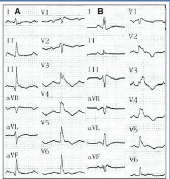
A 20-year-old woman (Patient 1) presented with near syncope after exercise. During admission, her mono-zygotic twin sister (Patient 2) was also invited for di-agnostic testing. Echocardiography (ECG) showed T wave inversions in lead V1 to V6 in the absence of a complete right bundle branch block (RBBB);
dura-tion of the QRS com-plex was 110-120 ms in lead V1 and 80 ms in lead V6 in both pa-tients (Figure 1).
On ECG, both
pa-tients had severely dilated right ventricles (RV) with reduced ejection fraction (EF); left ventricles were normal. Cardiac magnetic resonance imaging (MRI) confirmed severe RV dilation with thin free walls, and also showed fatty tissue in the left ventricle in both patients (Figure 2). These findings fulfilled the criteria for a diagnosis of ARVC.[3]
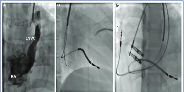
Electrophysiological (EP) testing failed to induce ventricular arrhythmias in either patient. Due to the positive family history and LV involvement, ICD im-plantation was planned in both patients for primary prevention of sudden death. During the initial left subclavian puncture, Patient 2 was seen to have a per-sistent left superior vena cava (PLSVC) draining into a dilated coronary sinus. An active fixation lead with
Abbreviations:
ARVC Arrhythmogenic right ventricular cardiomyopathy ECG Echocardiography
PLSVC Persistent left superior vena cava RV Right ventricles
standard stylets was advanced through the PLSVC and coronary sinus into the RV without any complica-tions (Figure 3).
DISCUSSION
Although significant progress has been made in re-cent years in our knowledge of ARVC, its etiology is still uncertain. In familial cases of ARVC, autosomal dominant inheritance with different penetrance and expression has been reported, and accounts for ap-proximately 50% of cases.[4]
The most common thoracic venous anomaly is
PLSVC, which occurs in 0.3% of the population.[5] To
the best of the authors’ knowledge, this is the first re-port of PLSVC in a case with ARVC in monozygotic twin sisters. This condition is usually asymptomatic, and may be detected incidentally by venography dur-ing device implantation, as in our case. Transvenous advancement of the lead through the PLSVC, coro-nary sinus and right atrium to the right ventricle can be technically challenging, as the tip of the lead tends to deflect away from the tricuspid annulus. This lim-ited maneuverability adds to the difficulty in finding a right ventricular implantation site with sufficient volt-age in ARVC. Manual reshaping of the stylet into a U-shape and forming a loop in the right atrium using the right atrial free wall for support with some specific maneuvers usually works well in these circumstances. As there is no reported specific link between genetic disorder and PLSVC, only one of the twin sisters had this anomaly.
The inflammatory process has been suggested as playing a role in cases of ARVC because of the com-Arrhythmogenic right ventricular cardiomyopathy in monozygotic twin sisters 645
Figure 1. Sisters IA and IB electrocardiograms. Normal si-nus rhythm. T wave inversions in V1 to V6. rSr’ in lead V1-V4. Duration of QRS complex: ~110 ms in lead V1 and 80 ms in lead V6.
Figure 2. FIESTA (Fast Imaging Employing Steady State Acquisition) sequence cardiovascular magnetic resonance images from the sisters with ARVC indicate fat infiltration at the outflow tract, free wall, and apex of the right ventricle and thinning of the underlying myocardial wall (black ar-rows) and a thinned lateral wall of the left ventricle (white arrows) due to fatty replacement.
Table 1. Similarities and differences of phenotype of monozygotic twin
Patient Symptoms/arrhythmia ECG RV morphology LV involvement Ventricular Differences in arrhythmia environmental
in EPS factors
1 Near syncope, PVBs Enlargement, + No No
dysfunction
2 Near syncope Enlargement, + No No
dysfunction PVBs: Premature ventricular beats.
Turk Kardiyol Dern Ars
646
mon finding of inflammatory infiltrates in the myo-cardium, suggesting that ARVC is a consequence of myocarditis.[6] Some cases have also been identified
with cardiotropic viruses in the myocardium.
How-ever, the role of inflammation in disease pathogenesis remains unknown, particularly as to whether these viruses contribute to disease or the diseased myocar-dium is more prone to virus infection.[7]
Figure 3. (A) AP projection demonstrating a LSVC. (B) RAO 30° view demonstrating lead advanced into the coronary sinus via a PLSVC, and an active fixation RV lead directed into the apex. (A) LAO 30° view demonstrating the RV lead placement.
A B C
Table 2. Summary of twins with ARVC reported in literature
Author Patient Symptoms/arrhythmia ECG RV morphology LV involvement
Non-Identical
Wlodorska IA VT, PVBs Wide QRS in V1 Enlargement, bulges –
IB Asymptomatic Normal Enlargement, bulges –
IIA Asymptomatic Wide QRS in V1 Enlargement –
IIB Asymptomatic Normal Enlargement, bulges –
Buja 1 Syncope, VT LP+ Enlargement, bulges
2 PVBs LP– Enlargement, bulges
Solenthaler 1 Syncope, HF, VT, SD Neg T in V1-3 Enlargement, bulges +
2 VT Neg T in V1-3 Enlargement –
Identical
Hiraoka[10] A PVBs Neg T in V1-3 Enlargement, bulges –
B PVBs Neg T in V1-3 Enlargement, bulges –
Wide QRS in V1
Indik[11] A HF, AFib, nsVT Neg T in V1-3 Enlargement, dysfunction –
Epsilon wave in V1
B HF, nsVT Neg T in V1-3 Enlargement, dysfunction –
Epsilon wave in V1
PVBs: Premature ventricular beats; AFib: Atrial fibrillation; HF: Heart failure; VT: Ventricular tachycardia; nsVT: Nonsustained VT; SD: Sudden death; LP: Late potentials.
The mechanism by which male gender results in greater risk of some expressions of ARVC is not known. The cardioprotective features of estrogens due to inhibition of myocardial cell apoptosis has been documented.[8] This theory would explain why
the disease is more common in males.
The arrhythmias are characteristically provoked by adrenergic stimulation such as physical exercise.
[9] The majority of patients with ARVC are susceptible
to adrenergic stimulation and the frequent induction of arrhythmia by isoprenaline infusion suggests a role for catecholamines.[10] The role of sympathetic
stimu-lation in provoking arrhythmias in ARVC possibly accounts for the high frequency of this situation in individuals who die during exertion.
In addition to a genetic cause of ARVC, hormonal, infectious or inflammatory, apoptotic and physical exercise theories have been proposed either as the cause of or as environmental factors facilitating gene expression. The relative influence of genetics and en-vironmental factors can be best assessed by studies of twins with the disease, when it is possible that differ-ent phenotypes are a result of environmdiffer-ental factors.
Our observations are compatible with those of Hi-raoka and Indik, in that findings and courses of the disease are similar in monozygotic twins.[11,12] Neither
of our patients had any history of myocarditis or ex-posure to toxins. The findings strongly suggest a ge-netic cause for ARVC.
In conclusion, this is the first report of PLSVC in a case with ARVC in monozygotic twin sisters. Limited maneuverability of the lead tip caused by PLSVC adds to the difficulty in finding an appropriate implantation site with good voltage in cases with ARVC. Specific techniques are required to overcome these challenges.
Conflict-of-interest issues regarding the authorship or article: None declared.
REFERENCES
1. Corrado D, Basso C, Thiene G. Arrhythmogenic right ven-tricular cardiomyopathy: diagnosis, prognosis, and treatment. Heart 2000;83:588–95.CrossRef
2. Tabib A, Loire R, Chalabreysse L, Meyronnet D, Miras A, Malicier D, et al. Circumstances of death and gross and mi-croscopic observations in a series of 200 cases of sudden death associated with arrhythmogenic right ventricular car-diomyopathy and/or dysplasia. Circulation 2003;108:3000–5. 3. Marcus FI, McKenna WJ, Sherrill D, Basso C, Bauce B,
Bluemke DA, et al. Diagnosis of arrhythmogenic right ven-tricular cardiomyopathy/dysplasia: proposed modification of the task force criteria. Circulation 2010;121:1533-41. CrossRef
4. Wyodarska EK, Konka M, Zaleska T, Lusawa T, Hoffman P, Rydlewska-Sadowska W, et al. Clinical profile of 30 families with arrhythmogenic right ventricular dysplasia/cardiomy-opathy. JACC 2002;39:42.
5. Morani G, Bergamini C, Toniolo M, Vassanelli C. How many leads through persistent left superior vein cava and coronary sinus? J Electrocardiol 2010;43:663–6. CrossRef
6. Thiene G, Corrado D, Nava A, Rossi L, Poletti A, Boffa GM, et al. Right ventricular cardiomyopathy: is there evidence of an inflammatory aetiology? Eur Heart J 1991;12:22–5. CrossRef
7. Bowles NE, Ni J, Marcus F, Towbin JA. The detection of cardiotropic viruses in the myocardium of patients with ar-rhythmogenic right ventricular dysplasia/cardiomyopathy. J Am Coll Cardiol 2002;39:892–5. CrossRef
8. Xi H, Shin WS, Suzuki J, Nakajima T, Kawada T, Uehara Y, et al. Dystrophin disruption might be related to myocardial cell apoptosis caused by isoproterenol. J Cardiovasc Pharmacol 2000;36 Suppl 2:25–9. CrossRef
9. Wichter T, Hindricks G, Lerch H, Bartenstein P, Borggrefe M, Schober O, et al. Regional myocardial sympathetic dysinner-vation in arrhythmogenic right ventricular cardiomyopathy. An analysis using 123I-meta-iodobenzylguanidine scintigra-phy. Circulation 1994;89:667–83. CrossRef
10. Leclercq JF, Potenza S, Maison-Blanche P, Chastang C, Cou-mel P. Determinants of spontaneous occurrence of sustained monomorphic ventricular tachycardia in right ventricular dys-plasia. J Am Coll Cardiol 1996;28:720–4. CrossRef
11. Hiraoka E, Koide M, Sakamoto S, Miki T, Ohga N, Suzuki S, et al. Identical twins with arrhythmogenic right ventricular dysplasia. Am J Cardiol 1995;76:1099–100. CrossRef
12. Indik JH, Smith DE, Sobonya RE, Marcus FI. Arrhythmogen-ic right ventrArrhythmogen-icular cardiomyopathy/dysplasia: a case report of identical twins with heart failure. Pacing Clin Electrophysiol 2002;25:1387–90. CrossRef
Key words: Arrhythmogenic right ventricular dysplasia/physiopathol-ogy; pacemaker, artificial; monozygotic twins.
Anahtar sözcükler: Aritmojenik sağ ventrikül kardiyomiyopatisi/fiz-yopatoloji; kalp pili, yapay; tek yumurta ikizleri.