Olgu sunumu/ Case report
The Treatment of Unruptured Ovarian Pregnancy (Op) Followıng Intrauterine Insemınation (Iuı): Case Report
Dokuzeylül Güngör Nur¹ , Tuğba Gürbüz ² , Erşahin Aynur¹ , Erşahin Suat Süphan ³, Yapıcıer Özlem4 1Department of IVF Center, Medical Park Göztepe Hosp., İst./ Turkey
2Department of Gynecology and Obstetric, Medical Park Göztepe Hosp., İst./ Turkey ³Department of Gynecology and Obstetric, Medical Park Bahçelievler Hosp., İst./ Turkey 4 Department of Pathology , Medical Park Göztepe Hosp., İst./ Turkey
ABSTRACT
Ovarian pregnancy (OP) seen quite rarely in ectopic pregnancy (EP). A 29-year-old multigravid woman came us for routine first ultrasound (US) control to see gestational sac (GS) after IUI. Beta-human chorionic gonadotropin (β hCG) amount (10396 mUI/mL) and ultrasound outcomes were doubtful for tubal pregnancy (TP). She has no complaints. Laparoscopy and incision of right ovarian EP were fulfilled. Ovarian pregnancy were approved histologically.
Key words: Ovarian pregnancy, ectopic pregnancy, intrauterin insemination
AŞILAMA TEDAVİSİ SONRASI OLUŞAN RÜPTÜRE OLMAMIŞ OVARYAN GEBELİK ÖZ.
Ovaryan gebelik, ektopik gebelikler içerisinde oldukça nadir görülür. Hiçbir şikayeti olmayan, aşılama sonrası gebe kalan 29 yaşındaki hastamızın, rutin yapılan ilk ultrason kontrolünde sağ tubal ektopik gebelikten şüphelenildi. Beta-human koryonik gonadotropin düzeyi (10396 mUI/mL) saptanan ve ultrasonografi ile tubal gebelik görülen hasta acil operasyona alındı. Laparoskopi esnasında sağ ovaryan ektopik gebelik saptandı. Ovaryan ektopik gebelik materyali over dokusu korunarak laparoskopik eksize edildi. Patolojik tanı ovaryan ektopik gebelik olarak onaylandı.
Anahtar Kelime: Ovaryan gebelik, dış gebelik, aşılama
Doi: 10.30569/adiyamansaglik. 482550
Yazışmadan Sorumlu Yazar Tuğba Gürbüz
Department of Gynecology and Obstetric , Medical Park Göztepe Hosp., İst./ Turkey
Tel : +90 5323283523
Email: [email protected]
Geliş Tarihi: 14.11.2018 Kabul Tarihi: 07.02.2019
Sayfa 1415 Introduction
Ovarian pregnancy (OP) seen quite rarely in ectopic pregnancy (EP) and its prevalence is ranging from 1:7000 to 1:70000. It accounts for almost 3% of all EP (1,2).
It is commonly a hard work to diagnose and manage it. Generally it is ended at first trimester causing internal hemorrhage and shock due to increased ovarian tissue (OT) vascularisation. We can use the Spiegelberg (3) criteria for diagnosis which include
a. The gestational pouch resides in the ovarian zone.
b. The EP is annexed to the womb via the connective tissue of ovary.
c. Histologic proof of the OT on the barrier of the gestational sac (GS) has been made. d. The tube on the related part is untouched.
Surgery is mostly preferred choice of treatment in these cases because of delayed clinical diagnosis. We notify the event of unruptured OP following intrauterin insemination (IUI) , which was diagnosed incorrectly as tubal pregnancy (TP) and was cured thanks to modest laparoscopy with success.
Case Presentation
A 29 years old multiparous female consulted to our clinic for routine control of pregnancy at sixth week of gestation after IUI. Her obstetric history included one caesarean section followed by two biochemical pregnancy. There was not any illness histories in her past that was passed on sexual ways or was pelvic inflammatory.
The Beta-human chorionic gonadotropin (β hCG) amount was 10396 mUI/mL. Ultrasound survey displayed a blank uterine cavity with 10 mm thickness and a GS with fetal heartbeat on the rightside adnexial region implying a TP with a minor quantity of free liquid in the sac of Douglas. Bodily checks showed blood pressure 100/75mmHg, pulsing 87 beats/minute, and body warmth 36.8◦C. At speculum checks there wasn't any blood and in bimanual checks it was seen that she had an ordinary dimension of uterus without cervical motion sensitivity. The right adnexa was painful while touching, yet any adnexal massif couldn't found. First laboratory outcomes she had: Hemoglobin (Hb): 12 g/dL, Hematocrit (Hct): 35.3%.
She was hospitalized to the gynecologic department with the doubt of TP. She was carried to the operating room and emergency laparoscopy was applied.
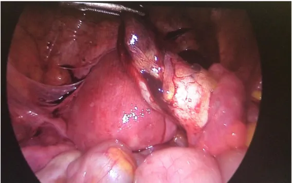
Throughout laparoscopy, 50mL clean blood was discharged from the abdominal cavity. Both fallopian tubes seemed like ordinary. There was a 4cm × 3 cm × 3.5 cmmass observed on the
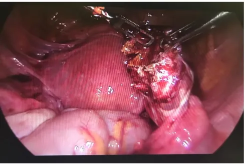
Sayfa 1416 right ovary, which is suggestive of EP. The GS was exracted from OT with ease. A wedge resection was performed. No suture was needed to control bleeding.
Figure 1.2.3 shown the 4cm × 3 cm × 3.5 cm mass observed on the right ovary
Sayfa 1417 Figure 3. shown that a wedge resection was performed
Figure 4 Figure 5
Figure 4 and 5 shown that pathological examinations verified a right ovarian gestation together with existence of necrotic chorion villus and a corpus luteum inside of the OT.
She experienced an eventless postoperative period and was allowed to leave following day. Weekly outpatient β hCG check was done till it became ≤5 mUI/mL.
Discussion
OP is an uncommon form of EP. Ovarian ectopics, which cause implantation over the surface of ovaries, occur by fertilisation of the ovums holded inside of the peritoneum cavity. Although the cause of such implantation anomalies remains unclear, current hypotheses
Sayfa 1418 include reflux of fertilised oocyte to the ovary, thickening of tunic albuginea, and abnormality of tubal functions (4).
The incidence of ovarian EP is increased by using artificial reproductive technology (ART) and intrauterine contraceptive devices (IUD). Delayed diagnosis of ovarian ectopic pregnancies can be fatal due to the massive haemorrhage and carry a risk of oophorectomy with reduced fertility. In the literature case series have pointed out preoperative diagnosis achieved in 11–28% of cases (5).
Comstock et al. (6) showed that although it was uncommon to see yolk sac or embryo, OPs were generally seen over or inside of the ovaries as a cyst with a large echogenic external circle. This can be distinguished from a corpus luteum (CL), which may also seem like a circle, as the greater part of the CL circles appear less echogenic than the ovaries themself, whereas for ovarian pregnancies it is greater. Therefore finding such a ring on imaging of suspected EP should alert the clinician for an OP.
Ipsilateral oophorectomy is definitive treatment for OP but this is becoming less common because of fertility preservation is preferred. These include partial ovariectomy (wedge resection), ovarian cystectomy, or blunt dissection of the trophoblastic tissue (7).
EP could generally be diagnosed by patient's history, indications and symptoms, β hCG amounts, and ultrasound survey. In this case the OP was diagnosed incorrectly as a TP, since ultrasound couldn't distinguish OP from TP. An exact diagnosis is important in EP to manage patient properly dependent upon the sort and position of the pregnancy (8).
After diagnosis of ovarian EP, if the patient is clinically stable without significant symptoms, clinicians may offer patients conservative or medical management. This may be suitable in those whomay carry operative risks. In those seeking future fertility, nonsurgical management may preserve ovarian tissue.
The quality of data is limited to case series and there is no proven superior method. In 2003 Mittal et al. (9) successfully treated an ovarian ectopic with laparoscopy-guided MTX injection. Juan and colleagues (10) described a case where etoposide (VP-16) was injected into the GS resulting in completeresolution of the ovarian EP and suggest that this in addition to systemic MTX is a potential option for medical treatment.
In patients who are clinically unstable or have significant symptoms or in whom intraoperative diagnosis is not clear, laparoscopy remains the preferred method of treatment. At the instance we examined, the patient was haemodynamically stable, and surgical
Sayfa 1419 management was the preferable treatment option. In this patient, medical treatment by using MTX was not a feasable choice because of the advanced serum β hCG amounts and existence of fetal heartbeat. The OP was laparoscopically extracted with ease. This modest method let the protection of the ovaries and reproduction capability of the patient.
Conclusion
Finally, OP is a very uncommon case that is hard to diagnose and a quantitative β hCG amount is not a reliable tool for diagnosis of EP. Although it is a rare occurrence, the rate of OP is rising because of ART. Preoperative diagnosis remains difficult; however US may assist in early detection. In proper cases, medical management should be considered, to preserve fertility of the patient.
Authorship declaration
Every author of the article meet the writership criterias in accordance with the last principles of the ICMJE, and every author agreed with the writing.
Informed consent
Written approval of the patients was acquired to publish this case report and any used images of it.
Conflict of interest
Every author declared that there are no potential conflicts of interest relating to research, authorship, and/or publication of this article.
Sayfa 1420
REFERENCES
1. Marcus SM, Brinsden PR. Primary ovarian pregnancy after in vitro fertilization and embryo transfer: report of seven cases. Fertil Steril. 1993;60:167–70.
2. Odejinmi F., Rizzuto MI., MacRae R., Olowu O., Hussain M. Diagnosis and laparoscopic management of 12 consecutive cases of ovarian pregnancy and review of literature,Journal ofMinimally Invasive Gynecology. 2009;16(3):354-59.
3. Spiegelberg O. Zur Cosuistik der Ovarialschwanger schalt, Arch Gynaekol. 1973;13:73-6.
4. Birge O.,Erkan MM., Ozbey EG., Arslan D. Medical management of an ovarian ectopic pregnancy: a case report, Journal ofMedical Case Reports. 2015;9(1):774.
5. Goyal LD.,Tondon R. , Goel P., Sehgal A. Ovarain ectopic pregnancy: a 10 years experience and review of literature, Iranian Journal of Reproductive Medicine, 2014;12:825-30.
6. Comstock C.,Huston K. , Lee W., The ultrasonographic appearance of ovarian ectopic pregnancies. Obstetrics &Gynecology 2005;105(1):42-5.
7. Gebeh A.,Amoako A., Joseph O., Banerjee A. Laparoscopic Surgery for Ovarian Pregnancy using Diathermy Hook with Conservation of Ovary: A Case Report and Literature Review,Journal of Clinical Medicine. 2013;2:214-19.
8. Jurkovic D.,Mavrelos D Ultrasound diagnosis of ectopic pregnancy,Ultrasound in Obstetrics andGynecology 2007;30(1):1-7.
9. Mittal S.,Dadhwal V., Baurasi P., Successful medical management of ovarian pregnancy,International Journal of Gynecology and Obstetrics.2003;80(3):309-10.
10. Juan Y.C.,Wang P.H., Chen C.H., Ma P.H., Liu W.M., Successful treatment of ovarian pregnancy with laparoscopy assisted local injection