KUWAIT MEDICAL JOURNAL December 2017 340
Kuwait Medical Journal 2017; 49 (4):
The First Reported Adult Case of Lichen Planus
following Rabies Vaccinatıon
ABSTRACT
Suzan Demir Pektas¹, Ela Kutucularoglu2, Pınar Ozoguz3
¹Department of Dermatology, Mugla Sitki Kocman University Faculty of Medicine, 48000, Mugla, Turkey
2Department of Dermatology, Aydin State Hospital, 09000, Aydin, Turkey
3Department of Dermatology, Afyon Kocatepe University Faculty of Medicine, 03000, Afyon, Turkey
Lichen planus (LP) is a chronic inflammatory dermatosis. Its etiology remains unknown, but it is regarded as being the result of reactions associated with cell-mediated immunity in the skin. A case of LP resulting from hepatitis B virus vaccination was described, and this was subsequently followed by the reporting of LP cases that
were due to human immunodeficiency virus, influenza, and diphtheria-pertussis-tetanus vaccination. Only one pediatric case with LP resulting from the rabies vaccine has been reported. In the present study, the development of LP in an adult male following rabies vaccination was described.
KEY WORDS: immunity, lichen planus, rabies vaccination
Address correspondence to:
Suzan Demir Pektas, M.D., Department of Dermatology, Mugla Sitki Kocman University Faculty of Medicine, 48000, Mugla, Turkey. Phone: +90 252 211 5219, Fax: +90 312 3116768. E-mail: [email protected]
INTRODUCTION
Lichen planus (LP), which is characterized by bright, itchy, polygonal papules and plaques, is a mucocutaneous inflammatory dermatosis. Cellular immunity is thought to play a role in the pathophysiology of the disease. Although the etiology of LP remains unknown, numerous presentations are associated with liver disease, hepatitis B virus (HBV) and hepatitis C virus (HCV) infections, autoimmune diseases, drugs, and vaccines[1]. A number of reports describing cases of LP associated with influenza, HBV, and diphtheria-pertussis-tetanus (DPT) vaccination have previously been published, and one pediatric case of LP that developed as a result of rabies vaccination has also been described[2]. However, there have been no previous reports of a diagnosis of LP induced by rabies vaccine in an adult. In this case report, we present the first case of LP diagnosed in an adult male following rabies vaccination.
CASE REPORT
A 50-year-old male with an itchy rash on his arms and legs for the previous 5 months was admitted to
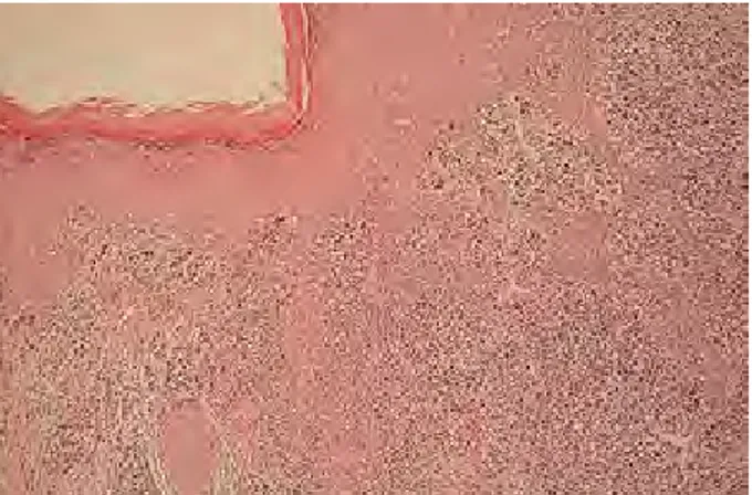
our hospital. We learned that the patient had a history of dog bites on the left hand 3 weeks prior to the beginning of the rash. Prophylactic rabies vaccine (Indirab, Gene) had been intramuscularly administered in the deltoid region at 0 and 7 days. It was learned that the final dose had not been given to the patient since the dog had lived. The patient had a 10-year history of hyperlipidemia, for which he received fenofibrate treatment. On physical examination, bright, erythematous, polygonal papules, commonly 0.5 – 2.5 cm in diameter, were observed on both the upper and lower extremities on the front and back of the body (Fig 1). There were no associated lesions in the oral mucosa, hair, scalp, and nails. A complete blood count and routine biochemical parameters including lipid levels were within normal limits. Hepatitis B surface antigen and anti-HCV antibody, human immunodeficiency virus serology, and syphilis tests were negative, and anti-HBV surface antibody tests were positive. A punch biopsy was taken from a lesion in the right lateral field of the femur of the patient. Histopathology revealed hyperkeratosis, and a focal increase of the granular layer and lichenoid band-style
KUWAIT MEDICAL JOURNAL 341 December 2017
infiltration (Fig 2, 3). As a result of the clinical and histopathological findings, LP was diagnosed. The patient was given photochemotherapy (PUVA) treatment 3 days per week. After 3 weeks, a reduction of itching and a decline in the hyperpigmentation of the lesions was observed.
Fig. 1: Bright, erythematous, polygonal papules, commonly 0.5 – 2.5
cm in diameter, were observed on both the upper extremities
Fig. 2: Hyperkeratosis, focal increase in the granular layer, and band
style lichenoid infiltration (H & E x40)
Fig. 3: Hyperkeratosis, focal increase of granular layer, lichenoid
band-style infiltration, apoptotic keratinocytes (H & E x100)
DISCUSSION
LP is a chronic inflammatory disease and it is thought that cellular immune reactions in the skin, such as cytotoxic T-cell activation, play a central role in the pathogenesis of the disease[1]. Cytotoxic T-cell infiltration into the epithelium results in apoptotis of the basal keratinocytes. The differentiation in the surface epitopes of keratinocyte is seen at the the onset of disease. It is thought that release of mediators from the stimulated cytotoxic T cells induces keratinocyte apoptosis. These epitopes may be viral or toxic in origin[3]. Although the exact mechanism of the relationship between vaccines and lichenoid rash remains unknown, it is primarily related to cell-mediated autoimmunity induced by viruses[4]. In an alternative mechanism, which is molecularly similar, cross-reactions develop in association with an epitope that is expressed on keratinocytes, such as graft versus host disease - an immunological response to some vaccine proteins[5]. In addition, it has been observed that the time between vaccination and onset of rash can vary from a few months to a few days, which may indicate the latent time required for the synthesis of autoantibodies. The time at which the rash begins may provide an important clue in the diagnosis of vaccine-associated reactions. In our case, LP developed three weeks after the second vaccination, which can be associated with the synthesis time of autoantibodies in cellular immunity A similar latent period till the formation of lesions in cases of LP development following hepatitis B virus, influenza and DPT vaccines has previously been shown, which also supports this theory[5-7]. It has also been asserted that various drugs are involved in the etiology of LP, the most common of which are anti-inflammatories, anticonvulsants and antidiabetics[1]. For almost 10 years, no licenoid skin signs have been detected in patients treated with fenofibrate in the literature, so there is no role of fenofibrate treatment in the development of the LP lesions in our patient. The fact that the lesions developed 3 weeks after vaccination was consistent with LP pathophysiology.
Human diploid cell vaccine (HDCV) derived from human haploid cell culture is inactivated rabies vaccine. It has been reported that local reactions occur in 30 – 74% of individuals, and systemic reactions occur in 5 – 40% of individuals following HDCV immunization. Advanced immune complex-like reactions developed in approximately 6% of individuals 2 – 21 days after administration of a booster dose of HDCV. This is associated with the presence of cultivated human albumin with β-propiolactone in HDCV, the development of Ig E antibodies to this allergen[8]. β-propiolactone is used with the aim of inactivating vaccines, and this vaccine
KUWAIT MEDICAL JOURNAL December 2017 342
contains trace amount of neomycin. There is no information, even a case report, to suggest that development of a skin lesion depends on propiolactone and neomycin.
Secondary materials are also used in developing vaccines, and new side effect profiles are exhibited, depending on these substances themselves or the vaccine. The first cases of LP resulting from the HPV vaccine were identified in 1990[9], and over 50 further cases have been reported since that time. The number of LP cases that are related to the influenza vaccine has recently increased[5]. The first DPT vaccine-induced cases of LP were published in 2011[7], and the first case of LP that developed following rabies vaccination was recently reported in 2015[2]. It may be difficult to distinguish histologically between vaccine-induced LP and classic LP. Although vaccine-induced LP rashes are widespread, mucosal and nail involvement is rare[7]. We diagnosed vaccine-induced LP in our patient via the amount of time that had passed between vaccination and the occurrence of rashes, accompanied by clinical and histopathological findings. Our patient is similar to the features of the case presented by Ozbagcivan et al[2] in terms of the presence of a latency period before the occurrence of lichenoid rash after rabies vaccine and the involment of the rashes across the entire body without mucosa and nail involvement. LP lichenoid rash may develop due to administration of the rabies vaccine, but it may also coincidentally develop. However, the lichenoid rash of our LP patient may not be a coincidence, given that the development of LP after rabies vaccination has been identified in a pediatric case[2]. This information highlights the fact that it is necessary to exert greater care with regard to the side effects of the vaccine.
CONCLUSION
In particular, LP cases that are far more aggressive after vaccination in immune-suppressed patients and dermatological cases will not be coincidental[10]. New studies to improve the understanding of the pathophysiology of LP and the relationship between vaccines are required.
ACKNOWLEDGMENT
The authors have nothing to disclose. We thank specialist Sada Tutar for the contribution in pathological examination of this case.
Three authors have contributed to this case report. Suzan Demir Pektas has helped in the diagnosis of this case and the preparation of this article. Ela Kutucularoglu has helped in the diagnosis of this case. Pinar Ozoguz has helped in the preparation of this article.
REFERENCES
1. Gorouhi F, Davari P, Fazel N. Cutaneous and mucosal lichen planus: A comprehensive review of clinical subtypes, risk factors, diagnosis, and prognosis. The Scientific World J 2014, Article ID 742826, 22 pages.
2. Ozbagcivan O, Akarsu S, Ilknur T, Ozer E, Fetil E. Lichen planus after rabies vaccination. Int J Dermatol 2015; 54:e558-559.
3. Drago F, Rebora A. Cutaneous immunologic reactions to hepatitis B virus vaccine. Ann Intern Med 2002; 136:780-781.
4. Limas C, Limas CJ. Lichen planus in children: a possible complication of hepatitis B vaccines. Pediatr Dermatol 2002; 19:204-209.
5. Sato NA, Kano Y, Shiohara T. Lichen planus occuring after ınfluenza vaccination: report of three cases and review of the literature. Dermatology 2010; 221:296-299.
6. Akay BN, Arslan A, Cekirge S, Erkin G, Anadolu-Brasie R. The first reported case of lichen planus following inactivated influenza vaccination. J Drugs Dermatol 2007; 6:536-538.
7. Ghasri P, Roehmholdt BF, Young LC. A case of lichen planus following Tdap vaccination. J Drugs Dermatol 2011; 10:1067-1069.
8. Orenstein WA, Wharton M, Bart KJ, Hinman AR. Immunization, In: Mandell GL, Bennett JE, Dolin R, eds. Principles and Practice of Infectious Diseases. 6th ed. Philadelphia: Elsevier Churchill Livingstone; 2005. p 3557-3589.
9. Tarakji B, Ashok N, Alakeel R, et al. Hepatitis B vaccination and associated oral manifestations: A non-systemic review of literature and case reports. Ann Med Health Sci Res 2014; 4:829-836.
10. De Golian EW, Brennan CB, Davis LS. Lichenoid drug reaction following ınfluenza vaccination in an HIV-positive patient: A case report and literature review. J Drugs Dermatol 2014; 13:873-875.