Research Article
Hakan Ayhan* and Fatma Ayhan
Water based PHEMA hydrogels
for controlled drug delivery
Kontrollu ilaç salımı için su bazlı PHEMA hidrojeller
https://doi.org/10.1515/tjb-2017-0250Received September 18, 2017; accepted September 20, 2017; previously published online October 31, 2017
Abstract
Objective: In the scope of presented work, synthesis of water
based acrylate hydrogels, characterization, and their usage in controlled drug release systems were aimed to investigate.
Methods: Synthesis of acrylate based hydrogels that have
different properties was carried out by free radical pho-topolymerization using photoinitiator. Because of its high biocompatibility, 2-hydroxyethyl metacrylate (HEMA) was used as monomer. Then drug release experiments were performed in pH 7.4 and 1.2 buffer solutions with certain ionic strength while the dynamic swelling behaviors were also determined. In the last part of the work, drug activi-ties of synthesized drug-loaded hydrogels were tested in mediums containing Staphylococcus aureus and
Pseu-domonas aeruginosa bacteria cultures.
Results: ATR-FTIR spectrums of all synthesized
hydro-gels were analyzed. The characteristic O-H, C-H, C=O, C-O tension vibrations bands were observed in the spectrums of the hydrogels. The rate of drug release in acidic pH 1.2 for two types of hydrogels was observed to be much faster than at pH 7.4. It was determined that hydrogel swelling ratio decrease with increasing monomer ratio. All drug loaded hydrogels were effective to inhibit the growth of both two bacterial strains.
Conclusion: Hydrogels synthesized were found to be
suit-able for the controlled drug delivery applications.
Keywords: HEMA; Photopolymerization; Hydrogels;
Con-trolled Drug Delivery.
Özet
Amaç: Sunulan araştırma kapsamında; su bazlı akrilat
hidrojellerin sentezi, karakterizasyonu ve bu hidrojellerin kontrollü ilaç salım sistemlerinde kullanımlarının ince-lenmesi amaçlanmıştır.
Yöntemler: Farklı özelliklere sahip akrilat bazlı
hidrojel-lerin fotopolimerizasyon yöntemi ile fotobaşlatıcı kulla-nılarak serbest-radikal fotopolimerizasyonuyla sentezi gerçekleştirilmiştir. Yüksek biyouyumluluğu nedeniyle 2-hidroksietil metakrilat (HEMA) monomeri kullanılmış-tır. Sonra ilaç salım deneyleri iyon şiddeti ayarlanmış pH 7,4 ve 1,2 tampon çözeltilerinde gerçekleştirilirken dinamik şişme davranışları da belirlenmiştir. Çalışmanın son bölümünde ise sentezlenen ilaç yüklü hidrojellerin ilaç aktiviteleri Staphylococcus aureus ve Pseudomonas
aeruginosa bakteri kültürlerinin kullanıldığı ortamda
test edilmiştir.
Bulgular: Sentezlenen bütün hidrojellerin ATR-FTIR
spektrumları analiz edilmiştir. Spektrumlarda HEMA hidrojelleri için karakteristik sayılabilecek O-H gerilme, C-H gerilme, C=O gerilme ve C-O gerilme titreşim bant-ları görülmüştür. Asidik değerde yani pH 1,2’de gözle-nen ilaç salım hızlarının her iki tip hidrojel için de pH 7,4’e göre oldukça hızlı olduğu gözlenmiştir. Monomer oranının artmasıyla hidrojel şişme derecesinin azal-dığı tespit edilmiştir. Tüm ilaç yüklü hidrojeller her iki bakteri grubunun üremesinin engellenmesinde etkili olmuştur.
Sonuç: Sunulan bu çalışmada sentezlenen tüm hidrojeller
kontrollu ilaç salımı için uygun bulunmuştur.
Anahtar Kelimeler: HEMA; Fotopolimerizasyon; Hidrojel;
Kontrollü İlaç Salımı.
Introduction
Today, there are many studies on controlled drug release systems because of the advantages such as the sustained
*Corresponding author: Hakan Ayhan, Muğla Sıtkı Koçman University – Chemistry, Biochemistry, BIOMATREG Biochemistry and Biomaterials Research Group, Muğla, Menteşe, Turkey,
e-mail: hayhan48@gmail.com
Fatma Ayhan: Muğla Sıtkı Koçman University – Chemistry, Biochemistry, BIOMATREG Biochemistry and Biomaterials Research Group, Muğla, Menteşe, Turkey
protection of the therapeutic level of medicines or the reduction of the harmful effects and drug amount in order to target the release to a specific cell type or tissue. When a drug is linked to a polymer or a lipid or encapsulated, the ability to provide drug safety and the desired effect of the drug can be greatly increased [1, 2]. Hydrogels, which can be used as controlled drug delivery systems in biomedi-cal field, are suitable for biomedibiomedi-cal applications because of their high water content, elastic properties similar to natural tissues, and their greater or lesser permeability to body fluids.
Hydrogels are hydrophilic polymeric network struc-tures made up of homopolymers or copolymers, which can swell up to thousands of folds from 10% to 20% of their dry weight in water [3, 4]. In addition, the hydrogels may respond to their surroundings in case of stimula-tions. In the study conducted in 2004 by the Court and friends, HEMA and MAA based copolymers were prepared by radiation polymerization and they observed that the swelling behaviors of hydrogels at pH 1–7.4 were different [5, 6]. Because of all these properties, the use of hydrogels as carriers in swelling-controlled release systems has been investigated for many years.
In swelling-controlled release systems, drugs or bio-molecules such as proteins are incorporated into the system and then released in response to changes in the environment. Depending on the nature of the hydrogel material, it swells or shrinks in response to changes in the surrounding pH or temperature. These systems can be produced in such a way that, the release of biomol-ecule outside of the gel occurs by diffusion from the hydrogel when swelling occurred under certain condi-tions. Physical entrapment, electrostatic interactions, physical adsorption, and chemical bonding methods can be used to immobilize the drug on or inside hydrogel. Each technique has advantages and disadvantages. The method used depends on the type of hydrogel and the drug.
Entrapment is one of the simplest and easiest ways to develop a controlled release system. One of the methods that can be used to entrap is to incorporate the drug into this network structure by performing hydro-gel polymerization in the presence of the drug. Diffusion of the drug out of the system occurs when the hydro-gel is placed in an environment that causes swelling and increases pore size. However, the drug to be used in this technique must be resistant to polymerization and should not react with the monomers in the system. In addition, polymeric system should not be too cross-linked in such a manner to prevent the diffusion of drug from the system [4].
In the biomedical field, one of the main methods used in the synthesis of hydrogels is by UV light polymerization [7]. The photopolymerization method has an increasing demand due to its broad application area. Nowadays, free-radical polymerisation with UV light is used in various applications such as coatings, information storage systems, films, contact lenses and biomaterials. In this method, polymerization occurs by converting the primary radicals generated from light absorption of photoinitiator at a suitable wavelength into highly cross-linked struc-tures of multifunctional acrylates [8].
There are many advantages of using photopolymeri-zation method especially in biomaterials. The method is generally not harmful to health and the polymers can be synthesized at temperatures and pH near physiological values and even in the presence of biologically active materials. At the same time, the process proceeds very quickly at these conditions for many monomers and con-ventional initiators. In addition, it is very advantageous for the formation of complex materials due to directing of exposure to UV light and providing temporary control of the process time. The photopolymerizable hydrogels used in biological systems generally consist of macro-molecular hydrogel precursors. These are water-soluble polymers with two or more reactive groups. Examples of such materials include PEG acrylate derivatives, PEG methacrylate derivatives, HEMA, polyvinyl alcohol (PVA) derivatives and modified polysaccharides such as hyalu-ronic acid derivatives, and dextran methacrylate [9, 10].
In this context, the synthesis of acrylate-based hydrogels with photopolymerization by free-radical photopolymerization by using photoinitiator, the char-acterization of the obtained hydrogels and their use in controlled drug release systems constitute the objectives of the presented work [11–14]. In the study, cross-linked HEMA monomers were selected for their high biocompat-ibility, no harmful effect, and three-dimensional struc-ture controlling drug, peptide or protein release. In the first part of the work, the hydrogels containing HEMA monomer were synthesized in the presence of crosslinker EGDMA and photoinitiator DMPA. At this stage, the effects using monomer, crosslinker and photoinitiator at different ratios on the hydrogel structure were examined. In addition, hydrogel structure was also investigated by changing light intensity. ATR-FTIR spectra were taken and dynamic swelling behaviors of the hydrogels were investigated on the basis of the characterizations of the obtained hydrogels.
In the last part of the study, the synthesis of drug-loaded hydrogels, drug release experiments and microbial activity assays were carried out.
Materials and methods
Synthesis of hydrogels
Black-Ray® B-100 (UVP, B 100 AP Upland, CA, USA) series
high intensity UV lamp and J-221 UV-meter (UVP, Upland, CA, USA) were used during the synthesis of acrylate based hydrogels with different properties. The synthesis of hydrogels by free radical polymerization using photoini-tiator by polymerization method, HEMA monomer (98%, Aldrich Chem. Co., USA) was used due to its high biocom-patibility, nontoxicity, and three dimensional structure that control drug-peptide or protein release.
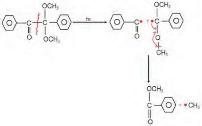
EGDMA (98%, Aldrich Chem. Co., USA), and DMPA (99%, Aldrich Chem. Co., USA) were used as cross-linking agent and photoinitiator, respectively (Figure 1).
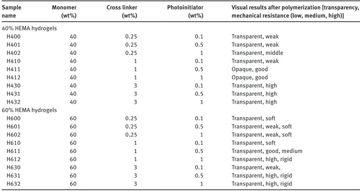
High reactive methyl groups are produced by the absorption of UV light when photoinitiator, DMPA, was used in free-radical polymerization of HEMA hydrogels synthesis. The formation of network, polymerization was started by the attack of methyl radicals to C=C double bounds in acrylate groups. The polymerization takes place when these primary radicals convert functional acrylates to cross-linked structures. The reaction of radical attain-ing from DMPA was schematically presented in Figure 2 [9]. Hydrogel synthesis was performed in the presence of monomer HEMA, cross-linker EGDMA, photoinitiator DMPA and distilled water in HEMA synthesis stage. In order to investigate the parameters that effect the syn-thesis of water based PHEMA hydrogels, monomer HEMA was added in 40% and 60% weight ratios. The composi-tion and naming of synthesized all HEMA hydrogels were given in Table 1. The ratio of cross-linker was changed as 0.25, 1 and 3 weight percent while photoinitiator, DMPA, ratio was adjusted to be at 0.1, 0.5 and 1 weight percent ratios. Polymerization experiments were conducted in glass petri dish of 10 mm diameter with fixed 400 μL
constant volume. Solutions with various compositions were firstly prepared by the solvation of photoinitiator in monomer/macromer at ratios given above and then dis-tilled water and cross-linker were added. Polymerization was performed by free radical photopolymerization after the treatment of solutions under 365 nm wavelength and 10 mW · cm−2 light intensity UV lump for 10 min.
The synthesized hydrogels were then washed with n-heptane to remove the unreacted monomers from poly-merization media.
It was also tried for UV light intensities changing between 3, 5, 7 and 10 mW · cm−2 in the early stage of
synthesis. The light intensity was excluded from vari-able parameter in the rest of the research since the best
Figure 1: Chemical structures.
(A) HEMA monomer; (B) photoinitiator DMPA; (C) cross-linking agent EGDMA.
Figure 2: Schematic representation of radicalic reactions of photoinitiator, DMPA.
Table 1: PHEMA hydrogels nomenclature. Monomer
HEMA (wt%) EGDMA (wt%)Cross linker Photoinitiator DMPA (wt%) Nomenclature
40 0.25 0.1 H400 40 0.25 0.5 H401 40 0.25 1.0 H402 40 1.00 0.1 H410 40 1.00 0.5 H411 40 1.00 1.0 H412 40 3.00 0.1 H430 40 3.00 0.5 H431 40 3.00 1.0 H432 60 0.25 0.1 H600 60 0.25 0.5 H601 60 0.25 1.0 H602 60 1.00 0.1 H610 60 1.00 0.5 H611 60 1.00 1.0 H612 60 3.00 0.1 H630 60 3.00 0.5 H631 60 3.00 1.0 H632 OCH~
I
-c-@
-
•
cH
,
II
0polymerization efficiency was obtained for 10 mW · cm−2
and all the remaining experiments were done at 10 mW · cm−2 light intensity value. Table 2 gives the
mor-phological characterization of 40% and 60% HEMA hydrogels for various cross linker and photoinitiator ratio.
Dynamic swelling behaviors of hydrogels
Dynamic swelling behaviors of synthesized PHEMA hydrogels were examined. For this purpose dried polymer discs with known weight amounts were incubated in 80 mL buffer solutions of pH 7.4 and adjusted with 0.13 M NaCl at 37°C, kept in incubator at constant stir-ring (Edmünd Mühler, TH 15-KS15). Then weighted on analytical balance at certain time intervals until equi-librium value was reached. The produced discs amount was 25 for each experimental sample. Synthesized discs were dried until constant weight was reached and used in experiments. Buffer solution of pH 7.4 that is necessary to determine dynamic swelling behaviors was prepared by using Na2HPO4 · 2H2O (Riedel-de Haen, Sigma-Aldrich, Germany), NaH2PO4 · 2H2O (Riedel-de Haen, Sigma-Aldrich, Germany), and NaCl (Merck KgaA, Germany). Ionic strength of prepared buffer solition was adjusted to 0.4 M. Wt value which was defined as swelling ratio was calculated with the Wt = [(Wt − W0)/W0] × 100 formula
according to the weighted values obtained after swell-ing experiments. In formula: Wt is the weight of swelled hydrogel at time t, the initial weight of dried hydrogel is shown by W0. The graphical change of Wt value, the swell-ing ration of hydrogel with time was obtained after calcu-lations for each hydrogel sample were done.
Characterization of hydrogels
Fourier transform infrared spectroscopy (Shimadzu FTIR-8000 Serisi, DR-8001) was used to characterize the structures of the synthesized PHEMA hydrogels with various properties. Synthesized hydrogel discs were dried in vacuum incubator (Nüve, EV 018 Vacuum Incuba-tor, Turkey) until constant weight values were obtained and then ATR-FTIR spectrums were taken. The ATR-FTIR spectrums obtained for HEMA polimers were organized as groups so that each group contain three samples by keeping constant the photoinitiator, DMPA ratio and the effect of the increase in cross-linker, EGDMA ratio to hydrogel stucture was investigated. In the scope of the research, surface and cross section images of the syn-thesized PHEMA hydrogels were taken by scanning elec-tron microscope for various magnification values. For the purpose, hydrogel discs that were dried in incubator until constant weights have been obtained were cut in to proper
Table 2: Morphology of 40% and 60% HEMA hydrogels. Sample
name Monomer (wt%) Cross linker (wt%) Photoinitiator (wt%) Visual results after polymerization [transparency, mechanical resistance (low, medium, high)]
40% HEMA hydrogels H400 40 0.25 0.1 Transparent, weak H401 40 0.25 0.5 Transparent, weak H402 40 0.25 1 Transparent, middle H410 40 1 0.1 Transparent, weak H411 40 1 0.5 Opaque, good H412 40 1 1 Opaque, good H430 40 3 0.1 Transparent, high H431 40 3 0.5 Transparent, high H432 40 3 1 Transparent, high 60% HEMA hydrogels H600 60 0.25 0.1 Transparent, soft
H601 60 0.25 0.5 Transparent, weak, soft
H602 60 0.25 1 Transparent, weak, soft
H610 60 1 0.1 Transparent, soft
H611 60 1 0.5 Transparent, good, medium
H612 60 1 1 Transparent, high, rigid
H630 60 3 0.1 Transparent, weak,
H631 60 3 0.5 Transparent, high, rigid
dimensions and put on circular sample discs. They were visualized with differently magnified SEM images after plating with gold.
Drug loading to hydrogels
For the purpose, gentamicin was selected due to its broad spectrum, resistance to heat and pH changes, and high hydrophilicity. Intravenous ampoules containing 80 mg gentamicin equivalent gentamicin sulfate in 2 mL (Gentamisin® 80 mg i.m/i.v/ ampoule, Deva Holding
A.Ş, Turkey) were used as drug for the synthesis of drug containing hydrogels. Various drug containing solutions were obtained by the addition of the same amount of gentamicin solution instead of distilled water in polym-erization dispersion media. Experiments were done in 400 μL constant volume, and 100 mm glass petri dishes at room temperature; first, photoinitiator was dissolved in monomer/macromer, then gentamicin solution, and cross-linker were added, respectively. The polymerization was performed by treatment of the final polymer solutions under 365 nm UV light with 10 mW · cm−2 light intensity
approximately 10 min by free-radical polymerization.
Drug release experiments
In vitro drug release experiments from drug loaded hydro-gels were carried out in this stage. For the purpose, the polymer discs with known weights and drug amounts were kept in a shaking incubator (Edmünd Mühler, TH 15-KS15) at 37°C constant temperature. Shaking speed was kept as 100 stroke/min in pH 7.4 and 1.2 buffer solu-tions, respectively, which ionic strenght was adjusted with 0.13 M NaCl. The volume of the drug released media was maintained by the addition of the same amount of solu-tions that taken to analyze the drug concentrasolu-tions in order to keep the release volume constant. Addition of 3 mL buffer solution was achieved each time when liquid samples of 3 mL was taken from release media until drug release rate completed. Initially, sampling frequency between readings were performed in shorter time intervals to investigate the burst effect if present, and then intervals were extended. The pH 7.4 buffer solution was prepared from Na2HPO4 · 2H2O (Riedel-de Haen, Sigma-Aldrich, Germany), NaH2PO4 · 2H2O (Riedel-de Haen, Sigma-Aldrich, Germany), and NaCl (Merck KgaA, Germany); sodium citrate (Aldrich Chem. Co., USA), HCl (30%, Merck KgaA, Germany), and NaCl (Merck KgaA, Germany) were used for pH 1.2 buffer solutions. In order to investigate the
released drug amount from gentamicin loaded hydrogel discs, quantity determination was carried out at 255 nm where maximum absorbans value of gentamicin observed. Gentamicin calibration graph and calibration curve of linear relationship was made. In the graph, x axis shows the gentamicin concentration amounts (mg · mL−1) while
related absorbans values at 255 nm were shown in y axis. The absorbance of the samples taken from release media at certion time intervals were measured in a UV-Vis spek-trophotometer (Labomed Inc., Spectro UV-VIS Double Beam PC and 8 Scanning Auto Cell) at 255 nm and gen-tamicin concentrations were calculated from the calibra-tion graph’s curve given below. Then cumulative addicalibra-tion method was applied to the measured released total gen-tamicin amount according to the method given in appen-dix and % released gentamicin amounts were calculated.
Analysis of drug activities
Disc diffusion method was used to analyze the activity of the drug loaded to hydrogel discs. Gentamicin loaded HEMA hydrogel discs were placed separatelly on the sur-faces of agar plates containing Pseudomonas aeruginose (ATCC 10145) and Staphylococus aureus (ATCC 25923) bac-terial strains spreaded from 0.1 mL standard suspensions. So, antibiotics diffused on agar surface and inhibited bac-terial growth in the case of effective amounts released. Cir-cular inhibition areas were formed around hydrogel discs that no bacterial growth was present after 24 h incubation time. Control experiments were performed by PHEMA hydrogels without drug. Commercial circular gentamicin discs containing 10 mg gentamicin equivalent gentamicin sulfate/10 mm diameter was also used to make additional control experiments.
Results and discussion
Synthesis of water based PHEMA hydrogels
In order to observe the effect of synthesis variables on HEMA hydrogel structure, the parameter values were determined according to the literature knowledge taken into consideration. Anseth and coworkers used 40% water and 0.1% photoinitiator ratios based on monomer weight, respectively in the synthesis of multilaminated poly(HEMA) hydrogels for controlled release [9]. The syn-thesis of HEMA/EGDMA hydrogels by photopolymeriza-tion were performed with 0%, 20%, 40%, 60% water and
1.0% photoinitiator ratios based on monomer weight as reported by Lee and Li [12]. Water soluble 2-hydroxyethyl metacrylate (HEMA) were added as 40% and 60% weight ratio by the direction of these literature value results. Thus, hydrogel synhesis were achieved in the presence of monomer HEMA, cross linker EGDMA, photoinitiator DMPA and distilled water.
Dynamic swelling behaviors of water based
PHEMA hydrogels
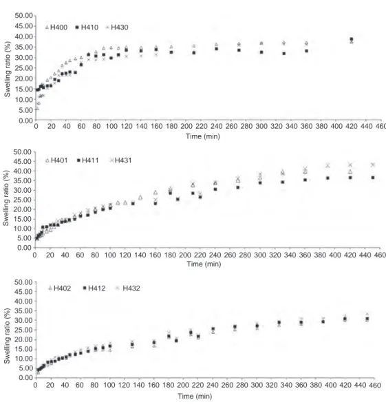
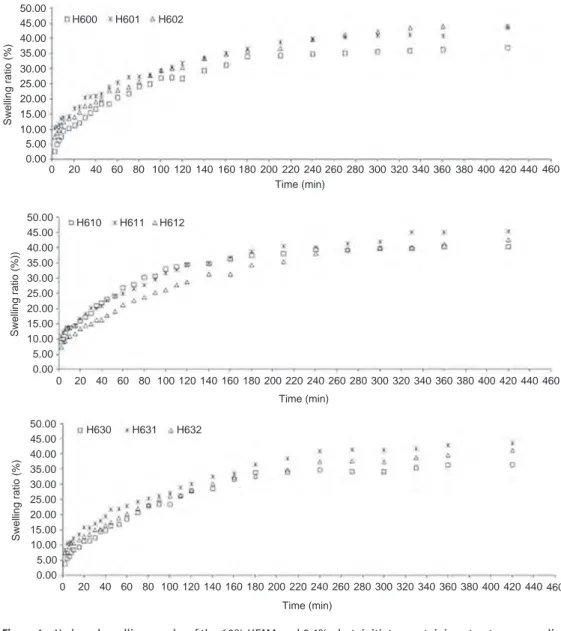
Hydrogel swelling ratios that show dynamic swelling behaviors of 40% and 60% HEMA hydrogels synthesized in the scope of research, Wt vs. time, were drawn in figures. Dynamic swelling behaviors of hydrogels were evaluated based on the cross linker and photoinitiator concentrations.
The swelling amounts for experimental results continued 30 days was given in Figure 3 which were observed to be between 30% and 42% swelling ratio. The swelling ratios did not change significantly as cross linker ratio increses (0.25%, 1.0% and 3.0%) in hydrogels with 40% HEMA when photoinitiator ratio was kept in the lowest value (0.1%) as Figure 3 shows.
Figure 4 gives the the swelling amounts of experi-mental results for 30 days. Figure 4 also indicates that the swelling amounts of 60% HEMA hydrogels showed approximately 40% swelling ratio in the case of the least photoinitiator ratio and swelling amount changes were small when cross linker ratio increased (0.25%, 1.0% and 3.0%). It was seen from the figüre that initial swelling ratio reaches to a certain value within 240 min, continuous with a slowly swelling ratio and then comes to equilibrium after 1 month time almost with the same
50.00 45.00 H400 H410 H430 Time (min) 0 20 40 60 80 100 120 140 160 180 200 220 240 260 280 300 320 340 360 380 400 420 440 460 Swelling ratio (%) 40.00 35.00 30.00 25.00 20.00 15.00 10.00 5.00 0.00 50.00 45.00 H401 H411 H431 Time (min) 0 20 40 60 80 100 120 140 160 180 200 220 240 260 280 300 320 340 360 380 400 420 440 460 Swelling ratio (%) 40.00 35.00 30.00 25.00 20.00 15.00 10.00 5.00 0.00 50.00 45.00 H402 H412 H432 Time (min) 0 20 40 60 80 100 120 140 160 180 200 220 240 260 280 300 320 340 360 380 400 420 440 460 Swelling ratio (%) 40.00 35.00 30.00 25.00 20.00 15.00 10.00 5.00 0.00
Figure 3: Swelling graphs of the 40% HEMA and 0.1% photoinitiator containing hydrogels with 0.25%, 1.0% and 3.0% weight percent cross-linker ratios.
•
•
,i ~·· !I • • ! h ~ 0 " • ii • ~..
,.
..
•
•
..
A•
•
•
•
li•
•
,.•
/!1•
! 9,•
~•
! *•
t
•
•
4•
0•
~•
!•
,.
., ,lo•
•
•
A Aamount. It was estimated that all the hydrogels of 60% HEMA coded as H6__ had swelling ratios between 35% and 45% values.
The swelling ratios of hydrogels with 40% and 60% HEMA contents did not show any statistically significant difference when cross linker amount increased at constant photoinitiator concentration for the amounts given above.
Characterization of hydrogels
Furthermore, synthesized HEMA hydrogels were charac-terized with FTIR spectrums and SEM images.
ATR-FTIR spectrometer was used for structural charac-terization of the hydrogels. The structural characcharac-terization
of the synthesized hydrogels was achieved by FTIR in order to determine the atomic groups present, and the effect of constituents on the hydrogels that were synthesized with different properties. Hydrogel surfaces were examined by the attenuated total reflectance FTIR spectrums of
50.00 45.00 H600 H601 H602 Time (min) 0 20 40 60 80 100 120 140 160 180 200 220 240 260 280 300 320 340 360 380 400 420 440 460 Swelling ratio (%) 40.00 35.00 30.00 25.00 20.00 15.00 10.00 5.00 0.00 50.00 45.00 H610 H611 H612 Time (min) 0 20 40 60 80 100 120 140 160 180 200 220 240 260 280 300 320 340 360 380 400 420 440 460 Swelling ratio (%)) 40.00 35.00 30.00 25.00 20.00 15.00 10.00 5.00 0.00 50.00 45.00 H630 H631 H632 Time (min) 0 20 40 60 80 100 120 140 160 180 200 220 240 260 280 300 320 340 360 380 400 420 440 460 Swelling ratio (%) 40.00 35.00 30.00 25.00 20.00 15.00 10.00 5.00 0.00
Figure 4: Hydrogel swelling graphs of the 60% HEMA and 0.1% photoinitiator containing structures cross linked with 0.25%, 1.0% and 3.0% w/w cross-linker ratios.
Table 3: The vibration bands estimated in hydrogels.
Wavelength (cm−1) Tensile vibration
900–1300 C-O 1600–1700 C=C 1600–1900 C=O 2700–3300 C-H 3000–3700 O-H n ;t.t"J.
'JP
• t. r:J~ 0 u • !•
~.
~000 • .6a
00..
a:,.
"
<, A ,. ,. • ~ 11 'J.
" 0 n 0 ID..
"
a
8 1 0.
0 ,."
'i ll..
I) n 0 I) Q•
0..
l 0 0 t. 0 6 n 0 6 0 0•
g..
0the samples. The vibration types of the bands present in HEMA hydrogels were given in Table 3.
ATR-FTIR spectrums of the hydrogels synthesized by using 40% and 60% 2-hydroxyethyl methacrylate arranged as groups to investigate (i) the effect of increas-ing cross linker ratios on hydrogel structure at constant DMPA ratio, and (ii) the photoinitiator effect at constant cross linker ratios.
Figure 5 shows the ATR-FTIR spectrums of 40% HEMA hydrogels for different cross linker amounts at constant photoinitiator ratio. The absorption in the region of about 3500 cm−1 was due to the stretching of O-H group,
and C-H streching was observed near 2900 cm−1 while
the band of carbonyl and C-O stretching vibrations were detected at 1750 cm−1 and 1175 cm−1, respectively. These
stretching vibrations designated after surface analysis of the polymeric structures obtained from HEMA were evaluated as characteristic vibrations. Especially, the presence and changeability of O-H band gives important information about the hydrophilicity of the structure [15–20].
ATR-FTIR spectrums of hydrogels with 60% HEMA ratio were given in Figure 6. In the spectrum, O-H stretching vibration was observed at 3400 cm−1 while the
band at 3000 cm−1 belongs to C-H stretching vibration,
and the absorption of the carbonyl C=O bond occurs at
1700 cm−1 and the C-O at 1150 cm−1 [21–25]. The bigger
O-H stretching vibration band in spectrum shows that HEMA content of these hydrogels is higher than 40% HEMA hydrogel since the O-H stretching vibration band is greater than O-H stretching vibration of the hydrogels structures containing 40% HEMA amount. Addition-ally, sharp carbonyl C=O peak in 60% HEMA hydrogels supports the high HEMA content. Figure 6 gives the in ATR-FTIR spectrums of 60% HEMA hydrogels (H601, H611, H631) which were grouped together for various cross linker concentrations. The increase of cross linker ratio as 0.25, 1.0, and 3.0 weight percent sequentially on hydrogel structure were investigated for 60% HEMA hydrogels in case when DMPA ratios were kept constant at 0.5 (a), 1.0 (b), 1.5 (c), respectively. The effect of cross linker weight ratio augmentation as 0.25%, 1.0% and 3.0% subsequently on hydrogel structure was investi-gated for 60% HEMA hydrogels at constant photoinitia-tor, DMPA amounts of 0.5, 1.0 and 1.5 weight ratios. It can be seen that the band intensities were almost the same in both three spectrums. This shows that enhancing cross linker ratios at fixed DMPA concentration did not cause any important change in hydrogel structure according to FTIR spectrums [26–28].
The surface and cross sectional images of the syn-thesized hydrogels of the research content were taken
Figure 5: ATR-FTIR spectrums of hydrogels containing 40% HEMA.
l
., n ··"'-!- - t ---i- --+---i---+---,t-=--+~=-J.~ii
· · -- - - < ~f - - - -i--v--a..-i---,,-,;,...,--- l--,,1-us•p111:i1
•••"'+ --1-11-f - - +- •\H --'---1-,,-~-11-Afi- - + - ---+-+...
...
...
...
fl.~...
,
"
: 'i n 1 'i ,;-
-l
::s~l1
iiiHI
.,
..
..
~ M. ..l L..l---'---1----1---'-'---+---t->'---J.---+---+--1-n.• ILl----l-:__4:___ +----l-- - + - - - 4 -- + - - - I - + IU *A 1'Ul •A~,
ti •1.a-J-+-,-,i~-...j--.\hch,---11---,-~+.-f',-;'.11 - --+--'-+-~•--• ji
~;.1d
=
1;~i-~!l
;
•1-9-i_,,~_,,,.'--"1F.:.:...:-+--:,..-if--+--+--,r-\l,,s:----r-h+- •·•Xg li-·
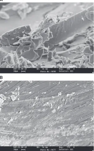
by scanning electron microscopy (SEM). SEM images of two hydrogels without drug were presented for example and as an idea. The surface structure of 1600 (800) times magnified H411 hydrogel was given in Figure 7A. A tightly blocked structure was seen in hydrogel section of the figure. The compact structure in images coincides also with the low swelling ratios of HEMA hydrogels. The SEM micrograph of gentamisin loaded hydrogel (HG411) cross section profile magnified 1600 times was given in Figure 7B. Gentamicin layers superimposed on another can be seen from hydrogel section.
Drug release assays
H411 and H611 coded hydrogels from hydrogels with HEMA content of 40% and 60% were selected for drug release experiments due to their high mechanical resistance and suitable swelling capacities. Gentamicin was loaded in a total of 5% considering the amount of monomer selected. Drug releases from HG411 and HG611 hydrogels at pH 1.2 and pH 7.4 were given in Figure 8A and B. As can be seen from figure, HG611 released 98.43% of the total loaded drug content after 160 min say about 2.5 h at pH 1.2 but it is lower than 80% for HG411 at the same pH.
At pH 7.4, the released drug percent from HG611 was 75.92% after 240 min or 4 h. At 480 min, drug release reached 79% and 98% release amount at 720 min (not
Figure 6: ATR-FTIR spectrums of hydrogels with 60% HEMA content.
Figure 7: SEM micrographs of water based PHEMA hydrogel samples. (A) H411; (B) HG411.
,..,
shown). Drug release rates of 40% HEMA containing hydrogel, HG411, were found as 45.37 after 240 min and at 480 min reaches to 65.60% release amount. Almost completely released amount like 99% was estimated after 720 min at pH 7.4 (not shown) [29–34].
The drug release amounts higher than 100% may come from the hydrogel degradation at low pH values.
Activity tests of drug loaded PHEMA
hydrogels
Gentamicin activity of drug-loaded hydrogels was deter-mined by using disk diffusion method. The results of the study using two different microorganisms were pre-sented in Table 4. It was observed that in experiments with non-drug loaded hydrogels for control purposes the discs only inhibited bacterial growth only in the area as small as their diameter (under hydrogels). This can be considered as proof that HEMA hydrogels do not show any harmful effect. It has been observed that all drug loaded hydrogels are effective in preventing the growth of both two bacterial groups. It can also be considered that the photopolymerization method used in the synthesis of drug-loaded hydrogels, in other words, UV light has no effect on gentamicin activity. While in the experiments
with 10 mg gentamicin discs purchased as standard, inhibition areas with 22 mm diameter for Pseudomonas
aeruginosa bacteria and 20 mm diameter for Staphylo-coccus aureus bacteria were observed, all of the
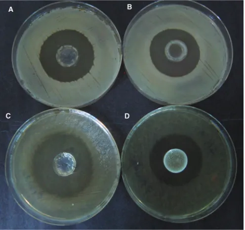
experi-ment results that were done with drug loaded hydrogels were found to have almost the same inhibition areas. In addition, when the experiments with the hydrogels con-taining the same amount of drug were compared, it was observed that the effect on growth was almost the same in both two groups. The visual results of some agar diffu-sion tests were given in Figure 9.
Table 4: Activity assay results by disk diffusion method of gentami-sin loaded hydrogels.
Hydrogel
code Hydrogel diameter
(mm)
Pseudomonas Inhibition area (mm)
aeruginosa Staphylococcus aureus
H411 10 10 10 HG411 18 40 – HG411 10 – 38 H611 10 10 10 HG611 19 32 – HG611 19 – 44
Gentamisin disc (10 μg): P.aeroginosa (ATCC) 22 mm, S. aureus (ATCC) 20 mm. Media: Brain Heart Agar (Fluca).
140 120 100 80 60 Dr ug release amount (%) 40 20 0 0 10 20 30 40 50 60 70 80 90 100 110 120 Time (min)
Drug release of HG611 hydrogels pH 1.2 pH 7.4 130 140 150 160 170 180 190 200 210 220 230 240 250 140 120 100 80 60 Dr ug release amount (%) 40 20 0 0 10 20 30 40 50 60 70 80 90 100 110 120 Time (min)
Drug release of HG411 hydrogels pH 1.2 pH 7.4 130 140 150 160 170 180 190 200 210 220 230 240 250
A
B
Figure 8: Drug release graphs of hydrogel samples. (A) HG411; (B) HG611. A A A A A /l. /l. A /l. /l. /l. /l. 6 A ll, :i. ~ 'I;
,.
_( .( A /l. A A A A A A A.
.
Conclusion
The synthesis of water-based PHEMA hydrogels with pho-topolymerization method using photoinitiator by free radical photopolymerization was performed. Stress vibra-tions observed in surface analyzes of hydrogels were eval-uated as characteristic vibrations. The O-H band intensity in the hydrogel’s spectrum using 60% HEMA is higher than the O-H stretch band intensity in the hydrogel’s with 40% HEMA-using hydrogels, proved that these hydrogels have more HEMA content in their structure.
The hydrogels with the highest dynamic swelling quantities were again synthesized as drug loaded forms. In the drug release experiments carried out in the stomach and intestinal pH values, the release took place within 3–4 h for pH 1.2, while for pH 7.4 this took 12 h.
Additionally, the drug activities of drug-loaded hydrogels were tested in the media in which
Staphylo-coccus aureus and Pseudomonas aeruginosa bacterial
cultures were grown. It was found that both bacterial strains were inhibited and therefore it was also seen that no lost in drug activity occured. As a general conclusion, curing method for hydrogel synthesis in the presence of
antibiotics can be used in the preparation of drug deliv-ery vesicles.
Conflict of interest statement: Authors have no conflict of
interest.
References
1. Brannon-Peppas L. Polymers in controlled drug delivery. Med Plast Biomater 1997;34–44.
2. Çapan Y. Sürekli salım sağlayan tabletlerin özellikleri ve değerlendirilmesi. FABAD Farm Bil Der 1993;18:27–39. 3. Hoffman AS. Hydrogels for biomedical applications. Adv Drug
Deliv Rev 2002;43:3–12.
4. Peppas NA, Brazel CS. Modeling of drug release from swellable polymers. Eur J Pharm Biopharm 2000;49:47–58.
5. Allahverdipoor M, Mahkam M. Controlled release of biomolecules from pH-sensitive network polymers prepared by radiation polym-erization. J Drug Target 2004;12:151–6.
6. Mahkam M. Novel pH-sensitive hydrogels for colon-specific drug delivery. Drug Deliv 2010;17:158–63.
7. Peppas NA, Ward JH. Preparation of controlled release systems by free-radical UV polymerizations in the presence of a drug. J Control Release 2001;71:183–92.
Figure 9: Agar diffusion test results.
8. Mellott MB, Searcy K, Pishko MV. Release of proteinfrom highly cross-linked hydrogels of poly(ethylene glycol) diacrylate fabri-cated by UV polymerization. Biomaterials 2001;22:929–41. 9. Anseth KS, Lu S. Photopolymerization of multilaminated
poly(HEMA) hydrogels for controlled release. J Control Release 1999;57:291–300.
10. Arsu N, Davidson RS, Holman R. Factors affecting the photoyel-lowing which occurs during the photoinitiated polymerization of acrylates. J Photochem Photobiol A Chem 1995;87:169–75. 11. Nguyen KT, West JL. Photopolymerizable hydrogels for tissue
engineering applications. Biomaterials 2002;23:4307–14. 12. Lee JL, Li L. Photopolymerization of HEMA/DEGDMA hydrogels in
solution. Polymers 2005;46:11540–7.
13. Lee JL, Cao X, He H. Design of a novel hydrogel-based intel-ligent system for controlled drug release. J Control Release 2004;95:391–402.
14. Anseth KS, Bryant SJ. The effects of scaffold thickness on tissue engineered cartilage in photocrosslinked poly(ethylene oxide) hydrogels. Biomaterials 2001;22:619–26.
15. Caliceti P, Salmaso S, Lante A, Yoshida M, Katakai R, Martellini F, et al. Controlled release of biomolecules from temperature-sen-sitive hydrogels prepared by radiation polymerization. J Control Release 2001;75:173–81.
16. Changez M, Burugapalli K, Koul V, Choudhary V. The effect of composition of poly(acrylic acid)-gelatin hydrogel on gentamicin sulphate release: in vitro. Biomaterials 2003;24:527–36. 17. Garg S, Vermani K, Gupta P. Hydrogels: from controlled release to
pH-responsive drug delivery. Drug Discov Today 2002;7:569–79. 18. Gümüşderelioğlu M, Basan H, Orbey T. Diclofenac sodium
releasing pH-sensitive monolithic devices. Int J Pharm 2002;245:191–8.
19. Gümüşderelioğlu M, İmren D, Basan H. pH’ya duyarlı hidrojeller ve kontrollü ilaç salım sistemlerindeki uygulamaları. FABAD Farm Bil Der 2001;26:81–92.
20. Gürsoy A, Dortunç B, Pişkin E, Peppas NA. Kontrollü İlaç Serbestleştiren Sistemler, Marmara Üniversitesi Eczacılık Fakültesi Yayınları No:469/5, İstanbul. 1989.
21. Jean-Pierre F. Photoinitiation, photopolymerization, and photo-curing: fundamentals and applications. New York, NY: Hanser/Gardner, 1995.
22. Onuki Y, Hoshi M, Okabe H, Fujikawa M, Morishita M, Takayama K. Formulation optimization of photocrosslinked polyacrylic acid modified with 2-hydroxyethyl metacrylate hydrogel as an
adhe-sive for a dermatological patch. J Control Release 2005;108: 331–40.
23. Peppas NA, Shahar A, Ward JH. Kinetics of “living” radi-cal polymerizations of multifuctional monomers. Polymer 2002;43:1745–52.
24. Peppas NA, Scott RA. Highly crosslinked, PEG-containing copolymers for sustained solute delivery. Biomaterials 1999;20:1371–80.
25. Ratner BD, Hoffman AS, Schoen FJ, Lemons JE. Biomaterials science – an introductıon to materials in medicine. Amsterdam: Elsevier Academic Press, 1996:107–15.
26. Singh S, Bajpai SK. Analysis of swelling behavior of
poly(methacrylamide-co-methacrylic acid) hydrogels and effect of synthesis conditions on water uptake. React Funct Polym 2006;66:431–40.
27. Skoog DA, Holler FJ, Nieman TA. Principles of ınstrumental analy-sis. Brooks 5th ed. Philadelphia: Saunders College Publishing, 1998.
28. Wang CH, Lee DJ, Fu YC, Lew MD, Naraharisetti PK. Gen-tamicin-loaded discs and microspheres and their modifica-tions: characterization and in vitro release. J Control Release 2005;102:345–59.
29. Saul JM, Williams DF. Hydrogels in regenerative medicine. In: Modjarrad K, Ebnesajjad, editors. Handbook of polymer applications in medicine and medical devices. Amsterdam: Elsevier, 2014:279–302.
30. Karpushkin E, Dušková-Smrčková M, Šlouf M, Dušek K. Rheology and porosity control of poly(2-methacrylate) hydrogels. Polymer 2013;54:661–72.
31. Huang J, Ten E, Liu G, Finzen M, Yu W, Lee JS, et al. Biocompos-ites of pHEMA with HA/b-TCP (60/40) for bone tissue engineer-ing: swelling, hydrolytic degradation, and in vitro behavior. Polymer 2013;54:1197–207.
32. Dragan ES. Design and applications of interpenetrating polymer network hydrogels. A review. Chem Eng J 2014;243:572–90.
33. Zhang L, Zheng GJ, Guo YT, Zhou L, Du J, He H. Preparation of novel biodegradable pHEMA hydrogel for a tissue engineering scaffold by microwave-assisted polymerization. Asian Pac J Trop Med 2014;7:136–40.
34. Caló E, Khutoryanskiy VV. Biomedical applications of hydro-gels: a review of patents and commercial products. Eur Polym J 2015;65:252–67.