Ankara Üniv. Yet. Fak. Derg. 45: 181-184. 1998
RAPID DIAGNOSIS OF BOVINE ADENOVIRUS
SUBGROUP 1 INFECTIONS IN CATTLE WITH
ACUTE RESPIRATORY DISEASE BY DIRECT
IMMUNOFLUORESCENCE
TECHNIQUE
Feray ALKAN*
Akut
Solunum
Sistemi
Enfeksiyonu
Bulguları
Gösteren
Sığırlarda
Bovine
Adenovirus Subgrup 1 Enfeksiyonlarının
Direkt İmmunofloresan Testi ile
Çabuk Teşhisi
Özet: Bu çalişmada, 8farkli sürüde bulunan solunum sistemi enfeksiyonu bulgularına sahip 64
sığıra ait nasal swap materyalinden
hazırlanan
epitel hücre preparatlan
direkt
immunjloresan
tekniği ile Bavine Adenovirus subgrup
Jantijenleri yönünden kontrol edildi. Örneklenen 8 sürüden 2
adedinde ve
64sığırdan
3adedinde Bovine adenovirus enfeksiyonu saptandı
(BA V tesbiti bir sürüde
2/5. diğerinde 1/6).
Elde edilen bulgular Bovine adenoviruslarm
sığırlarm
solunum sistemi enfeksiyon/arında
önemli bir etiyolojik
ajan olabi/diğini
ve nasal swap örneklerinde
antijen
saptanması
esasına
dayanan
direkt
immunojloresan
tekniğinin
BAV'larm
neden
olduğu
solunum
sistemi
enfeksiyonlarmda
etiyolojik
ajanın
saptanmasmda
kısa
sürede
uygulanan,duyarli
bir yöntem
olduğunu ortaya koydu.
Anahtar Kelimeler:
Adenavirus enfeksiyonu, direkt immunojloresan.
sığır
Summary:
Nasal ceils extracted from nasal swabs obtained from sixty four eattle with signs of
respiratory disease ,from 8 different herds, were testedfor
Bavine adenoviruses
subgroup
Jantigens
using direct immunojluorescence
technique.
BAdV antigen positive samples were detected in two of eight herds examined. Of the
64individua!
diseased eatt!e, three were found positive for BAdV subgroup
Jviral antigen ( 2/5 samples from one
herd.
J/6 from the other).
The findings reveal that BAdVs may be an important causative agent in eattle respiratory disease and
direct immwzojluorescence
technique as a rapid method, based on the detection of antigen in nasal
swab samples. has been used to establish the viral aetiology of acute respiratory
disease caused
BAdVs subgroup
J .Key words: Adenovirus infections,direct
immunojluorescence
technique,catt!e
182
Introduction
Bovine adenoviruses (BAdY) are members vf the genus
Masfadenovirus
of the familyAdenovi;idae
.
Two distinet subgroups of these viruses have been identified in cattle. Subgroup i BAdY is represented by serotypesI, 2,
:i
and 9, and subgroup 2 by serotypes 4 to 8 (4). An additional isoIate from New Zealand has been designated boyine adenovirus-Lo
(i). Subgroup i BAdY possess a subgroup-specific antigen that is shared with other mammalianadenoviruses. These viruses replicate
extensively in epithelial cells of the mucous
membranes of conjunctiva, nasal cavity,
throat. bronchi or intestinal tract.
Bovine respiratory disease involves complex interactions between infectious
agents and various physiological and
environmental factors. Among the viruses,
boyine respiratory syncytial virus,
parainfluenza type-3 virus, infectious boyine rhinotraeheitis virus, and boyine adenoviruses have all been aseribed an important primary role in the pathogenesis of respiratory tract disease in cattle (3, 6, 14). The identification of the aetiological agent in pneumonia cases is of ten difficult. Traditional virus isolation technique in cell culture has proved useful (5.13) but time, maintenance costs, and the
need for eonfirmatory tests for further
identification of isolated viruses represent considerable disadvantages. Some viruses are difficult or impossible to grow in conventional eell eulture systems and can only be detected
by immunological methods.
iIl1munofluorescenee (iF) examination of
diseased tissue, eells aspirated from bronehial lavages,or eells extracted from nasal swabs is the most efficient method of detecting many respiratory viruses in sick animals ( 7, i
i,
12,ı
5). The method has also been extensively used in the human medical field ( 9, iO,
13).The purpose of this study was to
investigate the role of BAdYas aetiological agents in cattle with respiratory disease and to
determine the effieacy of the direct
IF
FERAY ALKAN
technique for detection of BAdY 111 nasal
epithelial cell smears.
Material and Method
Sampling
animals:
Sixty four cattle with signs of respiratory disease, from 8 different herds, were examined. Respiratory disease was defined as the presence as at least one of the following signs: nasal discharge, abnormal breathing, respiratory distress. inCl'eased respiratory rate, cough.Nasal swap samples:
Nasa! swab samples were collected in PBS during the acute phase of disease. They were vigorously vortexed before being centrifuged at 1000 rpm at +4 oC as soon as possible af ter collection. The eell pellets were used for immunofluoreseence technique.Immunofluorescence
technique:
The cell pellets were resuspended in a smail volume of phosphate buffered saline (PBS) and washed twice with PBS. One drop of the suspensionwas dried on a microscope slide at room
temperature for 30 min. Af ter the smears
were fixed with acetone for i
O
min, thesmears were washed three times in phosphate buffered saline over 15 minutes,then air dried and overlaid with a predetermined working dilution (1/20 in PBS, PH. 7.2) of BAdY-specifie f1uorescent antibody eonjugate (Yeterinary Laboratories Ageney, Weybridge, UK). This was incubated in a humidified chamber for 30 min. at 37°C. After washing three times in PBS and once in distilled water
the smears were air dried and mounted in
buffered glycerol. Smears showing one or
more cells stained with characteristie nueleie and cytoplasmic fluorcseenee were considered positive. As a control cell cultures infeetcd with BAdY type- i wcre used.
Results
BAdY antigen posıtıve samplcs were
detected in two of the eight herds examined. Of the 64
individual diseased cattle.
three wereRAPID DIAGNOSIS OF BOVINE ADENOVIRUS SıJBGROUP
ı
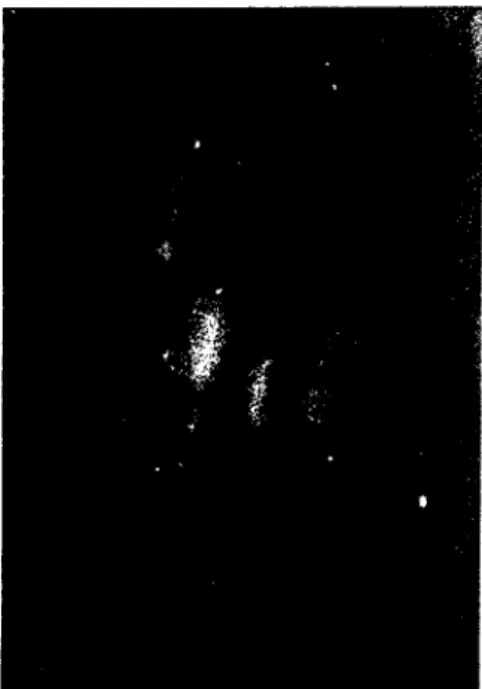
INFECTIONS IN CATTLE WITH AClJTE 183 RESPIRATORY DISEASE BY DIRECT IMMlJNOFLlJORESCENCE TECHNIQlJEantigens (2/5 samples from one herd, 1/6 from the other). The immunofluorescent staining in the nasal swab smears was for the most part dİffuse cytoplasmic. The cells were oval or round in shape, mainly epithelial ( Figure
I).
Figure i .Large clump of fluorescing nasal epithelial eells X
ı
60Discussion
Serological data suggest that bovine adenoviruses are widcspread allover the world and may be causally related to some outbreaks of respiratory disease in eaule. BAdY3 was isolated İn Turkey from caule with respiratory tract disease ( 5). Alkan et aL. (2) reported that serological prevalance rates for BAdY - I, BAdY- 2 and BAdY-3 type infections in caule
in Turkey were 23.7%, 35.2 % and 12.0 %
respectively, indicating that exposure to these vıruses LS common.
Although direct IF for adenovirus antigen İn clinical speeimens has been widely used in human medicine ( 9,
i
3) it is less frequently reported from veterinary diagnosticlaboratories, even though the method has
proved very successful for other bovine
respiratory pathogens (
i I,i
5) Edwards et ai. (7) reported that BAdY antigen positives hadbeen found by direct IF in LO.
i
% of 79respiratory disease outbreaks investigated by Yeterinary Investigation Centres in England
and Wales, while Haralambiev et aL. ( 8)
identified adenovirus antigen by IF in lung lavages from 3/42 pneumonic calves.
Few veterinary laboratories examıne
routinely for adenoviruses in bovine
respiratory disease. The results from this study
suggest it may be worthwhile adding
adenovimses to the range of antigens tested in diagnostic laboratories. By adopting such an
approach the role of adenoviruses in the
bovine respiratory disease complex may
become c1earer.
The other result from this study reveal that direct immunofluorescence technique as a
rapid method, based on the deteetion of
antigen in nasal swab samples, has been used to establish the viral aetiology of acute respiratory disease caused BAdYs subgroup i. The limitation of IF and other immunoassay methods for diagnosis by antigen detectİon is that theyonly the detect the organisım which are specifically sought.
Acknowledgement
i
am grateful to Dr. Steven Edwards forcomments. References
ı.
Adair, B. M., MC Killop, E. R., Smyth,J.
A., Curran, W. L. and MC Nulty, M. S.
e
ı
996). Boviııe adeııovims {'pe LO:properties of viruses isolated Foııı ('(Ises of boviııe haemorrhagic enterocolitis. Vet
Rcc 138. 250-252
2. Alkan,F., Özkul,A. Karaoğlu,M.T., Bilge,S.,
Akça,Y., Burgu,i., Yeşilbağ,K., ve
Oğuzoğlu,T.Ç.
e
1997). Sıp,trlardu Viral Nedenli Solunum Sistemi Enfeksıyonlamun Seroepidemiyolojisi, AÜ Vet Fak Dcrg 44(1),73-80.3. Baker,
J.
C., Werdin, R. E., Ames, T. R.,Markham, R.
J.
F. and Larson, V. L.e
1986) Stud)' 011 the etiologic role of bOi'ine184 4. 5. 6. 7. 8.
9.
LO.respiratoı)' syncytial virus in pneumonia of dair)' calves. JAVMA 189, 66-70
Benkö, M., Bartha, A. and Wadell, G.
(1988) DNA restriction enzyme analysis of hOl'ine adenoviruses. Interviro!ogy 29, 346
Burgu, I, Akça, Y and Şahal, M.( 1991 )
First isolation of bovine adenovirus type-3 in Turkey. ( Short Communication) Dtsch Tierarz Wschr 98,237
Caldow, G. L., Edwards, S., Peters, A. R., Nixon, P., Ibata, G. and Sayers, R. (1993) Associations between viral infections and respiratory disease in artifıcially reared calve.ı.. Vet Rec 133, 85-89
Edwards, S., White, H., Newman, R. H. and Nixon, P. (ı988) A Veterinary Service
Scheme for the rapid diagnosis of Viral infections İn Ruminant, Using
Immunofluorescence. State Vet J 42,
41-47.
Haralambiev, H., Mitov, B., Jarmin, P., Bostandjieva, R. and Simeonov, S.(1987) Examination for viruses of lung lavages from calves with bronchopneumonia: prefiminary results. Acta Vet Hung 35, 475-477.
Lehtomaki, K., Julkunen, 1., Sandelin, K., Saloncn, J., Virtanen, M., Ranki, M. and
Hovi, T. (1986) Rapid diagnosis of
respiratory adenavirus infectimıs in young adult men. J Clin Microbio! 24, I08-11
ı.
\1innich, L. and Ray, C. G. (1980)
Comparison of direct immunofluorescent staininf!, of clinical specimens for respiratory virus antif!,ens with convemional isolation techniqııes. J Clin Microbiol 12, 391-394
FERAY ALKAN
11- Nettleton,P.F., Herring,J.A and
Herring,A.J (1983). Evaıııation of an immunofluorescent test for the rapid diagnosis of field infections of infectious hovine rhinotracheitis Vet Rec 112: 298-300.
12- Pospisil,Z., Rozkosny ,V and M ensik,J.
(1972). Diaf!,nosis of infectious hovine rhinotracheitis by immuno/lııorescence.
Acta Vet Bmo 41: 281-286.
13- Salomon,H.E., GrandienjM., Avila,M.M., Pettersson,C.A. and Weissenbacher,M.C.
(1989) Comparison of three techniques for detection ofr respiratoı)' viruses in nasoplıaryngeal aspirates from children with lower acute respiratoı)' infectioııs. J Med
Virol 28: 159-
ı
62.!4 Stott,E.J., Thomas,L.U., ColIins,A.P.,
Crouch,S.,Jebbet,J., Smith,G.S.,
Luther,P.O. and Caswell,R. (ı 980). A survey of virus infectiollS of the respiratoı)' tract of cattle and their association with disease. J Hygiene (Cambridge) 85, 257-279.
15- Thomas,L.H and Stott,E.J. (
ı
98i ) Diagnosis of respiratoı)' virus infectioıı in the bavine respiratory trael by immUllOjluorescence. Vet Rec 108,432-435Yazar adresi: Doç. Dr. Feray Alkan AÜ Veteriner Fakültesi Viroloji Anabilim Dalı