Murat Durdu,
Cansu Bozca*,
Nazım Emrah Koçer**
Filler Reactions: Case Report and Review
of the Literature
Dolgu Reaksiyonları: Olgu Sunumu ve Literatürün
Gözden Geçirilmesi
Abstract
A gradual increasing interest in cosmetic procedures and the implementation of these practices by non-physicians have led to the development of various complications. Some of these complications are transient, while others require severe systemic treatments, and some may be life-threatening. These complications cause depression and isolated from society. In this article, three different reactions in the light of literature data due to the application of vitamins E, which are not fillers, have been presented and reactions of filler substances and treatment approaches are reviewed.
Keywords: Filler reactions, review, vitamin E, scleredema, paraffinoma, lipogranuloma
Günümüzde kozmetik işlemlere ilginin artması ve bu uygulamaların hekim olmayan kişiler tarafından yapılmaya başlanması çeşitli komplikasyonların gelişimine neden olmuştur. Bu komplikasyonların bazıları geçici olmasına karşın diğerleri ağır sistemik tedaviler gerektirmektedir. Bazı hastalarda hayatı tehdit eden önemli semptomlar gelişebilmektedir. Oluşan bu komplikasyonlar hastalarda depresyona ve toplumdan izolasyona neden olmaktadır. Bu makalede dolgu maddesi olmayan vitamin E içeren ampüllerin uygulanmasına bağlı gelişen üç farklı reaksiyon sunularak literatür verileri ışığında dolgu maddesi reaksiyonları ve tedavi yaklaşımları derlenmiştir.
Anahtar kelimeler: Dolgu reaksiyonları, derleme, E vitamini, sklerödema, parafinoma,
lipogranüloma
Öz
Introduction
Interest in cosmetic procedures is gradually increasing today. Filler substance injection, used to reduce wrinkles and eliminate age-related volume loss, is a type of aesthetic application. Application of filler substances may lead to complications. The complication rate may increase if the procedure is conducted by inexperienced physicians or non-physicians (1). Our study exams complications which developed following filler injection, with potential complications and treatment approaches reviewed as well.
Case Reports
Case 1
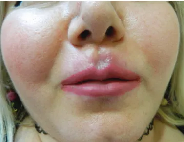
A 41-year-old female patient presented with thickening and hardening of the skin on the lip and perioral region. Her medical
history revealed that vitamin E was injected into her lip area as filler for lip augmentation three months prior by a coiffeur. Swelling, redness and stiffness developed in her lips ten days later, and then spread around her mouth. Oral systemic steroids (methylprednisolone 32 mg/day) were prescribed at another health center for the treatment of the swelling. However, the steroids were discontinued because of the onset of diabetes mellitus following the steroid treatment. Glucose levels returned to normal levels after the steroid therapy was discontinued. Dermatological examination showed non-pitting, indurated plaques in the lips and around the mouth (Figure 1a). The patient had difficulty with speaking and eating. Bloodwork revealed normal complete blood count, liver function tests, thyroid hormones, protein electrophoresis, C-reactive protein,
Başkent University Faculty of Medicine, Adana Hospital, Clinic of Dermatology, Adana, Turkey *Akdeniz University Faculty of Medicine, Department of Dermatology, Antalya, Turkey **Başkent University Faculty of Medicine, Adana Dr. Turgut Noyan Application and Research Center, Clinic of Pathology, Adana, Turkey
©Copyright 2017 by Turkish Society of Dermatology Turkish Journal of Dermatology published
by Galenos Publishing House.
Murat Durdu, Başkent University Faculty of Medicine, Adana Hospital, Clinic of Dermatology, Adana, Turkey Phone: +90 322 327 27 27 E-mail: [email protected] ORCID ID: orcid.org/0000-0003-1247-3932 Submitted/Geliş Tarihi: 14.06.2017 Accepted/Kabul Tarihi: 16.06.2017 Correspondence/ Yazışma Adresi:
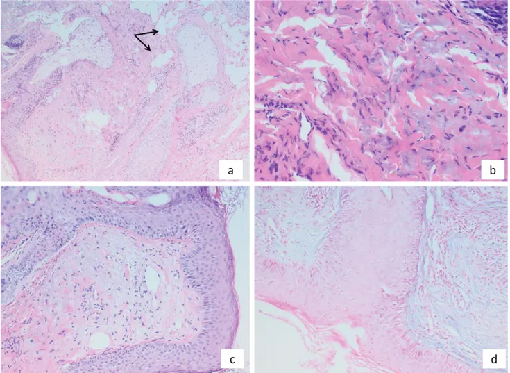
erythrocyte sedimentation rate, anti-streptolysin O and complement levels. Antinuclear antibody, anti-Sjögren’s syndrome-A/Ro, anti- Sjögren’s syndrome-B/La, anti-Sm, anti-Scl-70, rheumatoid factor, anti-ribonucleoprotein and hepatitis serology tests were negative. Histopathological examination showed coarsened and fenestrated collagen bundles in the dermis, mature adipose tissue located in the mid dermis and surrounding the sweat glands, and the presence of perivascular lymphocytic infiltration. In histochemical study, alcian blue pH 2.5 stain revealed acid mucopolysaccharide mucin material separating thickened collagen bundles (Figure 2). A Lupus band test yielded negative results with a direct immunofluorescence test. These clinical and histopathological findings were consistent with the diagnosis of scleredema, and the patient was administered oral methotrexate (15 mg/week) and colchicine 0.5 mg TID. Within a month of initiating this treatment, lip function improved. At the end of the third
month of treatment, the sclerosis completely resolved (Figure 1b).
Case 2
A 33-year-old female patient was admitted with erythematous nodules on her face and thigh of six days’ duration. The patient had injected herself with vitamin E after attending a platelet rich plasma course, and developed nodular lesions 3 weeks after the injection. Dermatological examination revealed multiple erythematous firm nodules on her cheek, forehead and on the anterior surface of her thigh (Figure 3, 4). A punch biopsy was obtained from the plaques. Histopathologic findings were consistent with lipogranuloma. Systemic steroids (methylprednisolone 32 mg/d) and topical steroid treatment were initiated. Facial lesions resolved within one month and the lesions on the thigh within two months. Intralesional steroid injection was applied to a small number of remaining lesions. All lesions developed post-inflammatory pigmentation.
Case 3
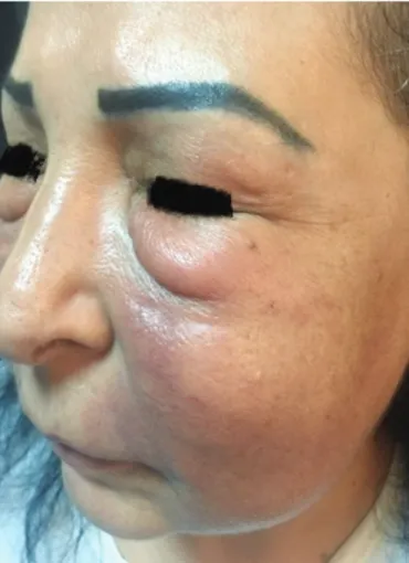
A 51-year-old female patient was admitted with swelling, erythema and stiffness of her face (Figure 5, 6). Her medical history revealed that vitamin E and co-enzyme Q was injected to her lip area as filler one year prior by a coiffeur. Botulinum toxin injection was applied around the eyes at two-week intervals at a different center two months before her current admission. She had experienced burning, tingling and redness in her eyes one week after the last application, and symptoms did not resolve despite hot-cold applications. Redness, swelling and firmness developed in her face, especially in her cheeks. The swelling in her right cheek spontaneously ruptured and drained; however, the swelling increased again thereafter. She was hospitalized, and a punch biopsy obtained from the right malar region revealed foreign body granuloma. She was started on systemic methyl-prednisolone (48 mg/d), ampicillin-sublactam, linezolid, and metronidazole treatment. Erythrocyte sedimentation rate, C-reactive protein levels and edema regressed with these treatments and she was discharged with ciprofloxacin (500 mg bid PO), an analgesic and a tapering dose of steroids. Upon admittance to our clinic, bilateral malar erythema, edema and wood stiff subcutaneous nodules were detected on her physical examination. Laboratory examination revealed a mild leukocytosis (13.800/μL), mild neutrophilia (90.9%), lymphopenia (7.3%), and elevated total IgE (386 IU/mL). Anti-nuclear antibody was negative and C1 inhibitor level was normal. The results were debated at dermatopathology council and systemic methyl-prednisolone (0.75 mg/kg/d), cyclosporine (5 mg/kg/d) and azithromycin (1500 mg/m) treatments were started. Soft tissue magnetic resonance imaging revealed thickness in subcutaneous tissue neighboring the maxillary and mandibular regions bilaterally, in the left preseptal zygomatic region, and hypo-intense regions and contrast in T1-T2. Malar edema and firmness began to regress at day 15 of treatment.
Discussion
Interventional cosmetic procedures administered by physicians other than dermatologists and plastic surgeons, and even
Figure 1a. Sclerotic plaques restricting mouth movements in a patient with scleredema due to vitamin E injection
Figure 1b. Complete recovery of indurated plaques after treatment
Figure 2. a) Histopathology shows fat tissue surrounding the sweat glands in mid dermis (arrows), b,c) thickened and fenestrated collagen bundles in the dermis, d) Alcian blue positive mucin structures (a, H&E x100; b and c, H&E x200; d, Alcian blue x200)
Figure 3. Multiple erythematous nodules on the thigh of a
by non-physicians, lead to significant medical complications. For patients who are seeking to improve their appearances, these complications may lead to depression, suicide attempts and even death (2). A literature review revealed that 77.5% of foreign body reactions associated with cosmetic application developed due to illegal applications (3). We presented three cases with complications secondary to vitamin E injection by a non-physician, with two cases developing complications following treatment by a hairdresser, and another after the patient self-administered vitamin E.
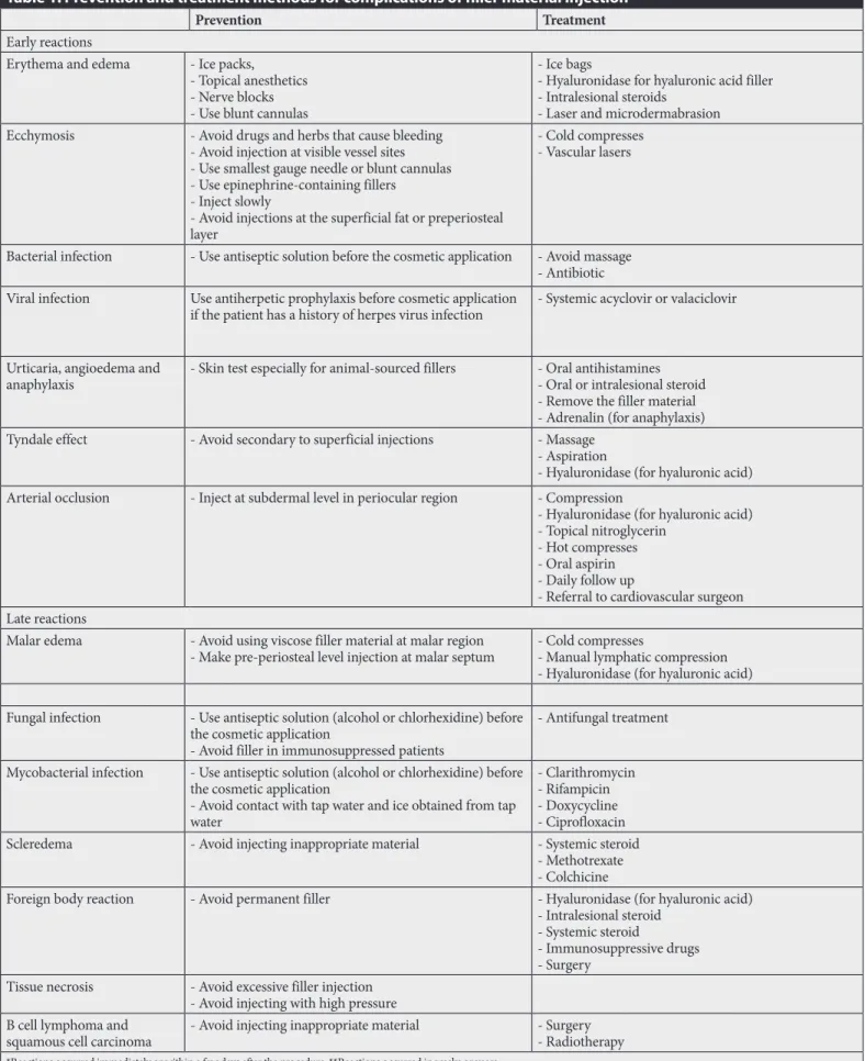
Filler materials are known to cause various cutaneous reactions (Table 1) (4). Temporal erythema, edema and tenderness develop in many patients. These acute reactions are related to application technique rather than the injected substance. These local reactions develop in 93% of the patients who used hyaluronic acid and in 90% of the patients who used collagen (5). Applying ice to the affected region is usually beneficial if a reaction develops. Green tinted makeup may be used to camouflage erythema (4).
Filler injections not only lead to temporal erythema but also cause malar edema, which lasts for days or even months. Malar edema usually develops due to compression on lymphatic vessels as the result of the applications in the
infraorbital region. This complication may be treated with hyaluronidase when hyaluronic acid filler has been utilized. However, treatment is quite difficult with other fillers. Cold compresses or manual lymphatic massage may be beneficial (4).
Ecchymosis due to blood extravasation may develop during filler injection. This reaction is usually self-limiting within 5-10 days. Ecchymosis occurs frequently in the perioral region, the inferior eyelid and the nasolabial sulcus. This complication may be reduced through cessation of analgesics, aspirin, and some vitamins (vitamin E, ginseng, garlic, ginger, gingko, morina liver fat) 10 days before the application, by avoiding injections in regions with visible vessels, by using fine needles or blunt cannulas, and by applying the injections to the superficial fat layer and pre-periosteal levels (4,6). Applying compression with or without ice may reduce ecchymosis. There is no definite evidence that vitamin K and bromalin reduce ecchymosis. Hemoglobin-targeting vascular laser and light sources may be used if ecchymosis persists (4).
Acute erythema, edema and severe itching at filler site are signs of a type 1 allergic reaction. These acute hypersensitivity reactions are quite rare. Local pain, swelling and erythema are seen in mild reactions. A skin test is recommended prior to bovine collagen injections due to the high risk of reaction
Figure 5. Facial swelling, erythema and stiffness on face of
Table 1. Prevention and treatment methods for complications of filler material injection
Prevention Treatment
Early reactions
Erythema and edema - Ice packs, - Topical anesthetics - Nerve blocks - Use blunt cannulas
- Ice bags
- Hyaluronidase for hyaluronic acid filler - Intralesional steroids
- Laser and microdermabrasion Ecchymosis - Avoid drugs and herbs that cause bleeding
- Avoid injection at visible vessel sites - Use smallest gauge needle or blunt cannulas - Use epinephrine-containing fillers - Inject slowly
- Avoid injections at the superficial fat or preperiosteal layer
- Cold compresses - Vascular lasers
Bacterial infection - Use antiseptic solution before the cosmetic application - Avoid massage - Antibiotic Viral infection Use antiherpetic prophylaxis before cosmetic application
if the patient has a history of herpes virus infection - Systemic acyclovir or valaciclovir Urticaria, angioedema and
anaphylaxis - Skin test especially for animal-sourced fillers - Oral antihistamines- Oral or intralesional steroid - Remove the filler material - Adrenalin (for anaphylaxis) Tyndale effect - Avoid secondary to superficial injections - Massage
- Aspiration
- Hyaluronidase (for hyaluronic acid) Arterial occlusion - Inject at subdermal level in periocular region - Compression
- Hyaluronidase (for hyaluronic acid) - Topical nitroglycerin
- Hot compresses - Oral aspirin - Daily follow up
- Referral to cardiovascular surgeon Late reactions
Malar edema - Avoid using viscose filler material at malar region
- Make pre-periosteal level injection at malar septum - Cold compresses- Manual lymphatic compression - Hyaluronidase (for hyaluronic acid) Fungal infection - Use antiseptic solution (alcohol or chlorhexidine) before
the cosmetic application
- Avoid filler in immunosuppressed patients
- Antifungal treatment Mycobacterial infection - Use antiseptic solution (alcohol or chlorhexidine) before
the cosmetic application
- Avoid contact with tap water and ice obtained from tap water
- Clarithromycin - Rifampicin - Doxycycline - Ciprofloxacin Scleredema - Avoid injecting inappropriate material - Systemic steroid
- Methotrexate - Colchicine
Foreign body reaction - Avoid permanent filler - Hyaluronidase (for hyaluronic acid) - Intralesional steroid
- Systemic steroid
- Immunosuppressive drugs - Surgery
Tissue necrosis - Avoid excessive filler injection - Avoid injecting with high pressure B cell lymphoma and
squamous cell carcinoma - Avoid injecting inappropriate material - Surgery- Radiotherapy
(3%). For individuals with a negative skin test result, risk of allergic reaction drops to less than 0.5%. Allergic reaction to hyaluronic acid is quite rare. These reactions usually develop against the proteins in the filler. Most reactions regress with antihistamines, although adrenalin and systemic steroids may be required for individuals who develop angio-edema and anaphylaxis. Emergency kits should be available at the facilities where filler is administered, though these severe reactions are rare (4). Widespread urticarial reaction, serum reaction-like fever, arthralgia and distant rash are reported infrequently in patients who develop a foreign body reaction (7).
Nodules may develop in filler sites in the early or late reaction period. Painless and non-erythematous nodules, seen shortly after filler application, usually develop due to excessive filler substance injection. Hyaluronidase may be used for treatment if hyaluronic acid was initially used as filler. If the nodule has developed due to the use of the other filler materials, the area may be drained through a small incision. Drainage may not be possible if there are extensive nodules (8). For these cases, intralesional 5-fluoro-uracil, a radiofrequency method, is utilized when surgery cannot be performed (4). The risk of developing cutaneous nodules increases with some filler materials. Five millimeter or smaller nodules may develop, particularly after poly-L-lactic acid injection, or rarely after the use of other materials. Filler material should be diluted further (minimally 5 mL), held for 24 h or longer (preferably 72 h) after dilution, kept at room temperature until applied, and applied to the subcutaneous fat layer instead of the dermis to eliminate these reactions (6).
Type 4 hypersensitivity reactions are another cause of nodular lesion development. These foreign body reactions vary depending on the injected filler material. While intradermal collagen and hyaluronic acid fillers may lead to cystic granulomas, subdermal silicon and acrylamide applications form Swiss cheese-like, honeycomb-shaped lipogranulomas (9). Sclerosing type granulomas develop due to subdermal injection of fillers like artecol (10).
A slowly developing erythema and induration is seen in patients who develop cystic-type foreign body reactions. Unlike sclerosing types of lesions, fluctuation develops in all cystic granulomas within 1-3 months. Induration usually resolves spontaneously within one year. The characteristic histopathological finding of cystic type foreign body reactions is foreign body-type giant cells, neutrophils, lymphocytes and macrophages surrounding the necrobiotic implant material. These granulomas may also be defined as collagen cells swimming in a sea of neutrophils (10).
Erythema is seen in patients who develop edematous granulomas. Usually, lymphocytes and macrophages are seen around the foreign material, and giant cells are rarely present. Intralesional steroid treatment is usually recommended for these cases (10).
Sarcoidosis-like or xanthomatous-type sclerosing granulomas develop within 6-24 months following filler injection. Inflammation lasts for years unless these types of granulomas are treated. Slowly growing erythema, swelling and firmness
develop in the whole injection site within a couple of weeks. Histopathologic examination reveals increased fibroblasts and macrophages between microspheres. Finger-type extensions are seen extending into the tissue surrounding the infiltration. The injection material may be identified using a polarized microscope (10).
Filler injection may lead to pigmentation changes in the skin. Post-inflammatory pigmentation may develop following ecchymosis in particularly dark-skinned individuals. Topical hydroquinone and tretinoin cream can be used in addition to sunscreen. A white color change may be observed when bovine collagen is injected superficially as a filler, while superficial injection of hyaluronic acid leads to a blue color change (Tyndale effect). This color change may be treated with massage, aspiration or hyaluronidase (11).
Bacterial, fungal or viral infection may develop secondary to filler injection. These infections may be polymicrobial. Herpes virus infections are the most common viral infection. Acyclovir or valaciclovir prophylaxis should be recommended for the patients who have a history of recurrent herpes virus infection. Additionally, as herpes infections are often secondarily infected with bacteria, antibiotics are usually used in conjunction with the antiviral treatment. Infection-related abscesses may be seen as a solitary lesion or in multiples. Candida infections, which develop in immune-compromised patients, are often difficult to differentiate (8). Infections may initially be confused with local non-infectious reactions. To reduce infection risk, all injection sites should be cleaned with an antiseptic solution like alcohol and chlorhexidine prior to treatment. With any suspicious lesion, antibiotics should be recommended. Any abscess requiring draining should be cultured, with antibiogram results determining the antibiotic selected. Another factor in treating a secondary infection is biofilm formation. Released by some bacteria, a biofilm’s protective film layer can hinder the effect of antibiotics by serving as a protective shield. Atypical mycobacteria should also be eliminated in persistent infections despite two weeks of antibiotic treatment (12).
Filler materials may also lead to vascular complications, which may be local or distant to the injection site. Local reactions may occur from intravascular injection of the filler material, causing thrombosis as well as secondary to excessive compression. Distant vascular complications develop due to intravascular injection of the filler material (4).
Severe pain at the injection site is the most important sign of a local vascular reaction. The skin pales, and capillary filling time prolongs. The skin becomes livedoid spotty before turning cyanotic. Necrosis occurs secondary to permanent ischemia. Ulcers develop with the sloughing of the necrotic layer. Hyperbaric oxygen, systemic antibiotic treatment, massage, vasodilators and supportive treatments are required for cases with vascular complications. Hyaluronidase may be used if hyaluronic acid had been injected. Ocular vessels may become obstructed after injection of filler material into the vessels around the eyes (13). Corneal edema, hyphema and visual impairment may subsequently develop. To prevent
these important complications, the following points should be strictly followed: Vascular structures around the eye should be carefully considered, blunt cannulas should be used preferentially, epinephrine-containing local anesthetics should be utilized for a vasoconstrictive effect, whether the tip of the syringe has entered the vessel should be verified prior to injecting, filler material should be applied gently and slowly with less than 0.1 mL in each region, and hard compression should be avoided after the injection (14). Intravascular injection of the filler material may lead to systemic complications in addition to the local effects. Progressive dyspnea and confusion were reported secondary to facial injection of hyaluronic acid-containing filler material. A subsequent chest tomography revealed a ground glass appearance, but lung biopsy results were consistent with a foreign body granuloma. This complication developed as a result of a non-thrombotic pulmonary embolism and was thought to be due to intravascular and excessive volume injection, as well as inappropriate massage afterwards (15).
Injection of inappropriate materials also leads to complications. The complications seen in our three cases developed due to vitamin E injection. Vitamin E injection-related scleredema was first reported in our case although vitamin E injection-related lipogranuloma and paraffinoma development had previously been reported (1). All three patients required long-term steroid use to resolve their complications. The first patient developed steroid-related diabetes, so methotrexate and colchicine treatment were started and the scleredema lesions completely recovered within three months.
Lipogranulomas may develop due to the traumatic effect of some filler materials on endogenous lipids or from exogenous application of paraffin, mineral oil, vaseline, silicon, vitamin E and autologous fat (16). These exogenous materials may lead to early or late lipogranuloma formation. As seen in our first case, early reactions generally develop within a few weeks of treatment and respond well to steroids. Conversely, reactions to exogenous paraffin develop months or even years (paraffinoma) after treatment. Initially, pain and tenderness develop at the injection site. Depigmentation or a dark yellow color change is observed in the skin. Fibrosis-related induration and restriction in movement develops in advanced stages, and cutaneous necrosis and ulceration may also develop. Sometimes this oily material reaches the lymph nodes and the lungs through lymphatic and hematogenous routes, and death has even been reported. Histopathological examination of affected tissues reveals destruction in subcutaneous fat, with hyaline necrosis in stromal septa. Fibrotic tissue causes the formation of fat globules of different sizes. Endothelial cells, foreign body-type giant cells, macrophages and eosinophilic collagen surround the fatty cysts. Definite treatment of paraffinoma is complete excision, as B-cell lymphoma and squamous cell carcinoma has been reported due to long-term paraffin presence in the skin (17,18). However, most patients do not want surgery due a lack of comesis afterwards. Magnetic resonance imaging (MRI) may be used for exact identification
of the surgical site (19,20). Spotty mass appearance (60%), nodules in fat density (100%), nodular (100%) and ring (80%) calcification are detected on tomography (20). Fibrous plaques gave iso-intensity in T1 weighted images and hypo-intensity in T2 weighted images, while the fluid paraffin component gave hypo-intensity in T1 and T2 weighted images (21). It was reported that hyper-intensity could be observed on an MRI obtained within 3 years of treatment and that hyper-intensity could be noted more than 20 years later. Hypo-intense areas were detected in our paraffinoma case 1.5 years after the initial treatment. Intralesional steroids, systemic corticosteroids, oral tetracycline, CO2 laser, 100% trichlor acetic acid and radiofrequency treatments may be used to reduce the inflammation (16).
Conclusion
Filler material injection is an elective procedure. These cosmetic procedures should be performed by dermatologists or plastic surgeons who can also manage potential complications from these procedures.
Ethic
Informed Consent: A consent form was completed by all
participants.
Peer-review: Internally peer-reviewed.
Authorship Contributions
Surgical and Medical Practices: M.D., C.B., Consept: M.D., C.B., Design: M.D., Data collection or processing: M.D., C.B., Analysis or interpretation: M.D., C.B., Literature search: M.D., C.B., N.E.K., Writing: M.D., C.B.
Conflict of Interest: No conflict of interest was declared by
the authors.
Financial Disclosure: The authors declared that this study
received no financial support.
References
1. Kamouna B, Darlenski R, Kazandjieva J, et al. Complications of injected vitamin E as a filler for lip augmentation: case series and therapeutic approach. Dermatol Ther 2015;28:94-7.
2. Omranifard M, Taheri S. Filler augmentation, safe or unsafe: A case series of severe complications of fillers. J Res Med Sci 2011;16:1627-31.
3. Park TH, Seo SW, Kim JK, et al. Clinical outcome in a series of 173 cases of foreign body granuloma: improved outcomes with a novel surgical technique. J Plast Reconstr Aesthet Surg 2012;65:29-34.
4. Chiang YZ, Pierone G, Al-Niaimi F. Dermal fillers: pathophysiology, prevention and treatment of complications. J Eur Acad Dermatol Venereol 2017;31:405-13.
5. Narins RS, Brandt F, Leyden J, et al. A randomized, double-blind, multicenter comparison of the efficacy and tolerability of Restylane versus Zyplast for the correction of nasolabial folds. Dermatol Surg 2003;29:588-95.
6. Tunca M. Kozmetik uygulama komplikasyonlar. Turkderm 2009;43:17-20. 7. Sorensen EP, Urman C. Cosmetic complications: rare and serious events
following botulinum toxin and soft tissue filler administration. J Drugs Dermatol 2015;14:486-91.
8. DeLorenzi C. Complications of injectable fillers, part I. Aesthet Surg J 2013;33:561-75.
9. Requena L, Requena C, Christensen L, et al. Adverse reactions to injectable soft tissue fillers. J Am Acad Dermatol 2011;64:1-34.
10. Lemperle G, Gauthier-Hazan N, Wolters M, et al. Foreign body granulomas after all injectable dermal fillers: part 1. Possible causes. Plast Reconstr Surg 2009;123:1842-63.
11. Douse-Dean T, Jacob CI. Fast and easy treatment for reduction of the Tyndall effect secondary to cosmetic use of hyaluronic acid. J Drugs Dermatol 2008;7:281-3.
12. Rodriguez JM, Xie YL, Winthrop KL, et al. Mycobacterium chelonae facial infections following injection of dermal filler. Aesthet Surg J 2013;33:265-9. 13. Chen Q, Liu Y, Fan D. Serious vascular complications after nonsurgical
rhinoplasty: A Case Report. Plast Reconstr Surg Glob Open 2016;4:e683. 14. Li X, Du L, Lu JJ. A novel hypothesis of visual loss secondary to cosmetic
facial filler injection. Ann Plast Surg 2015;75:258-60.
15. Jang JG, Hong KS, Choi EY. A case of nonthrombotic pulmonary embolism after facial injection of hyaluronic acid in an illegal cosmetic procedure. Tuberc Respir Dis (Seoul) 2014;77:90-3.
16. Kim MW, Park HS, Yoon HS, Cho S. Late-Onset Complication of Fillers: Paraffinoma of the Lower Eyelids Clinically Mimicking Xanthelasma. Ann Dermatol 2016;28:753-6.
17. Cha JA, Kim B, Lee KA. B cell lymphoma underlying paraffinoma of glabella. J Craniofac Surg 2017;28:798-800.
18. Ko CJ, Sarantopoulos GP, Bhuta S, et al. Scalp paraffinoma underlying squamous cell carcinoma. Arch Pathol Lab Med 2004;128:1171-2.
19. Cormio L, Di Fino G, Scavone C, et al. Magnetic resonance imaging of penile paraffinoma: case report. BMC Med Imaging 2014;14:39.
20. Gu DH, Yoon DY, Chang SK, et al. CT features of foreign body granulomas after cosmetic paraffin injection into the cervicofacial area. Diagn Interv Radiol 2010;16:125-8.
21. Erguvan-Dogan B, Yang WT. Direct injection of paraffin into the breast: mammographic, sonographic, and MRI features of early complications. AJR Am J Roentgenol 2006;186:888-94.