135
©Copyright 2020 by Turkish Society of Nuclear Medicine Molecular Imaging and Radionuclide Therapy published by Galenos Yayınevi.
Interesting Image
Mol Imaging Radionucl Ther 2020;29:135-138
Address for Correspondence: Seval Erhamamcı MD, Baskent University Faculty of Medicine, Ankara; Baskent University İstanbul Hospital, Department of
Nuclear Medicine, İstanbul, Turkey Phone: +90 216 554 15 00 E-mail: [email protected] ORCID ID: orcid.org/0000-0001-5016-4650 Received: 26.04.2020 Accepted: 29.06.2020
Hepatoselüler karsinomun (HSK) ilk belirtisi olarak, kemik metastazının neden olduğu spinal kord kompresyonu ile başvuran 69 yaşında bir erkek hastayı tanımladık. Başlangıç evreleme için, hastaya 18F-florodeoksiglukoz (FDG) pozitron emisyon tomografisi/bilgisayarlı tomografi (PET/BT)
görüntüleme yapıldı. Görüntüleme çok sayıda ekspansil osteolitik kemik lezyonlarında hafif 18F-FDG tutulumu gösterdi ve düşük doz BT’deki
karaciğer lezyonunda kayda değer atipik 18F-FDG tutulumu göstermedi. HSK’da dahil olmak üzere çeşitli tümörlerde potansiyel bir biyolojik marker
olduğu bildirilen prostat spesifik membran antijen (PSMA) ekspresyonunu değerlendirmek için ek bir PET/BT görüntüleme yapıldı. Hem metastatik kemik lezyonlarında hem de primer karaciğer lezyonunda/tümöründe yüksek PSMA tutulumu 68Ga-PSMA PET/BT ile saptandı.
Anahtar kelimeler: Hepatoselüler karsinom, kemik metastazı, PET/BT, 68Ga-PSMA, 18F-FDG
Öz
We have reported here the case of a 69-year-old man who presented with spinal cord compression due to bone metastases as the first manifestation of hepatocellular carcinoma (HCC). For the initial staging, the patient underwent 18F-fluorodeoxyglucose (FDG) positron emission tomography/
computerized tomography (PET/CT) imaging, which demonstrated mild 18F-FDG uptake in the multiple expansile osteolytic bone lesions, but no
remarkable atypical 18F-FDG uptake in the liver lesion on low-doses CT. An additional PET/CT scan was performed to evaluate the prostate-specific
membrane antigen (PSMA) expression, which has recently been reported to be a potential biological marker in a variety of tumors including HCC. High PSMA uptake was recorded in both the metastatic bone lesions and the primary liver lesion/tumor by the 68Ga-PSMA PET/CT.
Keywords: Hepatocellular carcinoma, bone metastases, PET/CT, 68Ga-PSMA, 18F-FDG
Abstract
Seval Erhamamcı
1, Nesrin Aslan
21Başkent University Faculty of Medicine, Ankara; Başkent University İstanbul Hospital, Department of Nuclear Medicine, İstanbul, Turkey 2Neolife Medical Center, Clinic of Nuclear Medicine, İstanbul, Turkey
Hepatoselüler Karsinomda
68Ga-PSMA ve
18F-FDG PET/BT ile Karşılaştırmalı Bulgular
Comparative Findings Between
68
Ga-PSMA and
18
F-FDG PET/CT for
Hepatocellular Carcinoma
136
Erhamamcı and Aslan. PET/CT in Hepatocellular Carcinoma Mol Imaging Radionucl Ther 2020;29:135-138
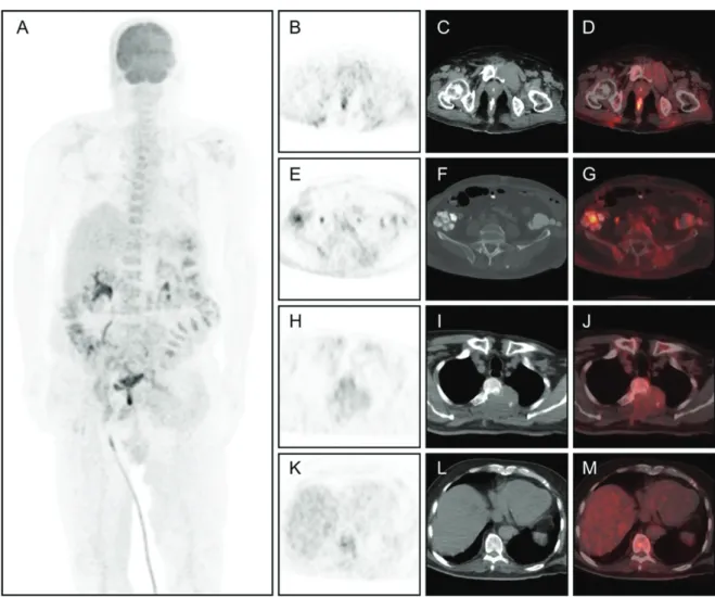
Figure 1. The case patient was a 69-year-old man who presented with the complaint of back pain. His magnetic resonance imaging revealed multiple metastatic lesions of the thoracic vertebral with spinal cord compression and bilateral iliac bones. Excisional biopsy of the T2-3 vertebral lesion due to spinal cord compression was also performed. Histopathological examination demonstrated metastatic malign tumor, which was consistent with the signs of hepatocellular carcinoma (HCC) metastases. The patient accordingly underwent 18F-fluorodeoxyglucose (FDG) positron emission tomography/ computerized tomography (PET/CT) imaging for initial staging. The scan MIP (A), transaxial PET (B, E, H, K), CT (C, F, I, L), and fused (D, G, J, M) images revealed mild uptake [maximum standardized uptake value (SUVmax)= 4.8] in the multiple osteolytic bone lesions in the thoracal vertebra, iliac bones, and sacroiliac joints, most of which also showed remarkable soft tissue components. On the other hand, no significant atypical uptake was noted in the primary liver tumor in the corresponding low-dose CT
137
Erhamamcı and Aslan. PET/CT in Hepatocellular Carcinoma Mol Imaging Radionucl Ther 2020;29:135-138
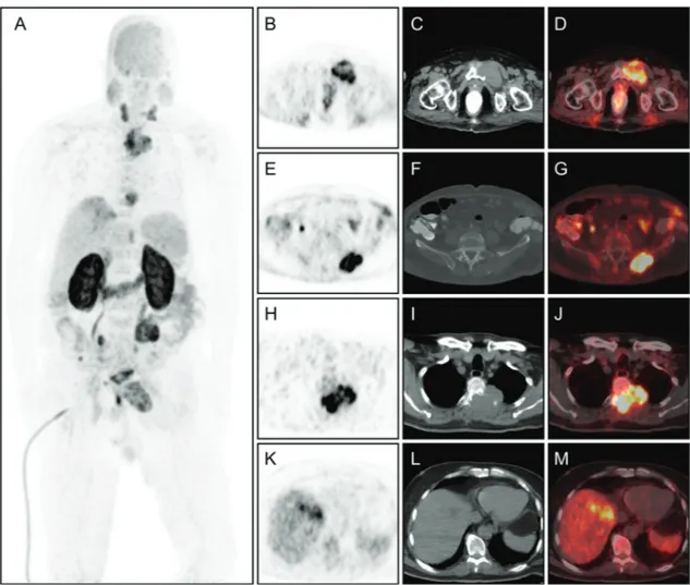
Figure 2. As an alternative PET/CT imaging, 68Ga-prostate-specific membrane antigen (PSMA) PET/CT was performed the same day. Corresponding to
the lesions in 18F-FDG PET, 68Ga-PSMA PET/CT MIP (A), transaxial PET (B, E, H, K), CT (C, F, I, L), and fused (D, G, J, M) images demonstrated high
PSMA expression in the metastatic bone lesions (SUVmax=23.9) and in the primary liver tumor (SUVmax=12.1). No other findings showed metastatic disease elsewhere in the body.
PET/CT imaging with 18F-FDG has low diagnostic accuracy in assessing HCC patients because of its low metabolism (1). 68Ga-PSMA PET/CT is a new
diagnostic technique to image recurrent prostate cancer (2). However, increased PSMA expression has been reported for different non-prostate malignancies, including HCC (3,4,5,6,7,8). There are only a few published documents on the merits of PSMA-PET for HCC (3,4,5,6,7,8). In fact, a few case reports and only 2 studies involving a small sample size has been reported in the recent past. In one of these studies, 68Ga-PSMA was reported to
be superior relative to 18F-FDG for imaging HCC patients (7). However, in another study on advanced HCC patients, the PSMA expression was detected
by 68Ga-PSMA PET, but it was not superior to that by 18F-FDG PET (8). In the current case, 68Ga-PSMA uptake was extremely high as compared to 18F-FDG uptake for bone metastases, and without 18F-FDG uptake in the primary tumor. Therefore, it is suggested that PET imaging with 68Ga-PSMA is
helpful in HCC patients with low FDG affinity. Moreover, we believe that the existence of PSMA expression may act as a guide for radioligand therapy targeting PSMA in the future
Ethics
Informed Consent: Informed consent was taken.
Peer-review: Externally and internally peer-reviewed.
Authorship Contributions
Concept: S.E., Design: S.E., Data Collection or Processing:
S.E., N.A., Analysis or Interpretation: S.E., N.A., Literature
Search: S.E., Writing: S.E.
Conflict of Interest: No conflict of interest was declared
by the authors.
Financial Disclosure: The authors declared that this study
138
Erhamamcı and Aslan. PET/CT in Hepatocellular Carcinoma Mol Imaging Radionucl Ther 2020;29:135-138
References
1. Sacks A, Peller PJ, Surasi DS, Chatburn L, Mercier G, Subramaniam RM. Value of PET/CT in the management of primary hepatobiliary tumors, part 2. AJR Am J Roentgenol 2011;197:260-265.
2. Afshar-Oromieh A, Avtzi E, Giesel FL, Holland-Letz T, Linhart HG, Eder M, Eisenhut M, Boxler S, Hadaschik BA, Kratochwill C, Weichert W, Kopka K, Debus J, Haberkorn U. The diagnostic value of PET/CT imaging with the (68)Ga-labelled PSMA ligand HBED-CC in the diagnosis of recurrent prostate cancer. Eur J Nucl Med Mol Imaging 2015;42:197-209. 3. Erhamamci S, Aslan N. Primary Hepatocellular Carcinoma With Intense
68Ga-PSMA Uptake But Slight 18F-FDG Uptake on PET/CT Imaging Clin Nucl Med 2020; 45:e176-e177.
4. Perez PM, Flavell RR, Kelley RK, Umetsu S, Behr SC. Heterogeneous Uptake of 18F-FDG and 68Ga-PSMA-11 in Hepatocellular Carcinoma. Clin Nucl Med 2019;44:e133-e135.
5. Taneja S, Taneja R, Kashyap V, Jha A, Jena A. 68Ga-PSMA Uptake in Hepatocellular Carcinoma. Clin Nucl Med 2017;42:e69-e70. 6. Sasikumar A, Joy A, Nanabala R, Pillai MRA, Thomas B, Vikraman KR.
68Ga-PSMA PET/CT imaging in primary hepatocellular carcinoma. Eur J Nucl Med Mol Imaging 2016;43:795-796.
7. Kesler M, Levine C, Hershkovitz D, Mishani E, Menachem Y, Lerman H, Zohar Y, Shibolet O, Even-Sapir E. 68Ga-labeled prostate-specific membrane antigen is a novel PET/CT tracer for imaging of hepatocellular carcinoma: a prospective pilot study. J Nucl Med 2019;60:185-191. 8. Kuyumcu S, Has-Simsek D, Iliaz R, Sanli Y, Buyukkaya F, Akyuz F,
Turkmen C. Evidence of Prostate-Specific Membrane Antigen Expression in Hepatocellular Carcinoma Using 68Ga-PSMA PET/CT. Clin Nucl Med 2019;44:702-706.