ORIGINAL RESEARCH
Medicine Science 2018;7(2):378-82
Total hip arthroplasty performed in secondary hip osteoarthritis caused by hartofilakidis
type 2 and type 3 developmental dysplasia of the hip; evaluation of outcomes and
comparison of clinical scores of these two types
Kemalettin Gulbahce1, Aydin Arslan2, Ali Utkan3, Cem Cuneyt Kose3 ,Bulent Ozkurt4
1Ozel Medibafra Hospital, Orthopedics and Traumatology Clinic, Samsun
2 Istanbul Gelişim University, Special Avcılar Anadolu Hospital, Orthopedics and Traumatology Clinic, Istanbul 3Ankara Numune Training and Research Hospital, Orthopedics and Traumatology Clinic, Ankara
4Ankara Numune Training and Research Hospital Orthopedics and Traumatology Clinic, Health Sciences University, Ankara
Received 27 September 2018; Accepted 14 January 2018 Available online 27.04.2018 with doi: 10.5455/medscience.2018.07.8787
Copyright © 2018 by authors and Medicine Science Publishing Inc. Abstract
The aim of this study was to evaluate the outcomes of Total hip arthroplasty (THA) performed for hip osteoarthritis caused by Hartofilakidis type 2 and 3 developmental dysplasia of the hip (DDH) and to compare these two types in terms of clinical scores. Thirty-six hips of thirty patients who suffered from hip osteoarthritis caused by DDH and who underwent THA operations from January 2005 to May 2010 were included in the study. In the case of insufficient coverage of the acetabular cup, roof reconstruction was performed using femoral head autografts. Subtrochanteric transverse femoral osteotomy was performed when required. Clinical evaluations were performed using Harris Hip Score (HHS). 3 male and 27 female patients were included in this study. Mean age was 57.2 (25-76) years. Mean follow up time was 34.3 (12– 65) months. 24 hips were classified as Hartofiladikis type 2, and 12 as type 3. HHS was 43.2 (38-54) points and 90.1 (76-96) points at the preoperative and postoperative last control visits, respectively (p<0.001). Hartofilakidis type 2 hips scored 42.5(38-46) points and 89.9(78-96) points at preoperative and postoperative last control visits, respectively. Hartofilakidis type 3 hips scored 44.7 (38-54) points and 90.4 (76-96) points at preoperative and postoperative last control visits, respectively. There was no difference between the type 2 and type 3 in terms of clinical outcomes (p=0.23, p=0.81). If a proper acetabular reconstruction can be performed by taking into account the restoration of abductor force and the acetabular coverage also by considering acetabular cup position, and if a proper femoral reconstruction can be performed when required via the use of subtrochanteric transverse osteotomy and by considering anatomical changes, successful and similar clinical outcomes can be achieved in hip osteoarthritis secondary to Hartofilakidis type 2 or type 3 DDH.
Keywords: Hip, dysplasia, arthroplasty, hartofilakidis
Medicine Science International Medical Journal
Introduction
In Turkey, secondary osteoarthritis caused by DDH is observed in a considerable number of individuals due to the apparent failure of scanning programs for DDH. THAs performed in hip osteoarthritis secondary to DDH have become a challenge for orthopedic surgeons over the course of many years throughout the world. Although there is a consensus about the indications and techniques associated to the primary osteoarthritis of hip, a debate still continues regarding these topics related to THA in secondary osteoarthritis of hip caused by DDH [1]. Numerous procedures have been defined to treat hip osteoarthritis caused by ignored or insufficiently treated DDH such as osteotomies associated with the pelvis or femur, acetabular roof reconstructions, arthrodesis etc.
*Coresponding Author: Aydin Arslan, Istanbul Gelisim University, Ozel Avcılar
Anadolu Hospital, Orthopedics and Traumatology Clinic, Istanbul
E-mail: [email protected]
However, Total hip arthroplasty (THA) comes to the fore when excessive pain and restriction of range of motion (ROM) are in question [2]. Only the limping or shortness of extremity requires no THA [3].
THA operation in hip osteoarthritis caused by DDH is more difficult than in primary hip osteoarthritis due to anatomical changes in DDH [2,4]. These anatomical changes differ according to the formation of the disease as well as the operations the patient has undergone [2,5]. In case of subluxation; acetabulum is shallowed, wide and elliptic; while anteromedial wall of acetabulum is insufficient, there is a good bone stock in the posterior wall. In the case of dislocation, affected pelvic side is thinner than the other side, all the acetabular walls are weak and quite a few cases demonstrate increased acetabular retroversion [2,6,7]. Significant femoral changes are short femoral neck, small femoral head, increased femoral neck angle and anteversion, posterior transposition of great trochanter and femoral medullar narrowing in isthmus region
doi: 10.5455/medscience.2018.07.8787 Med Science 2018;7(2):378-82
[8]. To regain hip function, the center of hip rotation should be transferred medially, and abductor forces should be transferred laterally [2,5,9-11]. It has been reported that when placing the acetabular cup, an uncovered space of up to 30-40% is acceptable [5]. The aim of this study was to evaluate outcomes of THA in hip osteoarthritis secondary to Hartofilakidis type 2 and 3 DDH and compare these two types in terms of clinical scores.
Material and Method
Thirty-six hips of thirty patients who suffered from hip osteoarthritis caused by DDH and who underwent THA operations from January 2005 to May 2010 were included in the study. ROM of lower extremities, muscle strength, length of the distance between anterior superior iliac spine and medial malleolus were noted. Trendelenburg test was performed. Pelvis, hip, knee and lumbosacral standard radiographies were performed. To decide prosthesis type and operation technique; femoral medulla, femoral cortical thickness, and acetabulum were evaluated through these images. Computerized tomography (CT) was performed in some cases to evaluate the configuration of acetabulum.
Patients were evaluated by using Hartofilakidis classification system [12]. According to this system classification is as follows; type 1: although there is some subluxation femoral head is in acetabulum, there are segmental deficiency of the superior wall and inadequate true acetabulum depth; type2: there are a a false acetabulum superior to the true acetabulum caused by femoral head, a complete absence of the superior wall, inadequate depth of the true acetabulum; type3: femoral head is completely uncovered by the true acetabulum and has migrate superiorly or posteriorly. There is a complete deficiency of the acetabulum and excessive anteversion of the true acetabulum. In the present study Hartofilakidis type 2 and 3 were included.
Low molecular weight heparin (LMWH) was used for thromboembolism prophylaxis. LMWH administration was continued until the 21st postoperative day. Infection prophylaxis was performed using Cefazolin 1000 mg beginning from 45 minutes preoperatively and continued to be administered three times a day until the third postoperative day.
Anterolateral Watson-Jones approach was used in all operations. In the case of insufficient coverage of the acetabular cup, roof reconstruction was performed using femoral head autografts (Figure 1). When operating on hips which necessitated >4 cm femoral lengthening, subtrochanteric transverse femoral osteotomy was performed (Figure 2). Acetabulum was placed in a higher position only when the true acetabular bone stock was determined to be weak as a result of CT.
Ankle pump exercises and isometric quadriceps exercises were commenced on the first postoperative day. After the 2nd postoperative day straight leg raises, sitting on bedside and isotonic quadriceps exercises were performed. Patients were mobilized after the third postoperative day by using a walker, permitting partial weight bearing.
Clinical evaluations were performed using Harris Hip Score (HHS) in the preoperative period and during the last postoperative control visit. 90 to 100 points was assumed as excellent, 80 – 89
points good, 70–79 points medium, and <70 points poor.
In terms of radiography, the acetabular cup was evaluated in terms of localization and medialization. Acetabular angle was measured. Roof reconstructions, coverage of the acetabular cup and femoral component localization were evaluated. Whether there was loosening of components or not was evaluated using Charnley/ DeLee’s [13] zones. Heterotopic ossification was evaluated via Brooker [14] classification.
Figure1. thirty-eight years old woman’s right hip secondary osteoartritis caused by
Hartofilakidis type 2 DDH, (a) preoperative and (b) postoperative anteroposterior radiographs, performed acetabular roof reconstruction using femoral head allograft
Figure2. twenty-five years-old woman’s bilateral hip oste arthritis caused by
Hartofilakidis type 3 DDH (a) anteroposterior radiographs of preoperative and (b) postoperative six months after second operation, subtrochanteric osteotomy fixed via cable and plate
Statistical analyses
SPSS Statistics for Windows, Version 17.0. (Chicago: SPSS Inc) was used for statistical evaluations. Shapiro-Wilk test was used to determine whether the data were normally distributed or not. If the data was normally distributed, the paired sample t-test was utilized. Two related sample t-test was used in the case of abnormally distributed data. A p-value of <0.05 was considered to be statistically significant.
Results
There were 3 male and 27 female patients included in this study. Mean age was 57.2 (25-76) years. Mean follow up time was 34.3 (12– 65) months. 24 hips were classified as Hartofiladikis type 2, and 12 hips were classified as type 3. Mean hospitalization time was 10.5 (5-22) days.
Mean HHS was 43.2 (38-54) points and 90.1 (76-96) points at preoperative and postoperative last control visits, respectively. (p<0.001) Hartofilakidis type 2 hips scored 42.5(38-46) points and 89.9(78-96) points at preoperative and postoperative last control visits, respectively. Hartofilakidis type 3 hips scored 44.7 (38-54) points and 90.4 (76-96) points at preoperative and postoperative
last control visits, respectively. There was no difference between the two types in terms of clinical outcomes (preoperative comparision; p=0.23, postoperative comparison; p=0.81) (Table 1). Considering all hips; 24 (66.6%) excellent, 10 (27.7%) good, 2 (5.7%) medium results were obtained.
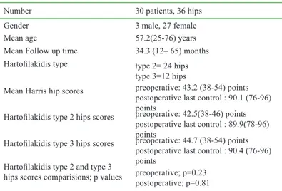
Table 1. Patients’ characteristics and comperative outcomes
Number 30 patients, 36 hips Gender 3 male, 27 female Mean age 57.2(25-76) years Mean Follow up time 34.3 (12– 65) months Hartofilakidis type type 2= 24 hips
type 3=12 hips
Mean Harris hip scores preoperative: 43.2 (38-54) points postoperative last control : 90.1 (76-96) points
Hartofilakidis type 2 hips scores preoperative: 42.5(38-46) points postoperative last control : 89.9(78-96) points
Hartofilakidis type 3 hips scores preoperative: 44.7 (38-54) points postoperative last control : 90.4 (76-96) points
Hartofilakidis type 2 and type 3
hips scores comparisions; p values preoperative; p=0.23postoperative; p=0.81
28 acetabular cups were placed at the anatomic localization. However, 8 acetabular cups were placed at a higher position. Distance between the acetabular cup distal edge and the tear drop figure of these eight hips was 34 (23-45) mm. There were seven hips which had insufficient coverage of acetabular cup. Roof reconstruction was established using femoral head autograft and screws for these hips. Five of these seven hips were classified as Hartofilakidis type 2, and the remaining two were type 3. During the follow up period there was no complication about these roof reconstructions.
Mean acetabular cup medialization was 22.1 (0-38) mm. Mean acetabular cup angle was 46.4° (34-64). Mean coverage of the acetabular cup was %69 (55-95). Femoral subtrochanteric transverse osteotomy was applied to 7 (19%) hips. All these hips were type 3. Mean femoral shortening was 3.8 (3-5) cm. All these osteotomies healed without complications. Mean leg length discrepancy was 2.8 cm preoperatively. At the last control visit this mean difference was 1.5 cm. Trendelenburg test was positive in all patients preoperatively, but at the postoperative period only three (10%) of patients displayed Trendelenburg positivity.
Twelve patients had complications. Four of them had complications during operation and eight of them had complications during the postoperative period. Femoral fissure occurred in three hips and was fixed with cable. Weight bearing was prohibited for these patients for six weeks. Neurapraxia of the peroneal branch of the sciatic nerve occurred in one patient and was found to have healed at the three-month follow up visit. Deep vein thrombosis was observed in two patients. Pulmonary thromboembolism occurred in one of these patients. These two patients were treated successfully. Heterotrophic ossification was observed in 3 (8%) hips. According to Brooker classification, two hips were type 2, and one was type 3. Deep infection was not observed in any of the hips. Superficial infection was observed in two patients. These patients were healed using antibiotics and dressing. One hip dislocated on the 50th postoperative day. This was treated via closed reduction and immobilization. With reference to Charnley’s/De Lee’s zones;
two hips demonstrated radiolucent line in zone I and one hip in zone III. Femoral loosening was not observed in any of the hips. Discussion
It has been reported that there is a positive correlation between the dysplasia grade and clinical outcomes [15]. However, Sochart and Porter [16] reported that there was no correlation between the dysplasia grade and clinical outcomes. Ermis et al. [10] reported that the average HHS of 27 hips classified as Hartofilakidis type 2 was 48.4 points preoperatively and 87.4 points postoperatively; the average HHS of 18 hips classified as Hartofilakidis type 3 was 45.2 points preoperatively and 83.2 postoperatively. These authors suggested that there was no difference in terms of clinical outcomes between these groups. In the present study, the average HHS of 24 hips classified as Hartofilakidis type 2 was 45.2 points preoperatively and 89.9 points postoperatively, and the average HHS of 12 hips classified as Hartofilkakidis type 3 was 44.7 points preoperatively and 90.4 points postoperatively. There was no difference between the two types in terms of preoperative and postoperative HHS.
Stability and sufficient coverage of the acetabular cup are important technical issues for these patients. Generally, the anatomical acetabular localization is the best place in terms of bone stock [17]. Current techniques for acetabular reconstruction are as follows: standard or custom-made metal augments, roof reconstruction with vascularized fibula or pedicled iliac graft, autologous or homologous bone, reinforcement rings, cranial positioning of the acetabulum, medial protrusion techniques (cotyloplasty) [2]. It has been reported that an 80% intact bone coverage is required for a stable acetabular reconstruction [18,19]. However, 30-40% uncovarage was reported acceptable in other studies [2,5]. In the present study, the mean coverage of acetabular cup was 69% (55-95). 28 acetabular cups were placed at the anatomical localization and the remaining 8 were placed at a higher position. Tatar et al. [9] placed the acetabular cup at a higher position in 6 of 53 patients. These authors found the mean distance between the tear drop figure and the distal edge of the acetabular cup to be 23 (14-36) mm. In the present study, this mean distance was 34 (23-45) mm. It has been reported that the acetabular cup angle that is assumed to be safe about prevention of hip dislocation is 47° [20]. In the current study, the mean acetabular cup angle was 46.4° (34-64). Tatar et al. [9] performed roof reconstructions with autologous bone graft in 6 of 53 patients. These authors reported that all bone grafts had healed at the six-month control visit. In the present study, 7 acetabula were reconstructed with femoral head autograft and screws and all of them had healed at the 6 months follow up visit.
Restoration of abductor forces is another critical issue. Although the hip rotational center is transferred to medial and inferior, there may be a residual limping due to pelvic tilt and/or leg length discrepancy [21,22]. In the current study, average medialization of the acetabular cup was 22,1(0-38) mm. Trendelenburg test results were positive for all patients preoperatively. At the last follow up visit, only 10% of all patients demonstrated positivity.
It has been reported that a lengthening of above 4 cm increases the risk of sciatic nerve lesion [23]. Hence, femoral osteotomy techniques are used to prevent sciatic nerve lesions. In addition,
using these techniques allows the restoration of femoral anteversion and abductor mechanism [2]. The current techniques for femoral reconstruction include distraction by external fixator, trochanteric osteotomies, Paavilainen osteotomy, osteotomy of the lesser trochanter, double chevron osteotomy, subtrochanteric osteotomies, diaphyseal osteotomies, osteotomies of the distal femur etc. [9]. In the present study, the measure of lengthening was under 4 cm. Subtrochanteric transverse osteotomy was performed on 7 hips and these were fixed using cable and plates. We prefer subtrochanteric transverse osteotomy due to the following advantages: it allows good fixation at the metaphyseal region; in some patients there is a narrowed intramedullary canal, with this osteotomy this portion can be removed; blood supply of the proximal fragment is preserved due to protected abductor insertions; generally, no nonunion is observed and sufficient metaphyseal support to keep prosthesis is supplied [2,24].
Complication rate of THA in secondary osteoarthritis caused by DDH is higher than the complication rate in primary osteoarthritis [25]. Due to femoral anatomical changes in dysplastic hips, fissures or fractures can occur more easily. In the present study, femoral fissures occurred in three hips and these were fixed using cables. After THA, sciatic nerve lesion is observed at a rate of 0.5-2% [26,27]. This risk increases up to 3-15% in the DDH cases [28]. In the current study, peroneal branch of sciatic nerve was damaged in one patient. At the first year follow up visit this patient had healed. Infection rate of THA in secondary osteoarthritis caused by DDH is higher than the infection rate in primary osteoarthritis. In the current study two superficial infections were observed and treated using dressing and antibiotics. Hip dislocation rate of THA in secondary osteoarthritis caused by DDH is about 5-11%. In the case of first time dislocation, successful results were reported with closed reduction [29]. In the current study, one dislocation that occurred on the 50th day was treated successfully via closed reduction. Incidence of heterotopic ossification is higher in the male sex. Using cementless implants and DDH increases the risk of heterotopic ossification. In the current study there were 3 heterotopic ossification cases. Two were classified as Brooker type II and one was Brooker type III.
We have faced two major limitations in this study. Firstly, the small number of patients in this study due to the fact that only hartofilakidis type 2 and 3 DDH patients were included made the statistical evaluations difficult. Secondly, the present study does not demonstrate long-term results.
Conclusion
In conclusion, if a proper acetabular reconstruction can be performed by taking into account the restoration of abductor forces and the acetabular coverage also by considering acetabular cup position, and a proper femoral reconstruction can be performed when required via the use of subtrochanteric transverse osteotomy and by considering anatomical changes, successful and similar clinical outcomes can be achieved in secondary hip osteoarthritis caused by Hartofilakidis type 2 or type 3 DDH.
Competing interests
No conflict of interest was declared by the authors Financial Disclosure
The financial support for this study was provided by the investigators themselves.
References
1. Anwar MM, Sugano N, Masuhara K, et al. Total hip arthroplasty in the neglected congenital dislocation of the hip. A five to 14-year follow-up study. Clin. Orthop. 1993;295:127-34
2. Yang S, Cui Q. Total hip arthroplasty in developmental dysplasia of the hip: Review of anatomy, techniques and outcomes. World J Orthop. 2012;3:42-8 3. Bicanic G, Barbaric K, Bohacek I, et al. Current concept in dysplastic hip
arthroplasty: Techniques for acetabular and femoral reconstruction. World J Orthop. 2014;5:412-24
4. Ishida K. Prevention of the development of the typical dislocation of the hip. Clin Orthop 1977;126:167-9
5. Delimar D, Bicanic G, Korzinek K. Femoral shortening during hip arthroplasty through a modified lateral approach. Clin Orthop Relat Res. 2008;466:1954-8.
6. Kumar JN, Kumar JS, Wang VT, et al. Medium-term outcome of total hip replacement for dysplastic hips in Singapore. J Orthop Surg. 2010;18:296-302.
7. Steppacher SD, Tannast M, Werlen S, et al. Femoral morphology differs between deficient and excessive acetabular coverage. Clin Orthop Relat Res. 2008;466:782-90.
8. Li PL, Ganz R. Morphologic features of congenital acetabular dysplasia: one in six is retroverted. Clin Orthop Relat Res. 2003;416:245-53.
9. Noble PC, Kamaric E, Sugano N, et al. Three-dimensional shape of the dysplastic femur: implications for THR. Clin Orthop Relat Res 2003;417:27-40.
10. Tatar O, Ersen O, Bilgic S, ark. Gelişimsel kalça displazisine bağlı koksartrozlu hastalarda total kalça artroplasti sonuçlarımız. Gulhane Med J. 2015; 57:361-66.
11. Ermiş MN, Dilaveroğlu B, Erçeltik O, et al. Intermediate-term results after uncemented total hip arthroplasty for the treatment of developmental dysplasia of the hip. Eklem Hastalik Cerrahisi. 2010;211:15-22.
12. Bombelli R, Santore RF, Poss R. Mechanics of the normal and osteoarthritic hip. A new perspective. Clin. Orthop. 1984;182:69-78.
13. Yiannakopoulos CK, Xenakis T, Karachalios T, et al. Reliability and validity of the Hartofilakidis classification system of congenital hip disease in adults. International Orthopaedics. 2009;33:353-8.
14. DeLee JG, Charnley J. Radiological demarcation of cemented sockets in total hip replacement. Clin Orthop Relat Res. 1976;121:20-32.
15. Brooker AF, Bowerman JW, Robinson RA, et al. Ectopic ossification following total hip replacement. Incidence and a method of classification. J Bone Joint Surg Am. 1973;55:1629–32.
16. Cameron HU, Botsford DJ, Park YS. Influence of the Crowe rating on the outcome of total hip arthroplasty in congenital dip dysplasia. J Atrhrolasty. 1996;11:582-7.
17. Sochart DH, Porter ML. The long-term results of Charnley low-friction arthroplasty in young patients who have congenital dislocation, degenerative osteoarthrosis or rheumatoid arthritis. J Bone Joint Surg Am. 1997;79:1599-617. 18. Mendes DG, Said MS, Aslan K. Classification of adult congenital hip
dysplasia for total hip arthroplasty. Orthopaedics 1996;19:881-7.
19. Yoo JJ, Yoon HJ, Yoon PW, et al. Medial placement of the acetabular component in an alumina-on-alumina total hip arthroplasty: a comparative study with propensity score matching. Arch Orthop Trauma Surg. 2013;133:413-9.
20. Harris WH, Crothers O, Oh I. Total hip replacement and femoral head bone grafting for severe acetabular deficiency in adults. J Bone Joint Surg Am.1977;59:752-9.
21. Sharp IK. Acetabular dysplasia: the acetabular angle. J Bone Joint Surg Br. 1961;43:268–72.
22. Kobayashi S, Saito N, Nawata M, et al. Total hip arthroplasty with bulk femoral head autograft for acetabular reconstruction in developmental dysplasia of the hip. J Bone Joint Surg Am, 2003;85:615-21.
23. Rozkydal Z1, Janícek P, Smíd Z. Total hip replacement with the CLS expansion shell and a structural femoral head autograft for patients with congenital hip disease. J Bone Joint Surg Am. 2005;87:801-7.
24. Lewallen DG. Neurovascular injury associated with hip arthroplasty. Instr Course Lect. 1998;47:275-83.
25. Masonis JL, Patel JV, Miu A, et al. Subtrochanteric shortening and derotational osteotomy in primary total hip arthroplasty for patients with severe hip dysplasia: 5-year follow-up. J Arthroplasty. 2003;18:68-73.
26. Pagnano W1, Hanssen AD, Lewallen DG, et al. The effect of superior placement of the acetabular component on the rate of loosening after total hip arthroplasty. J Bone Joint Surg Am. 1996;78:1004-14.
27. Schmalzried TP, Amstutz HC, Dorey FJ. Nerve palsy associated with total hip replacement. Risk factors and prognosis. J Bone Joint Surg Am. 1991;73:1074-80.
28. Nercessian OA, Macaulay W, Stinchfield FE. Peripheral neuropathies following total hip arthroplasty. J Arthroplasty. 1994;9:645-51.
29. Boos N, Krushell R, Ganz R, et al. Total hip arthroplasty after previous proximal femoral osteotomy. J Bone Joint Surg Br. 1997;79:247-53. 30. Leichtle UG, Leichtle CI, Taslaci F, et al. Dislocation after total hip
arthroplasty: risk factors and treatment options. Acta Orthop Traumatol Turc. 2013;47:96-103.