OCULOPLASTICS AND ORBIT
Intraocular pressure and superior ophthalmic vein blood
flow velocity in Graves’ orbitopathy: relation
with the clinical features
Onur Konuk&Zafer Onaran&Suna Ozhan Oktar&
Cem Yucel&Mehmet Unal
Received: 10 March 2009 / Accepted: 27 June 2009 / Published online: 16 July 2009
# Springer-Verlag 2009
Abstract
Background The aim of this study is to evaluate the association of intraocular pressure (IOP) and superior ophthalmic vein blood flow velocity (SOV-BFV) with the clinical features of Graves’ orbitopathy.
Methods During the 2002-2007 period, 66 eyes of 34 Graves’ orbitopathy cases were classified as mild, moderate and severe orbital disease, and evaluated according to their clinical features as: i)type 1 vs type 2 cases, and ii) cases with or without dysthyroid optic neuropathy. In all patients, a full ophthalmic examination including IOP and Hertel
measurements was performed. SOV-BFV was analyzed with color Doppler sonography.
Results The Hertel value, IOP in primary and upgaze position were higher, and SOV-BFV was lower in moderate and severe Graves’ orbitopathy cases that showed statistical significance from mild cases, and controls (p = 0.001). Moderate and severe Graves’ orbitopathy cases showed comparable Hertel measures and IOP in primary and upgaze position (p = 0.39); however, SOV-BFV was significantly lower in severe cases when compared to moderate cases (p = 0.001).This study demon-strated statistically significant negative correlation between IOP in both primary (r = 0.43,p = 0.008) and upgaze position (r = 0.51,p = 0.002), and SOV-BFV. Additionally, statistically significant positive correlation was detected between Hertel values and SOV-BFV(r = 0.402,p = 0.007).There was a statistical difference between type 1 and 2 cases in Hertel values(p = 0.006), IOP in upgaze position (p = 0.026) and SOV-BFV (p = 0.003). SOV-BFV of the eyes showing dysthyroid optic neuropathy was statistically lower than eyes without dysthyroid optic neuropathy (p = 0.006).
Conclusions IOP and SOV-BFV have significant associa-tion with the clinical features of Graves’ orbitopathy. The decrease in SOV-BFV increases the severity of Graves’ orbitopathy, and may have a role in the clinical course of dysthyroid optic neuropathy.
Keywords Graves’ orbitopathy . Intraocular pressure . Superior ophthalmic vein blood flow velocity . Dysthyroid optic neuropathy
Introduction
The characteristic clinical features of Graves’ orbitopathy include eyelid retraction, exophthalmos, strabismus and
None of the authors has a financial or proprietary interest in any method or material mentioned.
The authors have full control of all primary data ,and they agree to allow Graefe’s Archive for Clinical and Experimental Ophthalmology to review their data upon request
This study was presented in the 25th European Society of Ophthalmic Plastic and Reconstructive Surgery Meeting, 13 - 16 September, 2007, Ljubljana, Slovenia
O. Konuk
:
M. UnalDepartment of Ophthalmology, Gazi University School of Medicine, Ankara, Turkey
Z. Onaran
Department of Ophthalmology,
Kirikkale University School of Medicine, Kirikkale, Turkey
S. Ozhan Oktar
:
C. YucelDepartment of Radiology, Gazi University School of Medicine, Ankara, Turkey
O. Konuk (*)
441. cadde 437. sokak 3-3 Birlik Mah, TR-06610 Cankaya, Ankara, Turkey e-mail: [email protected]
optic neuropathy. In cases demonstrating severe Graves’ orbitopathy, additional findings such as elevated intraocular pressure (IOP) and visual field defects may be encountered. These clinical findings are common both in primary open-angle glaucoma and Graves’ orbitopathy, causing diagnos-tic and therapeudiagnos-tic challenges between two diseases [1–7]. In most of the cases with Graves’ orbitopathy, the rise in IOP is gaze-dependent, but sustained elevations of IOP may require further evaluation for treatment [1,6]. The rise in IOP is attributed to restriction and compression of the globe by enlarged extraocular muscles, accumulation of muco-polysaccharide deposits in the trabecular meshwork, and elevated episcleral venous pressure due to reduction of orbital venous drainage [8–10]. However, the relationship between IOP, SOV-BFV and the clinical features of Graves’ orbitopathy is still unclear. The aim of this study is to evaluate the association of the IOP levels and superior ophthalmic vein blood flow velocity (SOV-BFV) with clinical features of Graves’ orbitopathy.
Materials and methods
During the 2002-2007 period, 66 eyes of 34 Graves’ orbitopathy cases (12 male, 22 female) were consecutively evaluated according to the European Group on Graves’ Orbitopathy (EUGOGO) study and classified as mild, moderate and severe orbital disease. In this study, mild disease was defined as minimal to moderate soft-tissue swelling, proptosis <25 mm, no or only intermittent diplopia, no corneal or optic nerve involvement. Moderate disease was defined as marked soft-tissue swelling, and/or proptosis ≥25 mm, and/or inconstant diplopia, and/or punctate staining of the cornea, but no optic nerve involvement. Severe eye disease was defined as constant diplopia and/or optic nerve involvement [11]. In all patients, basic clinical data including age, gender, disease duration, history of steroid treatment and smoking habit were investigated. A full ophthalmic examination, includ-ing pupillary responses, best corrected visual acuity, color vision with Ishihara color plates, biomicroscopy, fundo-scopy, and visual field analysis with Humphrey automated visual field analyzer (program 30-2) were performed. Axial proptosis was measured with Hertel exophthalmometry, and IOP was measured with the same Goldmann applanation tonometer at 9 a.m. The disease activity of the cases was evaluated by the Clinical Activity Score (CAS) [12]. The CAS consists of seven items, and one point is added for each item present: spontaneous pain behind the globe, pain on attempted upgaze, redness of the conjunctiva, redness of the eyelid, chemosis, swelling of the caruncule, and eye lid swelling. A CAS score≥4 was classified as active disease. The cases demonstrating moderate or severe Graves’
orbitopathy were further evaluated in detail according to their clinical features as: i) type 1 vs type 2 cases [13] (type 1 cases included higher degrees of proptosis with orbital fat volume increase, and type 2 cases included cases with optic neuropathy and limited extraocular muscle functions), and ii) cases with or without dysthyroid optic neuropathy. The patients demonstrating the following conditions were regarded as dysthyroid optic neuropathy: decreased best-corrected visual acuity <0.6 on Snellen chart associated with changes in the visual field examination compatible with optic neuropathy, and/or decreased color vision with the Ishihara color test. Presence of optic disc edema on fundoscopic examination and/or apical crowding on com-puterized tomography (CT) or magnetic resonance imaging (MRI) of the orbit supported the diagnosis [14].
All cases were euthyroid in both clinical and laboratory examinations (free T3and free T4were within the normal
range, TSH was low or within the normal range) for at least 6 months before examinations. The cases demonstrating hyperthyroidism or hypothyroidism were excluded from the study, since uncontrolled hormonal status might affect the IOP and SOV-BFV. Similarly, the cases who demon-strated acute orbital inflammation and necessitated early corticosteroid treatment or who had received steroid treatment during the last 6 months were also excluded, since orbital inflammation and steroid treatment might also change SOV-BFV and IOP levels, regardless of the severity of the orbital disease. Twenty eyes of ten patients (five male, five female) without any history of thyroid or autoimmune disease were studied as controls.
The radiological evaluations of the orbits were done by CT and/or MRI, and SOV-BFV was analyzed by color Doppler sonography. Sonograms were obtained with an HDI 5000 SonoCT system (Philips, Bothell, WA, USA) using a 5-12 MHz multifrequency linear array transducer. The eyes were imaged with the patient lying supine, eyes closed and gaze directed toward ceiling. The transducer was applied to the closed upper eyelid using a thick layer of acoustic gel by resting the examiner’s hand on the orbital margin to minimize the pressure on the globe. Color Doppler sonography was performed with low pulse repetition frequency, high Doppler gain, low wall filter settings to allow detection of low velocities. At least three readings were obtained at the same sitting, and the average measure was regarded as the final value. To obtain Doppler spectra, a fixed sample volume of 2 mm within the vessel was chosen while examining the color flow image. Spectral sampling throughout the vessel was performed, and maximum systolic velocity was recorded after appropriate angle correction. The sonograms were digitally recorded in the hard disc of the sonographic unit. All examinations were performed by a single radiologist in a blinded fashion. The statistical significance between groups was evaluated
by the Kruskal–Wallis test and Mann–Whitney U-test, and the correlations were evaluated with the Spearman test. Statistical significance was set at the 0.05 level for the corrected p-value. We certify that all applicable institutional and governmental regulations concerning the ethical use of human volunteers were followed during this research. The data accumulation was in conformity with the Institutional Review Board of Gazi University Medical School, and the study was in adherence to the tenets of the Declaration of Helsinki.
Results
Demographic data
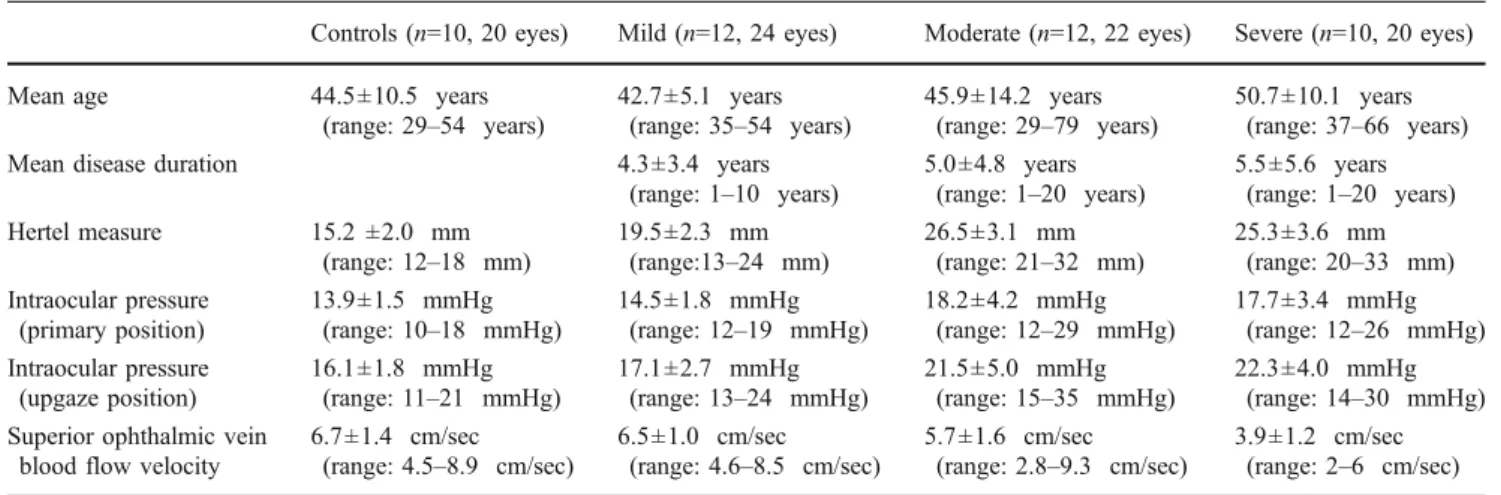
In the study group, 12 cases (24 eyes) showed clinical features of mild, 12 cases (22 eyes) showed moderate, and the remaining ten cases (20 eyes) showed severe orbitop-athy. The mean ± SD ages and disease durations of the cases did not show statistically significant difference (p = 0.47) (Table1). All of the cases demonstrated clinical features of inactive disease, and the mean ± SD CAS was 1.71±1.11 (range: 0–3) at the time of the study. Of the cases with moderate Graves’ orbitopathy, 17 eyes (77.2%) showed clinical features of type 1 disease, and five eyes (22.7%) showed type 2 disease. All cases with severe Graves’ orbitopathy showed features of type 2 disease. In the study cohort, 15 eyes (2.2%, 15/66 eyes) showed dysthyroid optic neuropathy.
None of the cases had a history of primary open-angle glaucoma, and they had not received antiglaucomatous treatment before the diagnosis of Graves’ orbitopathy. In the study group, 64.6% (19 of 34 cases) of the cases had a history of systemic steroid treatment, and the mean ± SD period between the discontinuation of steroid therapy and clinical evaluation was 12.0±3.5 months (range: 9–15 months). None
of the patients had a history of orbital radiotherapy. All the patients had received anti-thyroid drugs, and two patients had received radioactive iodine for management of hyperthyroid-ism. Smoking history was positive for 64.7% (six cases in mild, seven cases in moderate and nine cases in severe group, total 22 of 34 cases, p = 0.52) of the cases, and 16 of them were current smokers.
Clinical data
The mean ± SD Hertel values, IOP values in primary and upgaze position, and SOV-BFV are given in Table 1. The mean ± SD Hertel value, IOP in primary and upgaze position were higher, and SOV-BFV was lower in moderate and severe Graves’ orbitopathy cases that showed statistical significance from mild cases, and controls (p = 0.001). Moderate and severe Graves’ orbitopathy cases showed comparable mean±SD Hertel value, IOP in primary and upgaze position (p:0.39), however SOV-BFV was significantly lower in severe Graves’ orbitopathy cases when compared to moder-ate cases (p = 0.001). These data were not statistically significant between mild Graves’ orbitopathy cases and controls (p = 0.42). All but two cases showed symmetrical orbital involvement. Two cases showed unilateral moderate orbital disease. The IOP and SOV-BFV showed no statistical difference between two orbits of the patients having symmetrical orbital disease which demonstrated the predict-ability of SOV-BFV values regarding the clinical features of Graves’ orbitopathy (p = 0.43). This study demonstrated statistically significant negative correlation between IOP in both primary (r = -0.43, p = 0.008) and upgaze position (r = -0.51, p = 0.002) and SOV-BFV. Additionally, a statistically significant positive correlation was detected between Hertel values and SOV-BFV (r = 0.402, p = 0.007). There was no correlation between the Hertel values and the IOP in primary and upgaze position (p = 0.29).
Table 1 Demographic data of the study group
Controls (n=10, 20 eyes) Mild (n=12, 24 eyes) Moderate (n=12, 22 eyes) Severe (n=10, 20 eyes)
Mean age 44.5±10.5 years
(range: 29–54 years) 42.7±5.1 years(range: 35–54 years) 45.9±14.2 years(range: 29–79 years) 50.7±10.1 years(range: 37–66 years)
Mean disease duration 4.3±3.4 years
(range: 1–10 years) 5.0±4.8 years(range: 1–20 years) 5.5±5.6 years(range: 1–20 years) Hertel measure 15.2 ±2.0 mm
(range: 12–18 mm) 19.5±2.3 mm(range:13–24 mm) 26.5±3.1 mm(range: 21–32 mm) 25.3±3.6 mm(range: 20–33 mm) Intraocular pressure
(primary position)
13.9±1.5 mmHg
(range: 10–18 mmHg) 14.5±1.8 mmHg(range: 12–19 mmHg) 18.2±4.2 mmHg(range: 12–29 mmHg) 17.7±3.4 mmHg(range: 12–26 mmHg) Intraocular pressure
(upgaze position)
16.1±1.8 mmHg
(range: 11–21 mmHg) 17.1±2.7 mmHg(range: 13–24 mmHg) 21.5±5.0 mmHg(range: 15–35 mmHg) 22.3±4.0 mmHg(range: 14–30 mmHg) Superior ophthalmic vein
blood flow velocity
6.7±1.4 cm/sec
Type 1 vs type 2 cases
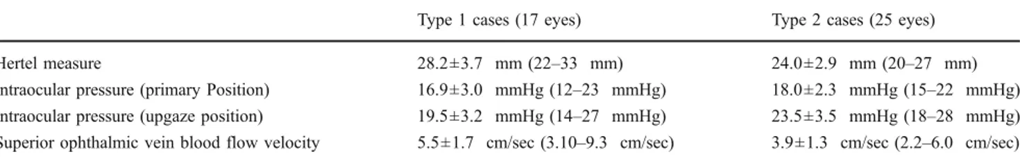
Preoperative Hertel values, IOP levels and SOV-BFV of cases with moderate and severe Graves’ orbitopathy were further evaluated as type 1 and 2 cases, and are given in Table2. Type 1 cases showed significantly higher measure-ments in the Hertel values (p = 0.006), and SOV-BFV (p = 0.003) than type 2 cases. Additionally, there was a statistical difference between type 1 and 2 cases in IOP in upgaze position (p = 0.026), whereas IOP in primary position showed no statistical difference (p = 0.56). The mea n± SD increase in IOP in upgaze position in type 2 cases was 5.5±2.8 mmHg (range: 3–11 mmHg), and this was statistically higher than Type 1 cases [(2.6 ±0.9 mmHg (range: 1–4 mmHg)] (p = 0.002).
Dysthyroid optic neuropathy
Preoperative Hertel values, IOP levels and SOV-BFV of cases with moderate and severe Graves’ orbitopathy were evaluated with regard to the presence of dysthyroid optic neuropathy; this evaluation is given in Table3. The Hertel and IOP values in primary and upgaze positions showed no statistical significance between the two groups (p = 0.47). However, the SOV-BFV of the eyes showing dysthyroid optic neuropathy was statistically lower than eyes without dysthyroid optic neuropathy (p = 0.006).
Discussion
This study showed an increase in IOP in Graves’ orbitopathy cases demonstrating moderate or severe orbital disease. Although the elevation of IOP in Graves’ orbitop-athy was first described by Wessely [15] in 1918, the exact
cause of this finding is still unclear. According to the Goldmann equation (IOP: aqueous inflow/outflow facility + episcleral venous pressure), the IOP is positively correlated with aqueous inflow and episcleral venous pressure, and negatively correlated with outflow facility. Elevated epis-cleral venous pressure values has been demonstrated in Graves’ orbitopathy, and raised retrobulbar pressure above normal venous pressure has been reported as a possible cause of reduced orbital venous drainage, which may increase the IOP [16–19]. In an animal model, Saber et al. [20]. demonstrated that orbital venous obstruction may cause the clinical and histological changes that were encountered in Graves’ orbitopathy. In the current study, a decrease in SOV-BFV showed a comparable reduction of orbital venous outflow facility, whereas a decrease in SOV-BFV was associated with an increase in IOP and severity of Graves’ orbitopathy.
An elevation of IOP up to 4–6 mmHg on upgaze position has been detected in healthy individuals [21,22]. However, it is more common in Graves’ orbitopathy, especially in cases demonstrating extraocular muscle infiltration, which can be caused by the tight and fibrotic rectus muscles compressing the globe. In our study, control and mild Graves’ orbitopathy cases showed a comparable increase in IOP in upgaze position. However, this was significantly higher than controls in cases demonstrating moderate or severe Graves’ orbitopathy. Additionally, elevation of IOP was statistically higher in type 2 cases when compared to type 1 cases.
In this study, type 2 cases showed significantly lower SOV-BFV than type 1 cases. This may explain the prominent congestive features of Graves’ orbitopathy in type 2 cases, including chemosis, enlarged extraocular muscles, and increased incidence of dysthyroid optic neuropathy. Similarly, cases demonstrating dysthyroid optic
Table 2 Type 1 and type 2 cases demonstrating moderate or severe Graves’ orbitopathy
Type 1 cases (17 eyes) Type 2 cases (25 eyes)
Hertel measure 28.2±3.7 mm (22–33 mm) 24.0±2.9 mm (20–27 mm)
Intraocular pressure (primary Position) 16.9±3.0 mmHg (12–23 mmHg) 18.0±2.3 mmHg (15–22 mmHg) Intraocular pressure (upgaze position) 19.5±3.2 mmHg (14–27 mmHg) 23.5±3.5 mmHg (18–28 mmHg) Superior ophthalmic vein blood flow velocity 5.5±1.7 cm/sec (3.10–9.3 cm/sec) 3.9±1.3 cm/sec (2.2–6.0 cm/sec)
Table 3 Moderate or severe Graves’ orbitopathy cases with or without dysthyroid optic neuropathy
Dysthyroid optic neuropathy (+) (15 eyes) Dysthyroid optic neuropathy (-) (27 eyes)
Hertel measure 26.0±4.1 mm (20–33 mm) 25.9±3.3 mm (20–32 mm)
Intraocular pressure (primary position) 16.7±3.5 mmHg (12–26 mmHg) 18.2±3.0 mmHg (12–29 mmHg) Intraocular pressure (upgaze position) 20.9±3.8 mmHg (14–30 mmHg) 21.9±4.9 mmHg (15–35 mmHg) Superior ophthalmic vein blood flow velocity 4.1±1.1 cm/sec (2.0–6.0 cm/sec) 5.5±1.7 cm/sec (2.8–9.3 cm/sec)
neuropathy showed lower SOV-BFV than cases without dysthyroid optic neuropathy. In those cases, the probable causes of optic nerve damage are: i) the elevation of IOP, or ii) compression of the optic nerve itself or its vascular components by congested extraocular muscles and orbital fibroadipose tissue. This study demonstrated that, Graves’ orbitopathy cases with or without dysthyroid optic neurop-athy have comparable IOP levels in both primary and upgaze position, but dysthyroid optic neuropathy cases have significantly lower levels of SOV-BFV. Although the number of evaluated cases was limited, this finding may support the importance of vascular congestion and venous outflow deficiency in the clinical course of dysthyroid optic neuropathy in Graves’ orbitopathy.
In this study, the Hertel values of type 1 cases were statistically higher than type 2 cases, and a positive correlation between Hertel values and SOV-BFV was detected. It is known that the extraocular muscle functions are usually preserved in type 1 cases, and proptosis is the prominent clinical feature of these cases. However, type 2 cases are characterized with enlarged and restricted extraocular muscles, with relatively limited protrusion of the globe and increased rate of dysthyroid optic neuropathy due to venous congestion of the fibroadipose connective tissue in the restrictive bony orbit. In these cases, the compression of the orbital soft tissues in the bony orbit may imitate an orbital compartment syndrome, which may explain the congestive clinical features of type 2 cases, with decreased rates of SOV-BFV and increased rates of dysthyroid optic neuropathy. In contrast, the ability of protrusion of the globe due to relatively less restrictive features of the extraocular muscles in type 1 cases may provide an escape from orbital compartment syndrome, causing less congestion in the orbit with higher values of SOV-BFV, and significantly lower rates of dysthy-roid optic neuropathy.
In conclusion, IOP and SOV-BFV showed significant associations with the clinical features of Graves’ orbitop-athy. The decrease in orbital venous outflow increases the severity of Graves’ orbitopathy, and may have a role in the clinical course of dysthyroid optic neuropathy. Additionally, the measurement of SOV-BFV may help to evaluate the contribution of venous congestion to the clinical features of Graves’ orbitopathy.
References
1. King JS, Netland PA (2002) Glaucoma in thyroid eye disease. In: Dutton JJ, Haik BG (eds) Thyroid eye disesase, Diagnosis and treatment. Marcel Dekker, Inc, New York, pp 319-324
2. Gamblin GT, Harper DG, Galentine P, Buck DR, Chernow B, Eil C (1983) Prevalence of increased intraocular pressure in Graves’ disease-evidence of frequent subclinical ophthalmopathy. N Engl J Med 308:420–424
3. Ohtsuka K, Nakamura Y (2000) Open angle glaucoma associated with Graves’ disease. Am J Ophthalmol 129:613–617, doi:10.1016/S0002-9394(99)00473-0
4. Kalmann R, Mourits MP (1998) Prevalence and management of elevated intraocular pressure in patients with Graves’ orbitopathy. Br J Ophthalmol 82:754–757, doi:10.1136/bjo.82.7.754
5. Cockerham KP, Pal C, Jani B, Wolter A, Kennerdell JS (1997) The prevalence and implications of ocular hypertension and glaucoma in thyroid associated orbitopathy. Ophthalmology 104:914–917
6. Goldberg I (2003) Thyroid eye disease and glaucoma. J Glaucoma 12:494–496, doi:10.1097/00061198-200312000-00010
7. Ohtsuka K (1997) Intraocular pressure and proptosis in 95 patients with Graves’ ophthalmopathy. Am J Ophthalmol 124:570–572 8. Piltz-Seymour JR, Stone RA (1996) Glaucoma associated with
systemic disease. In: Rich R, Shields MB, Krupin T (eds) The glaucomas. Mosby, St. Louis, pp 1157–1176
9. Weinreb RN, Karwatowski WS (1996) Glaucoma associated with elevated episcleral venous pressure. In: Rich R, Shields MB, Krupin T (eds) The glaucomas. Mosby, St. Louis, pp 1143–1155 10. Higginbotham EJ (2000) Glaucoma associated with increased episcleral venous pressure. In: Albert DM, Jakobiec FA, Azar DT, Gragoudas E, Power SM, Robinson NL (eds) Principles and practice of ophthalmology. WB Saunders, Philadelphia, pp 2781– 2792
11. Prummel MF, Bakker A, Wiersinga WM et al (2003) Multi-center study on the characteristics and treatment strategies of patients with Graves’ orbitopathy: the first European Group on Graves’ Orbitopathy experience. Eur J Endocrinol 148:491–495, doi:10.1530/eje.0.1480491
12. Mourits MP, Koornneef L, Wiersinga WM, Prummel MF, Berghout A, van der Gaag R (1989) Clinical criteria for the assessment of disease activity in Graves’ ophthalmopathy: a novel approach. Br J Ophthalmol 73:639–644, doi:10.1136/bjo.73.8.639
13. Nunery WR, Martin RT, Heinz GW, Gavin TJ (1993) The association of cigarette smoking with clinical subtypes of ophthalmic Graves’ disease. Ophthal Plast Reconstr Surg 9:77–82 14. McKeag D, Lane C, Lazarus JH et al (2007) Clinical features of dysthyroid optic neuropathy: a European Group on Graves’ Orbitopathy (EUGOGO) survey. Br J Ophthalmol 91:455–458, doi:10.1136/bjo.2006.094607
15. Wessely K (1918) Diskussion des weiteren Beitrags zur Lehre von Augendruck. Ber Zusammenkunft Dtsch Ophthalmol Ges 41:80–81 16. Otto AJ, Koornneef L, Mourits MP, Deen-van Leeuwen L (1996) Retrobulbar pressures measured during surgical decompression of the orbit. Br J Ophthalmol 80:1042–1045, doi:10.1136/bjo.80.12.1042
17. Jorgensen JS, Guthoff R (1988) The role of epicleral venous pressure in the development of secondary glaucomas. Klin Monatsbl Augenheilkd 193:471–475, doi:10.1055/s-2008-1050284
18. Somer D, Ozkan SB, Ozdemir H, Atilla S, Söylev MF, Duman S (2002) Colour Doppler imaging of superior ophthalmic vein in thyroid associated eye disease. Jpn J Ophthalmol 46:341–345, doi:10.1016/S0021-5155(02)00485-9
19. Alp MN, Ozgen A, Can I, Cakar P, Gunalp I (2000) Colour Doppler imaging of the orbital vasculature in Graves’ disease with computed tomographic correlation. Br J Ophthalmol 84:1027– 1030, doi:10.1136/bjo.84.9.1027
20. Saber E, McDonnell J, Zimmermann KM, Yugar JE, Feldon SE (1996) Extraocular muscle changes in experimental orbital venous stasis: some similarities to Graves’ orbitopathy. Graefes Arch Clin Exp Ophthalmol 234:331–336, doi:10.1007/BF00220709
21. Reader AL 3rd (1982) Normal variations of intraocular pressure on vertical gaze. Ophthalmology 89:1084–1087
22. Spierer A, Eisenstein Z (1991) The role of increased intraocular pressure on upgaze in the assessment of Graves’ ophthalmopathy. Ophthalmology 98:1491–1494