ANALYSIS OF CANDIDATE MOLECULAR TARGETS IN ADULT (CML) AND CHILDHOOD (AML, ALL) LEUKEMIAS
A THESIS SUBMITTED TO
THE DEPARTMENT OF MOLECULAR BIOLOGY AND GENETICS AND THE INSTITUTE OF ENGINEERING AND SCIENCE OF
BİLKENT UNIVERSITY
IN PARTIAL FULFILLMENT OF THE REQUIREMENTS FOR THE DEGREE OF DOCTOR OF PHILOSOPHY
By
CEMALİYE AKYERLİ BOYLU May, 2004
I certify that I have read this thesis and that in my opinion it is fully adequate, in scope and in quality, as a thesis for the degree Doctor of Philosophy.
Prof. Dr. Meral Beksaç
I certify that I have read this thesis and that in my opinion it is fully adequate, in scope and in quality, as a thesis for the degree of Doctor of Philosophy.
Prof. Dr. Uğur Özbek
I certify that I have read this thesis and that in my opinion it is fully adequate, in scope and in quality, as a thesis for the degree of Doctor of Philosophy.
Assoc. Prof. Işık G. Yuluğ
I certify that I have read this thesis and that in my opinion it is fully adequate, in scope and in quality, as a thesis for the degree of Doctor of Philosophy.
Assist. Prof. Cengiz Yakıcıer
I certify that I have read this thesis and that in my opinion it is fully adequate, in scope and in quality, as a thesis for the degree of Doctor of Philosophy.
Assoc. Prof. Tayfun Özçelik
Approved for the Institute of Engineering and Science
Prof. Dr. Mehmet Baray
ABSTRACT
ANALYSIS OF CANDIDATE MOLECULAR TARGETS IN ADULT (CML) AND CHILDHOOD (AML, ALL) LEUKEMIAS
Cemaliye Akyerli Boylu
Ph.D. in Molecular Biology and Genetics Supervisor: Assoc.Prof.Tayfun Özçelik
May 2004, 131 Pages
Candidate molecular targets were investigated in three different types of leukemias, chronic myeloid leukemia (CML), acute myeloid leukemia (AML) and acute lymphocytic leukemia (ALL). The first group of these molecular targets was identified through a cDNA based gene expression profile analysis in sixty-seven CML patients who were classified according to clinical parameters known as new prognostic score (NPS). CML patients can be divided into three groups of low-risk, intermediate-risk, and high-risk, based on NPS. Response of these risk groups to treatment is not uniform and the gene expression profiles associated with each risk group remain unknown. Seven genes were chosen from a cDNA microarray study in which two high versus two low-risk patients were analyzed. Semi-quantitative and real-time reverse transcription polymerase chain reaction (RT-PCR) analysis of these differentially expressed transcripts highly correlated with the microarray data. Expression levels of all genes, except PTGS1, were significantly different between the high (n=9) and low-risk (n=7) CML by semi-quantitative RT-PCR (IFITM1 and
CXCL3 p=0.001; CCNH p=0.012; RAB1A p=0.01, PRKAR2B p=0.016; UCP2
p=0.04; and PTGS1 p=0.315). Real-time RT-PCR analysis showed similar results for IFITM1 expression in thirty-four low and eleven high-risk patients (p=9.7976 x 10⁻¹¹). Higher IFITM1 or lower CXCL3 expression correlated with improved survival (p=0.01 and p=0.059 respectively). Gene expression profiling is a valuable tool to identify candidate risk group indicator genes for the development of a molecular classification system for CML, which may also predict survival.
Although the connection between DNA-repair gene mutations and hematological malignancies are now well established, germ-line mutations in the base excision repair (BER) pathway was only recently documented in an inherited cancer syndrome in human homologue of E. coli mut Y (MYH). Interestingly, the cancer associated MYH missense mutations Tyr165Cys and Gly382Asp have been documented with a high frequency (1 percent) in a control group of the British population. Therefore, we screened the above mentioned missense variants in two different childhood leukemias, AML (n=45) and ALL (n=140). Neither mutation was present in any of the patient samples and controls, except for one patient diagnosed with AML/M3. Tyr165Cys mutation in the heterozygous state was present in the sample obtained at the time of initial diagnosis. Further sampling, at remission, and the analysis of parental DNA, showed only the normal allele. Therefore, the mutation was considered to be specific for the leukemic blasts. Based on these results, an association between childhood leukemias and the MYH missense variants Tyr165Cys and Gly382Asp was not observed. Also, these variants appear to be absent -if not at a very low frequency- in the Turkish population, contrary to the British population.
ÖZET
ADAY MOLEKÜLER HEDEFLERİN YETİŞKİN (KML) VE ÇOCUKLUK ÇAĞI (AML, ALL) LÖSEMİLERİNDE İNCELENMESİ
Cemaliye Akyerli Boylu
Doktora tezi, Moleküler Biyoloji ve Genetik Bölümü Tez Yöneticisi: Doç.Dr.Tayfun Özçelik
Mayıs 2004, 131 Sayfa
Aday moleküler hedefler üç farklı lösemi tipinde incelenmiştir. Bunlar, kronik miyelositer lösemi (KML), akut miyelositer lösemi (AML) ve akut lenfositik lösemi (ALL)’ dir. İlk moleküler hedef grubu yeni prognostik skorla sınıflandırılmış altmış yedi KML hastasında cDNA’ya bağlı gen ifade profillerinin incelenmesi ile saptanmıştır. Hastalar, yeni skorlama ile yüksek, orta ve düşük riskli olarak sınıflandırılmıştır. Risk gruplarının tedaviye cevapları farklıdır ve her grubun gen ifade profilleri bilinmemektedir. cDNA mikrodizilimleri kullanılarak iki yüksek ve iki düşük riskli hasta karşılaştırılmış ve yedi adet gen seçilmiştir. Yarı-nicel ve gerçek zamanlı ters yazılımlı polimeraz zincir reaksiyonu (RT-PCR) yapılmış ve sonuçların cDNA mikrodizilimleri ile benzer olduğu gösterilmiştir. PTGS1 dışındaki tüm genlerin ifadeleri, yarı-nicel RT-PCR sonucuna göre, istatistiksel olarak yüksek (n=9) ve düşük (n=7) riskli grup arasında anlamlı farklılık göstermektedir (IFITM1 ve CXCL3 p=0.001; CCNH p=0.012; RAB1A p=0.01, PRKAR2B p=0.016; UCP2 p=0.04; ve PTGS1 p=0.315). Gerçek zamanlı RT-PCR düşük (n=34) ve yüksek (n=11) riskli hastalarda IFITM1 için benzer sonuçları vermiştir (p=9.7976 x 10⁻¹¹). Kaplan-Meier analizleri sonucunda yüksek IFITM1 veya düşük CXCL3 ifadelerinin sağ kalımla ilişkili olduğu saptanmıştır (sırası ile p=0.01 ve p=0.059). Sonuçlarımız, gen ifade profillerinin risk gruplarının tanımlanmasında kullanılabileceğini göstermekte ve ayrıca sağ kalımları belirlemekte yardımcı olabilmektedir.
DNA onarım genlerindeki mutasyonlarla hematolojik hastalıklar arasındaki ilişki iyi bilinmekle beraber, baz çıkarma onarım genlerinden insan E. coli mut Y homoloğu MYH’deki eşey hücre mutasyonu kalıtsal bir kanser hastalığında ilk kez geçtiğimiz yıl gösterilmiştir. Kanserle ilgili MYH mutasyonlarından Tyr165Cys ve Gly382Asp İngiliz kontrol populasyonunda yüksek sıklıkta (yüzde 1) gözlenmiştir. Bu bulgulara dayanarak, çocukluk çağı lösemileri olan AML (n=45) ve ALL (n=140) hastalarında yukarıda belirtilen mutasyonları kandan elde edilen DNA molekülünde inceledik. Mutasyonlar AML/M3 tanısı olan bir hasta hariç, diğer hasta örneklerinde ve kontrolde saptanmamıştır. Tyr165Cys mutasyonu tanı sırasında alınan örnekte heterozigot olarak bulunmaktaydı. Remisyonda alınan örnek ve ebeveyn DNA’ları incelendiğinde sadece normal alel gözlenmiştir. Bu nedenle, mutasyonun lösemik blastlara özgü olduğu düşünülmüştür. Bu sonuçlar çocukluk çağı lösemileri ile MYH mutasyonları Tyr165Cys ve Gly382Asp arasında bir ilşki bulunabileceğini göstermemiştir. Son olarak, bu mutasyonların kontrol grubunda gösterilememesi Türk toplumundan elde edilen sonuçların İngiliz toplumundan farklı olduğunu düşündürmüştür.
Bu çalışma
TÜBİTAK NATOA2 bursu ve Bilkent
Üniversitesi ile Cleveland Clinic araştırma
fonları tarafından sağlanan desteklerle
yürütülmüştür.
ACKNOWLEDGEMENTS
It is my pleasure to express my deepest gratitude to my advisor Assoc. Prof. Tayfun Özçelik for his guidance, encouragement, continuous support and invaluable efforts throughout my thesis work. I truley appreciate his humanity and friendship. Travelling, cooking and sitting in front of computer with him are incredibly
enjoying.
Prof. Dr. Meral Beksaç who supervised and coordinated the clinical studies, made invaluable contributions both to my thesis and to my perception of the
importance of clinical sciences. I would like to thank her cordially for her support and belief in me. The team of excellent hematologists including Professors Muhit Özcan, Günhan Gürman, Osman İlhan, and Dr. Klara Dalva at Ankara University; Prof. Dr. Uğur Özbek at İstanbul University made very valuable contributions to my studies by providing patient samples.
I would like to address my special thanks to Prof. Dr. Bryan RG. Williams, for providing opportunity to work in his laboratory as a real Williams lab member. His support, suggestions and encouragement always made me love science more and believe in what I am doing. He is a great boss.
I wish to express my thanks to Prof. Dr. Mehmet Öztürk for making available the research laboratories of our department and for his educational activities.
A special thanks goes to past and present MBG family and Willams Lab members for extending their helping hands whenever I needed and for their warm friendships and suggestions.
I would like to thank my parents, especially my sister Hatice Ataer for their support and interest. Having a sister like you who just couldn’t be loved more means so much.
My sincere thanks to my dearest husband Harun for his understanding, encouragement, unconditioned support and making life more enjoyable. What really makes you special is simply being you. I am very happy to be your wife.
TABLE OF CONTENTS page SIGNATURE PAGE ii ABSTRACT iii ÖZET iv ACKNOWLEDGEMENTS vi
TABLE OF CONTENTS vii
LIST OF TABLES xi
LIST OF FIGURES xii
ABBREVIATIONS xiv
CHAPTER 1. INTRODUCTION 1
1.1 Hematopoiesis: Genesis and differentiation of blood cells 1
1.2 Hematological malignancies 3
1.3 Leukemias 4
1.4 Chronic myeloid leukemia (CML) 5
1.4.1 Epidemiology and clinical characteristics of CML 5 1.4.2 Genetic and epigenetic factors in CML 7
1.4.3 Molecular biology of CML 8
1.4.3.1 Philadelphia chromosome 8
1.4.3.2 BCR-ABL signalling pathway 14 1.4.3.3 Diagnosis and monitoring of CML with BCR-ABL 15 1.4.3.4 Pathogenesis and transformation of CML 17
1.4.4 Treatment of CML 17
1.4.4.1 Minimal residual disease (MRD) 25
1.5 The childhood leukemias 26
1.5.1 Acute lymphocytic leukemia (ALL) 27 1.5.2 Acute myeloid leukemia (AML) 28
1.7 DNA repair and childhood leukemias 30
1.8 cDNA microarray technology 31
1.9 Microarray overview 32
1.10 Mutation screening 34
1.10.1 Restriction enzyme analysis 35 1.10.2 Amplification refractory mutation system (ARMS) 35
1.11 Aim and strategy 36
CHAPTER 2. MATERIALS AND METHODS 38
2.1 Materials 38
2.1.1 Patient samples 38
2.1.2 Cell lines and tissue culture reagents 38
2.1.3 Oligonucleotides 39
2.1.4 Enzymes 42
2.1.5 Thermal cyclers 42
2.1.6 cDNA clones 42
2.1.7 Chemicals, reagents and kits 42
2.1.8 Standard solutions, buffers and reagents 43
2.1.9 Nucleic acids 45
2.1.10 Fluorescent dyes 45
2.1.11 Electrophoresis and photography 46
2.2 Methods 46
2.2.1 Sample collection and clinical scoring 46 2.2.2 RNA isolation, DNaseI treatment and control RNA 47
2.2.3 BCR-ABL analysis 49
2.2.4 Treatment of K562 cells with Interferon-α and extraction
of total RNA 49
2.2.5 Agarose gel electrophoresis of nucleic acids 49 2.2.6 Quantification and qualification of nucleic acids 50
2.2.7 Construction of arrays 51
2.2.7.1 Preparation of target DNA 51 2.2.7.1.1 Isolation of clones from bacteria 51
2.2.7.1.2 PCR amplification and purification of clones 52 2.2.7.1.3 Preparation of spotting cDNAs 53 2.2.7.2 Printing of DNA target microarrays on glass slides 53 2.2.7.2.1 Treatment of glass slides (poly-L-lysine coating) 53
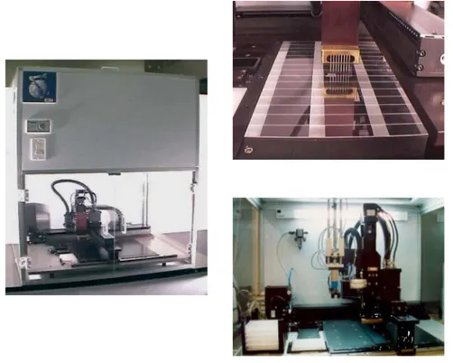
2.2.7.2.2 Spotting with arrayer 54
2.2.7.2.3 Test prints 56
2.2.7.2.4 Printing the real microarray 57
2.2.7.2.5 Blocking slides 57
2.2.8 Amplification of mRNA 58
2.2.8.1 First and second strand cDNA synthesis 59
2.2.8.2 Double strand cDNA clean up 59
2.2.8.3 In vitro transcription and aRNA purification 60
2.2.8.4 Second round amplification 60
2.2.9 Preparation of probe 61
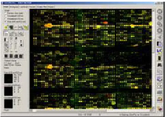
2.2.10 cDNA microarray hybridization 63 2.2.11 cDNA microarray washing and scanning 63 2.2.12 Data normalization and analysis 64 2.2.13 Expression analysis of a gene by semi-quantitative RT-PCR 65 2.2.13.1 First strand cDNA synthesis, fidelity and DNA
contamination control 65 2.2.13.2 Determination of optimal cycle of a gene for
semi-quantitative PCR 65 2.2.13.3 GAPDH normalization and semi-quantitative RT-PCR 66 2.2.14 Expression analysis of a gene by real-time RT-PCR 66
2.2.15 Statistical analysis 67
2.2.16 Polymerase Chain Reaction (PCR) 68 2.2.17 PCR and restriction enzyme digestion 68 2.2.18 PCR and amplification refractory mutation system (ARMS) 69
CHAPTER 3. RESULTS 70
3.1 Patient information 70
3.3 Array results of CML samples 73 3.4 Cluster analysis of two high and two low risk samples 84
3.5 Semi-quantitative RT-PCR analysis 92
3.6 Real-time RT-PCR analysis 96
3.7 Statistical analysis of data 97
3.8 Kaplan-Meier analysis for correlation of survival with gene expression 97 3.9 Detection of Gly382Asp MYH mutation by Bgl II digestion 99 3.10 Detection of Tyr165Cys MYH mutation by ARMS 100
CHAPTER 4. DISCUSSION 102
CHAPTER 5. FUTURE PERSPECTIVES 106
REFERENCES 108 APPENDICES 125
a. The patient information form 126
b. The informed consent form 127
c. The raw fluorescent data obtained from arrays of CML patients
and control 128
LIST OF TABLES
page
Table 1 List of primers and PCR conditions 40
Table 2 Patient samples with NPS values 70
Table 3 Genes at least 2 fold induced by IFNα-2b in K562 cell line 73 Table 4 Differentially expressed transcripts in high and low-risk
CML patients 86
Table 5 The top eleven differentially expressed genes in high-risk
compared to low-risk samples 88
Table 6 The top eleven differentially expressed genes in low-risk
LIST OF FIGURES
page
Figure 1 Differentiation of hematopoietic cells 3 Figure 2 Locations of the breakpoints in the ABL and BCR genes and
structure of chimeric mRNAs derived from the various breaks 12 Figure 3 Functional domains of p210 BCR/ABL 13 Figure 4 Signaling pathways of p210BCR/ABL 15
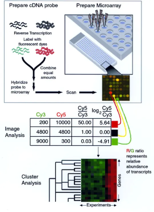
Figure 5 cDNA microaarray schema 33
Figure 6 SDDC-2 Microarrayer 56
Figure 7 K562 cell line array picture 73
Figure 8 Array with high background 75
Figure 9 Array with scratches 76
Figure 10 Array with Cy5 labeling problem 76
Figure 11 Array with spots running into each other 77
Figure 12 Array with not working samples 77
Figure 13 Array with fluorescent background 78
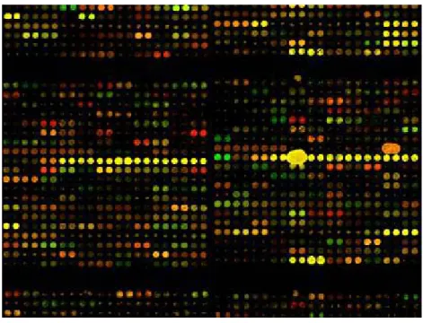
Figure 14 Array of CML 2 versus reference RNA 79
Figure 15 Array of CML 4 versus reference RNA 79
Figure 16 Array of CML 8 versus reference RNA 80 Figure 17 Array of CML 12 versus reference RNA 80 Figure 18 Scatter plot of CML 2 versus reference 82 Figure 19 Scatter plot of CML 4 versus reference 82
Figure 20 Scatter plot of CML 8 versus reference 83 Figure 21 Scatter plot of CML 12 versus reference 83
Figure 22 Gene clusters of four CML samples 84
Figure 23 Zoomed gene cluster images of four CML samples 85 Figure 24 Scatter plot of 58 risk group indicator genes in CML 2 89 Figure 25 Scatter plot of 58 risk group indicator genes in CML 4 89 Figure 26 Scatter plot of 58 risk group indicator genes in CML 8 90 Figure 27 Scatter plot of 58 risk group indicator genes in CML 12 90 Figure 28 Scatter plot of CML 2 versus CML 8 91 Figure 29 Scatter plot of CML 2 versus CML 4 91 Figure 30 Cycle optimization of GAPDH, IFITM1, UCP2 and CCNH
genes 92
Figure 31 Semi-quantitative RT-PCR results of seven genes in high, low & intermediate risk CML, and other hematological malignancies 94 Figure 32 Relative expression of IFITM1 by real-time PCR in low- and
high-risk CML 96
Figure 33 Kaplan-Meier analysis 98
Figure 34 Bgl II digestion profile in detection of Gly382Asp 100 Figure 35 Detection of Tyr165Cys MYH mutation by ARMS 101
ABBREVIATIONS
A absorbance
ALL acute lymphocytic leukemia AML acute myeloid leukemia ATP adenine triphosphate
bp base pair
BSA bovine serum albumin BER base excision repair
cAMP cyclic adenosine mono phosphate cDNA complementary DNA
CML chronic myeloid leukemia C-terminus carboxy terminus Cy3 indocarbocyanine Cy5 indodicarbocyanine
dATP adenosine deoxyribonucleoside triphosphate dCTP cytosine deoxyribonucleoside triphosphate ddH2O deionized water
ddNTP dideoxynucleotide triphosphate DEPC diethylpyrocarbonate
dGTP guanosine deoxyribonucleoside triphosphate DNA deoxyribonucleic acid
DNase deoxyribonuclease
dNTP deoxynucleotide triposphate
dTTP thymine deoxyribonucleoside triphosphate DTT dithiothretiol
EDTA ethylenediaminetetraacetic acid EtBr ethidium bromide
EtOH ethanol g gram
IVT in vitro transcription kb kilobase
LB Luria-Bertani medium LOH loss of heterozygosity LOI loss of imprinting M molar min minute ml milliliter mm milimeter mM milimolar µl microliter mRNA messengerRNA
NaOAc sodium acetate NaCl sodium chloride NaOH sodium hyrdoxide ng nanogram
nm nanometer
NPS new prognostic score N-terminus amino terminus OD optical density
Oligo(dT) oligodeoxythymidylic acid O/N over night
PBS Phosphate buffered saline PCR polymerase chain reaction pmol picomole
RNA ribonucleic acid rpm revolution per minute SDS sodium dodecyl sulphate
sec second
TAE tris-acetic acid-EDTA TBE tris-boric acid-EDTA
Tris Tris (hydroxymethyl)-methylamine U unit
UV ultraviolet
v volt
v/v volume for volume µg microgram
CHAPTER 1. INTRODUCTION
1.1 Hematopoiesis: Genesis and differentiation of blood cells
Hematopoiesis is the formation of blood cells (Fauci et al., 1998). The bone marrow, lymph nodes and spleen are all involved in hematopoiesis. These organs and tissues have traditionally been divided into myeloid tissue including the bone marrow and the cells derived from it-erythrocytes, platelets, granulocytes, and monocytes, and lymphoid tissue consisting of thymus, lymph nodes, and spleen (Cotran et al., 1989).
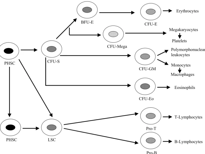
In 1924, Maximow postulated that blood cells were derived from a single class of progenitors. In 1938, Downey added the concept of hierarchies of pluripotent cells. The demonstration that single cells were capable of establishing nodules of hematopoietic growth in the spleen of irradiated mice and that such colonies displayed multilineage or pluripotent differentiation (erythroid, myeloid, megakaryocytic) came in 1961 by Till and McCulloch. These landmark experiments established that a stem cell existed for hematopoiesis. A stem cell has the ability of self-renewal and the production of progeny destined to differentiate (Lee et al., 1993). The common pluripotent hematopoietic stem cell (PHSC) gives rise to lymphoid and the trilineage myeloid stem cells (CFU-S) (Figure 1) (Cotran et al., 1989).
The lymphoid stem cell (LSC) is the origin of precursors of T-cells (pro-T cells) and B cells (pro-B cells). The former differentiates into mature T cells under the inductive influence of the thymus and the latter to mature B cells under the influence of bursa-equivalent tissue. An important difference between lymphoid and myeloid differentiation is that, there are no distinctive, morphological recognizable stages in lymphoid differentiation. From the multipotent myeloid stem cell, three
types of committed stem cell arise which differentiate along the erythroid / megakaryocytic, eosinophilic, and granulocyte / macrophage pathways (Cotran et al., 1989). Hematopoietic colonies could be grown in semisolid medium (Lee et al., 1993). Thus, the committed stem cells have been called the colony-forming units (CFU). As it is indicated in Figure 1, granulocytes and macrophages have a common precursor, CFU-GM. In the erythroid pathway, two distinct committed stem cells can be recognized. Based on the morphology of the colonies, BFU-E (burst-forming unit-erythroid) is primitive one. The later stage is CFU-E. From all these different committed stem cells, intermediate stages are derived, and finally the morphological recognizable precursors of the differentiated cell lines are formed. These are proerythroblasts, myeloblasts, megakaryoblasts, monoblasts and eosinophiloblasts, which in turn give rise to mature progeny. The mature blood elements have a finite life span and their numbers must be constantly replenished. Thus, self-renewal is an important property of stem cells. The pluripotent stem cells have the greatest capacity of self-renewal, but normally most of them are not in cycle. Self-renewal ability declines as commitment proceeds, but a greater fraction of the stem cells are found in cycle (Cotran et al., 1989).
Figure 1: Differentiation of hematopoietic cells (adapted from Cotran et al., 1989).
1.2 Hematological malignancies
Hematological malignancies arise as a clonal proliferation of one of the hematopoietic progenitor cells. The clinical manifestations of a particular malignancy depend on the stage of differentiation and lineage of the affected cell. The specific nature of the initial mutation and of subsequent mutations that may take place during the clonal evolution is also critical. The abnormal clone of cells must possess either a growth advantage or a block in apoptosis and/or differentiation over the normal cells. Clonal analysis of mature blood cells indicates that many myeloproliferative and/or myelodysplastic disorders generally arise in the pluripotent stem cell (Fauci et al., 1998)
PHSC PHSC CFU-S CFU-E BFU-E Erythrocytes LSC T-Lymphocytes B-Lymphocytes CFU-Mega Polymorphonuclear leukocytes Megakaryocytes Platelets Pro-T Pro-B CFU-GM Monocytes Macrophages CFU-Eo Eosinophils
1.3 Leukemias
The term “leukemia” was derived from the Greek word meaning “white blood” and was first described by John Hughes Bennett and Rudolf Vichow in 1845. The leukemias are a heterogeneous group of neoplasms that arise from the malignant transformation of hematopoietic cells (Wilson et al., 1991). This group of cancers arises in immature hematopoietic cells. They are characterized by the disturbance of normal hematopoiesis and failure in production of normally functioning cells. The associated clinical features appear to reflect the level and the lineage in the stem cell hierarchy at which malignant transformation has taken place. Leukemias account approximately 5% of all cancers (http://www.iarc.fr).
Leukemic cells proliferate primarily in the bone marrow and lymphoid tissues where they interfere with normal hematopoiesis and immunity. The accumulation of leukemic cells in the bone marrow is both due to excessive proliferation and to a defect in terminal maturation. These cells further enter the circulation and infiltrate into other tissues such as lymph nodes, liver, spleen, skin, viscera, and the central nervous system (Wilson et al., 1991).
Environmental toxins, cancer chemotherapy, radiation, and viruses such as human T-cell lymphotropic virus type-I (HTLV-I) in adult T-cell leukemia/ lymphoma are among the well-established leukomogenic factors (Wilson et al., 1991). Several hereditary factors have also been implicated as significant risk factors in leukemia, especially in childhood. Most of them appear to be related to either some form of immune deficiency or a syndrome of chromosomal instability (Wilson
et al., 1991).
A limited number of hematopoietic stem cells sequentially enter into cell cycle. These then differentiate into multiple lineages in the peripheral blood and lymphoid organs. According to the traditional concepts of hematopoietic development, progenitor cells differentiate into a single phenotype without the ability to switch lineage. However, some adult acute leukemias are “biphenotypic” in nature, with the expression of both lymphoid and myeloid linage cell surface antigens. In addition, in a subset of acute leukemias, lymphoid and myeloid lineage
markers fail to be expressed and these are known as acute undifferentiated leukemias. Such leukemic cells may represent leukemic expansion of the stem cell itself with no lineage markers (Sawyers et al., 1991).
The classification of leukemias is based on the cell type involved and the state of maturity of the leukemic cells. Thus, acute leukemias are characterized by the presence of very immature cells, blasts, and a rapid fatal course in untreated patients. On the other hand, chronic leukemias are at least initially associated with well-differentiated, mature leukocytes and with a relatively slow course. There are two major variants of acute and chronic leukemias known as lymphocytic and myelocytic. Therefore, a simple classification yields four patterns of leukemia: acute lymphocytic leukemia (ALL), chronic lymphocytic leukemia (CLL), acute myelocytic leukemia (AML) and chronic myelocytic (myeloid) leukemia (CML) (Cotran et al., 1989).
Most commonly leukemias are diagnosed by a blood test to count the number of red cells, white cells and platelets. A biopsy of the bone marrow may also be performed.
1.4 Chronic myeloid leukemia (CML)
1.4.1 Epidemiology and clinical characteristics of CML
CML is a clonal myeloproliferative disease that results from neoplastic transformation of primitive hematopoietic progenitor cells. It involves myeloid, monocytic, erythroid, megakaryocytic, B-lymphoid and occasionally T-lymphoid lineages (Cortes et al., 1996; Faderl et al., 1999a; Kabarowski et al., 2000).
CML accounts for 7-15% of leukemias in adults, and it has an incidence of 1-2 cases per 100,000 people per year (Cortes et al., 1996; Faderl et al., 1999a; Faderl
et al., 1999b). The median age at presentation is 45-55 years, but the disease can be
that there may be some correlation with HLA antigens CW3 and CW4. Therapeutic radiation has also been associated with increased risk of CML (Cortes et al., 1996).
CML is historically important in two aspects. It was the first human disease, in which a specific abnormality of the karyotype (Philadelphia chromosome (Ph)) could be linked to pathogenetic events of leukemogenesis (Faderl et al., 1999a; Deininger et al., 2000a). Second is at the therapeutic level. CML is one of the first neoplastic diseases in which the use of a biological agent interferon could suppress the neoplastic clone and prolong survival (Faderl et al., 1999a).
The disease is clinically divided into three phases as chronic, accelerated and blastic. The most of the cases (85%) are diagnosed at the chronic phase (CP) (Faderl
et al., 1999a; Chase et al., 2001). In this phase, the number of myeloid progenitor
cells increase and since differentiation continues, matured granulocytes are found in the peripheral blood. This period is approximately 3-5 years. The accelerated phase (AP) begins after several weeks to several years. Terminal differentiation is quickly lost; thrombocytosis, basophilia and multiple cytogenetic differences are added to the picture. The third phase known as blastic phase (blastic crisis (BC)) characterizes the last phase in which unmatured blast cells immediately increase. 30% or more leukemic cells are found in the peripheral blood or bone marrow. At this period, average survival is 3-6 months (Faderl et al., 1999a).
CML patients can be divided into three groups of low-risk, intermediate-risk, and high-risk, based on clinical parameters (age, spleen size, blast, platelet, eosinophil and basophil counts) known as new prognostic score (NPS) (Hasford et
al., 1998; Bonifazi et al., 2000). This scoring is the modified form of Sokal score,
which worked well as a prognostic discriminator for survival of three different risk groups (Sokal et al., 1984; Sokal et al., 1985; Hehlmann et al., 1997). Response of these risk groups to treatment is not uniform. Low risk patients respond better to treatment including interferon-alfa administration and their survival is much better compared to high-risk ones (Alimena et al., 1996; Kloke et al., 2000). The molecular characteristic of these three groups of patients is currently unknown.
1.4.2 Genetic and epigenetic factors in CML
Although CML was the first human disease in which a specific abnormality of the karyotype could be linked to pathogenetic events of leukemogenesis, genetic factors related to CML are not very well known. The specific chromosomal abnormality is known as Philadelphia chromosome (Ph) which results from reciprocal translocation of the long arms of chromosomes 9 and 22, t(9;22) (q34;q11). It is observed in 90-95% of all cases (Kurzrock et al., 1988; Faderl et al., 1999a; Warmuth et al., 1999a).
. The translocation breakpoint encompasses ABL on 9q34 and BCR on 22q11. As a result of this translocation, different numbers of BCR exons are added to chimeric BCR/ABL fusion gene. The BCR sequences in this mRNA are responsible for converting cellular proto-oncogene ABL to an oncogene (Heisterkamp et al., 1985). BCR causes ABL to become constitutively active as a tyrosine phosphokinase. The chimeric protein has higher tyrosine phosphokinase activity than the normal ABL protein(Faderl et al., 1999a; Chopra et al., 1999; Deininger et
al., 2000a; Deininger et al., 2000b). The main characteristics of this disease can be
listed as, adhesion independence, growth factor independence, and resistance to apoptosis (Di Bacco et al., 2000).
Cytogenetic and molecular changes occur in 50-80% of patients during transition to accelerated and blastic phases (Faderl et al., 1999a). Minor cytogenetic changes include monosomies of chromosomes 7, 17, and Y, trisomies of chromosomes 17 and 21, and translocation t(3;21)(q26;q22). Major changes include trisomies 8 and 19, isochromosome i(17q), and an extra Ph chromosome (double Ph) (Faderl et al., 1999a). Trisomy 8 is the most common, and i(17q) occurs almost exclusively in the blastic phase. In addition molecular abnormalities in p53, RB1,
c-MYC, p16INK4A, RAS and AML-EVI-1, a fusion protein resulting from translocation t(3;21)(q26;q22) have been documented (Neubauer et al., 1993; Faderl et al., 1999a). Alteration of p53 such as deletions, rearrangements, and mutations occur in 20-30% of patients with CML at blastic phase (Randhawa et al., 1998; Faderl et al., 1999a). Amplification of c-myc is found in 20% of patients (Randhawa et al., 1998). Other events occurring more rarely include mutations in RAS (6%) and rearrangements and
deletions of RB (14%) and p16 (15%) (Randhawa et al., 1998). Majority of these structural aberrations are seen at the accelerated and blastic phases of the disease.
Interestingly, a novel type of genetic alteration in CML appears to be loss of imprinting (LOI), which has been demonstrated for insulin-like growth factor-II gene (IGF2). Genomic imprinting is an epigenetic modification of a specific parental allele of a gene, or the chromosome on which it resides, in the gamete or zygote leading to differential expression of the two alleles of the gene in somatic cells of the offspring. LOI is a new disease mechanism in cancer which involves loss of parental origin-specific expression of genes which leads to activation of the normally silent copy of growth-promoting genes and/or silencing of the normally transcribed copy of tumor suppressor genes. LOI of IGF2 was documented in the accelerated and blastic phases of CML(Vogelstein et al., 1998; Randhawa et al., 1998).
1.4.3 Molecular biology of CML
1.4.3.1 Philadelphia chromosome (Ph)
The discovery of Ph chromosome in 1960 as the first consistent chromosomal abnormality associated with a specific type of leukemia was a breakthrough in cancer biology (Nowell et al., 1960). It took 13 years before it was appreciated that the Ph chromosome is the result of a t(9;22) reciprocal chromosomal translocation (Rowley J.D., 1973) and another 10 years before the translocation was shown to involve the
ABL proto-oncogene normally on chromosome 9 and a previously unknown gene on
chromosome 22, later termed BCR for breakpoint cluster region (bcr) (Groffen et al., 1984; Deininger et al., 2000a). Translocations have one of two effects. They may lead to the deregulation (overexpression) of oncogenes by their juxtaposition to enhancer or promoter sequences that are active in the cell type from which the tumor arises. The alternative molecular consequence of translocations is gene fusion, which results in a chimeric oncoprotein, the contribution to whose transforming ability is from both partners (Solomon et al., 1991).
The physiologic function of translocation partners
The ABL gene is the human homologue of the v-abl oncogene carried by the
Abelson murine leukemia virus (A-MuLV) (Deininger et al., 2000a). It is about 225 kb in size and is expressed as either 6 or 7 kb mRNA transcript, with alternatively spliced first exons, exon 1a and 1b respectively. The gene encodes a nonreceptor tyrosine kinase, which is higly conserved from Drosophila to humans (Laneuville P., 1995; Catherine et al., 1998). This protein is a ubiquitously expressed 145-kd protein in all tissues with several structural domains. Three SRC homology domains (SH1-SH3) are located toward the NH2 terminus. The SH1 domain carries the
tyrosine kinase function, whereas SH2 and SH3 domains allow for interaction with other proteins. The C-terminal part of ABL contains a DNA-binding domain, nuclear localization signals, and a binding site for actin (Catherine et al., 1998; Faderl et al., 1999a; Deininger et al., 2000a). Both the SH1 and SH2 protein are required for transformation. DNA-binding and tyrosine kinase activity of nuclear ABL is regulated in a cell cycle dependent manner. ABL is localized in both nucleus and cytoplasm. The normal protein is involved in the regulation of cell cycle, in the cellular response to genotoxic stress and in the transmission of information about the cellular environment through integrin signaling. Overall, ABL serves a complex role as a cellular module that integrates signals from various extracellular and intracellular sources and that influences decisions in regard to cell cycle and apoptosis. Mice with targeted disruptions in the gene have high neonatal mortality rates and increased susceptibility to infections suggesting a role in B-cell development (Raitano et al., 1997). However, ABL knockout mice failed to resolve most of the issues (Deininger et al., 2000a). Because of this, the normal function of this gene is not completely understood.
The BCR gene occupies a region of about 135 kb on chromosome 22. It is
expressed as mRNAs of 4.5 and 6.7, which encodes for the same cytoplasmic 160-kd protein. Like ABL, BCR protein is ubiquitously expressed and has several distinct domains. The first N-terminal exon encodes a serine-threonine kinase, which binds BCR-associated protein-1 (Bap-1), a member of the 14-3-3 family of proteins (Reuther et al., 1994) and possibly BCR itself. This has an important role in signal transduction and cell cycle regulation. A coiled-coil domain at N-terminus allows
dimer formation in vivo (McWhirter et al., 1993). The center of the molecule contains a region with DBL-like and pleckstrin-homology (PH) domains that stimulate the exchange of guanidine triphosphate (GTP) for guanidine diphosphate (GDP) on Rho guanidine exchange factors, which in turn may activate transcription factors such as NF-ĸB (Deininger et al., 2000a). The C-terminus has GTPase activity for Rac, a small GTPase of the Ras superfamily that regulates actin polymerization and the activity of NADPH oxidase in phagocytic cells (Diekmann et
al., 1995; Deininger et al., 2000a). In addition, BCR can be phosphorylated on
several tyrosine residues (Wu et al., 1998), especially tyrosine 177, which binds Grb-2 (growth factor receptor-bound protein Grb-2), an important adaptor molecule involved in the activation of the Ras pathway (Ma et al., 1997). Interestingly, ABL has been shown to phosphorylate BCR in COS1 cells, resulting in a reduction of BCR kinase activity (Ma et al., 1997; Deininger et al., 2000a). Although, these data argue for a role of BCR in signal transduction, their true biologic relevance remains to be determined. The fact that the BCR knockout mice are viable, fertile and have no obvious defects in hematopoietic cell development and also the fact that an increased oxidative burst in neutrophils is thus far the only recognized defect probably reflect the redundancy of signaling pathways (Raitano et al., 1997; Catherine et al., 1998; Deininger et al., 2000a). If there is a role for BCR in the pathogenesis of Ph-positive leukemias, it is not clearly discernible.
Molecular anatomy of the BCR-ABL translocation
The breakpoints within the ABL gene at 9q34 can occur anywhere over a large (greater than 300 kb) area at its 5’ end, either upstream of the first alternative exon Ib, downstream of the second alternative exon Ia, or more frequently between the two, and the breakpoints within the BCR gene at 22q11 occurs within a 5.8-kb area spanning BCR exons 12-16 (Deininger et al., 2000a; Salesse et al., 2002). Depending on the breakpoint in the BCR gene, three main types of BCR/ABL genes can be formed (Melo et al., 1996). Regardless of the exact location of the breakpoint, splicing of the primary hybrid transcript yields an mRNA molecule in which BCR sequences are fused to ABL exon a2. In contrast to ABL, breakpoints within BCR localize to 1 of 3 so-called bcr. The majority of patients with CML have
breakpoints in introns 1 and 2 of the ABL gene and in the major breakpoint cluster region (M-bcr) of the BCR gene, either between exons 13 and 14 (b2) or 14 and 15 (b3) (Figure 2). Because of alternative splicing, fusion transcripts with either b2a2 or b3a2 junctions can be formed. The final product of this genetic rearrangement is 210 kDa cytoplasmic fusion protein, p210 BCR/ABL , which is essential and sufficient for the malignant transformation of CML, and responsible for the phenotypic abnormalities of chronic phase CML (Salesse et al., 2002). Less frequent, the CML is caused by atypical BCR/ABL transcripts. The breakpoints are further upstream in the 54.4-kb region between the BCR exons e1 and e2, termed the minor breakpoint cluster region (m-bcr). The resultant e1a2 mRNA is translated into a 190 kDa protein (p190 BCR/ABL ). Recently, a third breakpoint cluster region (µ-bcr) was identified downstream of exon 19, giving rise to a 230 kDa fusion protein (p230 BCR/ABL ), associated with the rare Ph-positive chronic neutrophilic leukemia (Pane et
al., 1996; Deininger et al., 2000a).
In contrast to ABL, the BCR-ABL exhibits deregulated, constitutively active tyrosine kinase activity and is found exclusively in the cytoplasm of the cell, complexed with a number of cytoskeletal proteins (Salesse et al., 2002).
These features appear to underlie the ability of BCR/ABL to induce leukemic phenotype. However, the presence of the Ph chromosome in a hematopoietic cell is not in itself sufficient to cause leukemia, because, the fusion transcripts are detectable at low frequency in the blood of many healthy individuals (Biernaux et al., 1996; Bose et al., 1998). It is unclear why Ph-positive leukemia develops in a tiny minority of these persons. It may be that the translocation occurs in cells committed to terminal differentiation that are thus eliminated or that an immune response suppresses or eliminates BCR/ABL expressing cells (Deininger et al., 2000a). Indirect evidence that such a mechanism may be relevant comes from the observation that certain HLA types protect against CML (Posthuma et al., 1999). Another possibility is that, BCR/ABL is not the only genetic lesion required to induce chronic-phase CML. Indeed, a skewed pattern of G-6PD isoenzymes has been detected in Ph-negative Epstein-Barr virus transformed B-cell lines derived from patients with CML, suggesting that a Ph-negative pathologic state may precede the emergence of the Ph chromosome (Fialkow et al., 1981).
Figure 2: Locations of the breakpoints in the ABL and BCR genes and structure of chimeric mRNAs derived from the various breaks (Salesse et al., 2002).
Functional domains of BCR/ABL
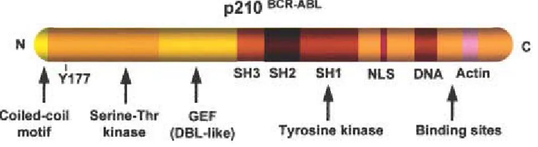
Several functional domains have been identified in the BCR/ABL protein that may contribute to cellular transformation (Figure 3). In the ABL portion, these domains are the SH1 (tyrosine kinase), SH2 and actin-binding domains; in the BCR portion, they include the coiled-coil oligomerization domain comprised between amino acids 1-63, the tyrosine at position 177 (Grb-2 binding site) and the phosphoserine/threonine rich SH2 binding domain.
Figure 3: Functional domains of p210 BCR/ABL (Salesse et al., 2002). Some of the important domains are illustrated, such as coiled-coil oligomerization domain, the tyrosine 177 (Grb-2 binding site), the phosphoserine/threonine rich SH2 binding domain and the rho-GEF (DBL-like) domain on BCR portion, and the regulatory src-homology regions SH3 and SH2, the SH1 (tyrosine kinase domain), the nuclear localization signal (NLS), and the DNA-and actin-binding domains in the ABL portion.
Physiological properties of BCR/ABL
The physiological properties demonstrated for BCR/ABL are: the induction of neoplastic transformation and cell proliferation, the induction of growth factor independence and inhibition of apoptosis in growth factor dependent hematopoietic cell lines and the inhibition of adhesion of chronic myeloid cells to marrow stroma (Chopra et al., 1999). These processes involve multiple and redundant intracellular pathways.
Ph negative CML
About 9-16% of CML patients are Ph-negative without apparent rearrangement of chromosomes 9 and 22 (Kantarjian et al., 1985; Hild et al., 1990; Chase et al., 2001). In 30-50% of these cases, however, the BCR/ABL fusion gene is detectable by molecular analysis (Cortes et al., 1995; Cross, 1997). There is believed to be no prognostic difference between BCR/ABL-positive cases that do or do not have a visible Ph chromosome at diagnosis. The term Ph-negative CML should
therefore be avoided or qualified to indicate whether patients have the BCR/ABL fusion or not. Cases that are Ph-negative and BCR/ABL-negative are heterogeneous but usually have atypical clinical features, respond less well to treatment and show earlier disease progression (Chase et al., 2001).
1.4.3.2 BCR-ABL signaling pathway
The mechanisms implicated in the pathogenesis of CML are altered adhesion to stroma cells and extracellular matrix (Gordon et al., 1987), constitutively active mitogenic signaling (Puil et al., 1994) and inhibition of apoptosis (Bedi et al., 1994; Deininger et al., 2000a).
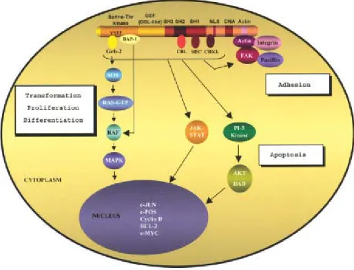
Increased tyrosine kinase activity of p210BCR/ABL results in the phosphorylation of several cellular substrates and in autophosphorylation of p210BCR/ABL , which in turn induces recruitment and binding of a number of adaptor molecules and proteins. Activation of a number of signaling pathways by p210BCR/ABL , leads to malignant transformation by interfering with basic cellular processes, such as control of cell proliferation and differentiation (Sawyers, 1993; Afar et al., 1994; Puil et al., 1994; Jiang et al., 2000), adhesion (Gordon et al., 1987; Bhatia et al., 1999) and cell survival (Bedi et al., 1994; McGahon et al., 1994; Cortez
et al., 1995; Cotter, 1995) (Figure 4).
The structure of p210BCR/ABL , allows multiple protein-protein interactions and suggests the involvement of diverse intracellular signaling pathways. In brief, it activates signal transduction pathways such as RAS/MAPK (RAS/mitogen activated protein kinases), PI-3 kinase (phosphotidylinositol 3 kinase), c-CBL (casitas B-lineage lymphoma protein) and CRKL (CRK-oncogene-like protein) pathways, JAK-STAT (Janus kinase-signal transducers and activators of transcription) and the Src pathway. Of these, the RAS that is at the center of the most prominent signaling pathways in CML, Jun-kinase, and PI-3 kinase pathways have been demonstrated to play a major role in transformation and proliferation (Raitano et al., 1995; Sawyers et
al., 1995; Skorski et al., 1995, 1997). Inhibition of apoptosis is thought to result
AKT (serine/threonine kinase) of c-myc and BCL-2 (Raitano et al., 1995; Sawyers et
al., 1995; Skorski et al., 1995, 1997; Warmuth et al., 1999b). p210BCR/ABL , effects on CRKL, c-CBL, and on proteins associated with the organization of the cytoskeleton and cell membrane, such as paxillin, actin, talin, vinculin and FAK/PYK2 (focal adhesion kinase/ prolin-rich kinase 2) , result in adhesion defects and cytoskeletal abnormalities, characteristics of CML cells (Salgia et al., 1997; Sattler et al., 1998; Sattler et al., 2002).
Figure 4: Signaling pathways of p210BCR/ABL (Salesse et al., 2002).
1.4.3.3 Diagnosis and monitoring of CML with BCR/ABL
Most commonly all leukemias are diagnosed by a blood test to count the number of red cells, white cells and platelets. Bone marrow aspiration with
measurements of the percentage of blasts and basophils can be done. A biopsy of the bone marrow may also be performed (Faderl et al., 1999b).
The cytogenetic analysis is the gold standard diagnostic test in CML. It is also valuable in demonstrating additional karyotipic abnormalities that occur with disease resistant and transformation (Mitelman, 1993). However, cytogenetic analysis is time-consuming, and only 20-25 cells are examined per sample (Faderl et al., 1999b). In 10% of patients with CML, Ph can not be demonstrated by cytogenetic studies, but molecular analysis will detect BCR/ABL rearrangements in up to one half of patients. Genomic polymerase chain reaction (PCR) and Southern blot assay can determine the exact breakpoints of the fusion genes. Reverse transcriptase PCR (RT-PCR) and Northern blot analysis detect BCR/ABL transcripts at the RNA level. Western blot analysis or immunoprecipitation demonstrate p210 BCR/ABL protein by using monoclonal antibodies against N-terminal region of BCR and C-terminal region of ABL (Guo et al., 1991).
Monitoring patients who are receiving therapy is commonly done by PCR and fluorescence in situ hybridization (FISH) for BCR/ABL. Quantitative RT-PCR is used for follow up of patients after stem cell transplantation (Hochhaus et al., 1998). Despite its sensitivity, RT-PCR may miss Ph-positive cells that are not transcribing the relevant gene product at the time of analysis. FISH allows analysis of metaphase and non-dividing interphase cells and is easily quantifiable. Interphase FISH is performed on peripheral blood. This technique is fast, and it analyzes more cells than is possible with conventional cytogenetic methods. It is therefore, more reliable in assessing cytogenetic responses in CML (Muhlmann et al., 1998). However, it overestimates the degree of cytogenetic response at high Ph-positive percentage values. Because of a false-positive rate of 10%, interphase FISH is not useful once the number of Ph-positive cells decreases to less than 10%. Hypermetaphase FISH allows analysis of up to 500 cells in metaphase per sample in a time-efficient manner without false-positive results, but it requires bone marrow samples other than peripheral blood (Seong et al., 1995). Another FISH technique applicable to blood samples uses double-color probes to detect Ph-positive leukemias and has shown superior sensitivity and specificity (Dewald et al., 1998; Buno et al., 1998).
1.4.3.4 Pathogenesis and transformation of CML
Despite the central influence of BCR/ABL, on the initial development of CML, secondary genetic driving forces mentioned above would presumably important in disease progression (Wetzler et al., 1993; Kantarjian et al., 1997; Faderl
et al., 1999c). The disease transformation is often results by refractoriness to
treatment, leukocytosis with increases in blood and marrow blasts, basophilia, increases or decreases in platelet counts unrelated to therapy, and clinical manifestations such as unexplained fever, spleenomegaly, extramedullary disease, weight loss, and bone and joint pains.
1.4.4 Treatment of CML
The accumulation of leukemic blasts in bone marrow suppresses normal hematopoietic stem cells. Therapeutically, the aim is to decrease population of leukemic clone enough to allow the recovery of normal stem cells (Cotran et al., 1989).
In order to cure a malignancy, all of the cancer cells must be destroyed. Hormones, cytokines (interferons, interleukins, tumor necrosis factor), monoclonal antibodies coupled to tumoricidal agents, cells (lymphokine activated killer (LAK) cells, tumor infiltrating lymphocytes (TIL)), and chemotherapeutic drugs are among biologic and chemical agents, which are used in cancer treatment (Wilson et al., 1991).
To achieve a cure with chemotherapeutic drugs, (1) the cancer cells must be sensitive to the agent; (2) the drug must reach the malignant cell; (3) if the drug is effective only in a phase of the cell cycle, it must be given frequent enough that all the cancer cells enter this phase of the cycle in the presence of drug; and (4) the malignant cells must be destroyed before drug resistance emerges (Wilson et al., 1991).
The natural history of chronic myelogenous leukemia has changed significantly since the first treatment attempts with arsenicals (Fowler's solution) in 1856 by Lissauer. In the past, the median survival of patients with chronic myelogenous leukemia was 3 years; less than 20% of patients were alive 5 years after diagnosis. Survival duration has since doubled to 5 to 7 years; 50% to 60% of patients are alive at 5 years, and more than 30% are alive at 10 years (Kantarjian et
al., 1993). Several reasons account for this change: earlier diagnosis; better
supportive care; and more effective therapies, such as allogeneic stem-cell transplantation, interferon-α and tyrosine kinase inhibitors (Faderl et al., 1999b; Deininger et al., 1997). Prognostic models based on multivariate analysis allowed stratification of treatment options according to a patient's risk profile, thereby maximizing the chances for the best possible therapeutic outcome (Sokal et al., 1988; Kantarjian et al., 1990; Hasford et al., 1998).
Busulfan, an alkylating agent, controls hematologic variables. In most patients receiving busulfan, disease cannot be controlled safely at the low leukocyte levels needed to induce a complete hematologic response; therefore, partial hematologic response is maintained (Faderl et al., 1999b).
Hydroxyurea, a cell cycle-specific inhibitor of DNA synthesis, became available for the treatment of chronic myelogenous leukemia and it allows rapid but transient hematologic control, is well tolerated, and has few side effects (nausea, vomiting, diarrhea, mucosal ulcers, and skin manifestations) (Faderl et al., 1999b).
Hydroxyurea and busulfan produce complete hematologic remission in 50% to 80% of patients (Kantarjian et al., 1998). Cytogenetic remissions are rare, and both agents do not affect disease progression. Patients will inevitably experience transformation to the blastic phase and die of its complications after a median of 3 to 6 years. Hydroxyurea therapy is superior to busulfan therapy (Faderl et al., 1999b). When these two agents were compared in a randomized study of patients with early chronic-phase chronic myelogenous leukemia, median survival (56 months compared with 44 months) and median duration of chronic-phase disease (47 months compared with 37 months) were significantly longer in the patients receiving hydroxyurea (Hehlmann et al., 1993).
Allogeneic stem-cell transplantation has curative potential in chronic myelogenous leukemia. It produces long-term survival in 50% to 80% of patients and disease-free survival in 30% to 70%. Relapses occur in 15% to 30% of patients, and plateaus are reached at 5 years after transplantation. However, late relapses rarely occur beyond years after transplantation. Applicability of allogeneic stem-cell transplantation is limited by availability of matched siblings and age restrictions. Less than 30% of patients in Europe and North America receive stem-cell transplants from matched sibling donors (Horowitz et al., 1996).
Several factors influence the outcome of stem-cell transplantation. First, younger patients do best with this therapy: Disease-free survival is 60% to 70%, transplant-related mortality is 10%, and probability of relapse is 20% (Faderl et al., 1999b). Older patients do worse mainly because of an increase in treatment-related mortality. Disease-free survival rates are about 30% at 5 years after therapy (Horowitz et al., 1996). Second, disease phase determines outcome. Results are more favorable if patients undergo transplantation during the chronic phase instead of during transformation. Transplantation during advanced phases is characterized by increased rates of leukemia relapse and treatment-related mortality. Third, chemotherapy before transplantation affects disease-free survival. In one study of patients with chronic-phase disease, disease-free survival after transplantation was significantly higher among those who were pretreated with hydroxyurea than among those who were pretreated with busulfan (Goldman et al., 1993). Fourth, the preparative regimen and graft-versus-host disease prophylaxis affect outcome.
Relapse after transplantation occurs in as few as 10% to 20% of patients (those with chronic-phase disease) to as many as 70% to 80% (those with blastic-phase disease and T-cell depletion) (Mrsic et al., 1992; Arcese et al., 1993). Outcome with second transplantations from HLA-identical siblings depends on the time between transplant and relapse. Patients who experienced relapse within 6 months after transplantation had a disease-free survival rate of 7%, a treatment-related mortality rate of 69%, and a probability of relapse of 77%. For patients who experienced relapse more than 6 months after transplantation, the disease-free survival rate was 28%, the treatment-related mortality rate was 30%, and the probability of relapse was 59% (Mrsic et al., 1992).
Interferons are glycoproteins produced by eukaryotic cells in response to antigenic stimuli such as those that occur with viral infections and malignant diseases. They have pleiotropic effects, including antiviral, immunomodulatory, antiproliferative and antiangiogenic activities (Estrov et al., 1993). Interferon-α (IFN-α) administration is a promising therapeutic intervention in the chronic phase of CML (Sacchi et al., 1997). It suppresses the leukemic clone and prolongs survival (Faderl et al., 1999a). However, at the advanced stages of the disease IFN-α treatment does not offer a significant therapeutic advantage. Single-arm studies of IFN-α therapy in CML have consistently produced high rates of complete hematologic (46% to 80% of patients) and cytogenetic responses (complete response in 13% to 32% of patients and partial response in 11% to 16%) (Kolb et al., 1995; Kantarjian et al., 1996; Collins et al., 1997; Mahon et al., 1998). IFN-α treatment with chemotherapy confirmed better survival with IFN-α treatment than with either hydroxyurea therapy or busulfan therapy (Faderl et al., 1999b). Five-year survival rates were 57% with IFN-α therapy and 42% with chemotherapy.
Patients with CML who fail IFN-α therapy and who are not candidates for allogeneic stem cell transplantation have limited treatment options and a relatively poor prognosis. Cytosine arabinoside (ara-C) have shown single-agent anti-CML activity and have induced hematologic and cytogenetic responses in CML. The combination regimens with ara-C and IFN-α yielded substantially higher rates of complete hematologic and cytogenetic remission in good-risk patients (Thaler et al., 1997). This response translated into significantly longer survival (Guilhot et al., 1997). Combinations of IFN-α with other effective agents (hydroxyurea, busulfan) and intensive chemotherapy were not associated with better results than those seen with IFN-α alone (Kantarjian et al., 1991).
Despite encouraging results with matched unrelated donor transplantation (2-year disease-free survival rates, 14% to 43%) (McGlave et al., 1993; Hansen et
al., 1998) the procedure carries significant morbidity and mortality rates depending
Autologous bone marrow transplantation results in transient cytogenetic responses, but a survival advantage has not been proven (Reiffers et al., 1994). Relapse due to reinfused Ph-positive cells may occur (Deisseroth et al., 1994).
Homoharringtonine is a plant alkaloid derived from the Cephalotaxus
fortuneii tree. Used in a low-dose continuous infusion, homoharringtonine resulted
in complete hematologic response in two thirds of patients and cytogenetic responses in one third (half of whom had major responses). Combinations of homoharringtonine with IFN-α and ara-C are being tested, with promising results (Faderl et al., 1999b).
5-Aza-2'-deoxycytidine (decitabine) is a potent hypomethylating cytidine analogue (Issa et al., 1997). Decitabine produced response rates of 25% in patients with blastic-phase disease and 53% in patients with accelerated-phase disease (Faderl
et al., 1999b).
Polyethylene glycol (PEG) interferon is a modified IFN-α molecule that is covalently attached to polyethylene glycol. This interferon has a significantly longer half-life than its parent compound and can be given once weekly instead of daily. In addition to causing fewer side effects, PEG interferon produced a hematologic response in 50% of patients, including 4 of 13 patients who had been resistant to interferon (Faderl et al., 1999b). Preliminary results with PEG interferon are promising, and further investigations are required to validate its role in the treatment of CML.
Antisense oligonucleotides are short DNA sequences modified to bind target RNA sequences within the cell, preventing translation of RNA into functional proteins. BCR-ABL antisense oligonucleotides reduce the level of p210BCR/ABL in CML cells and slow the rate of growth and proliferation. Antisense sequences directed against BCR-ABL for ex vivo purging in autologous bone marrow transplants are of interest (Ratajczak et al., 1992; De Fabritiis et al., 1998; Maran et
al., 1998).
Adoptive immunotherapy: The role of a subset of T lymphocytes in the suppression of leukemic cells is increasingly appreciated. The idea that leukemic cell proliferation is controlled by the immune system is based on several observations. First, the frequency of disease recurrence is increased with T-cell-depleted stem-cell transplantation. Second, donor lymphocyte infusions reestablish cytogenetic remissions in many patients who experience relapse after allogeneic transplantation. Third, a positive correlation has been seen between graft-versus-host disease and reduced risk for relapse after transplantation. Finally, cytogenetic response correlates with interferon therapy-associated autoimmune phenomena (Faderl et al., 1999b).
Research is focusing on identification of specific T-cell clones that can eliminate leukemic progenitors and on proteins that can serve as tumor-specific targets. CML is a good model because p210BCR/ABL is uniquely associated with the Ph translocation (Lim et al., 1997; Moldremm et al., 1997; Choudhury et al., 1997). Identification of leukemia-specific antigens and stimulation of leukemia-specific T-cell responses may allow the use of immunogenicity of leukemic T-cells in such approaches as immune gene therapy and peptide vaccination (Smit et al., 1997).
Tyrosine kinases are enzymes that transfer phosphate from ATP to tyrosine residues on substrate proteins that in turn regulate cellular processes such as proliferation, differentiation, and survival (Druker et al., 2000). Therefore, tyrosine kinase inhibitors are very attractive molecules in CML therapy. Although many molecules along the signaling cascade can be targeted, inhibition of phosphotyrosine kinase activity has been studied most extensively (Levitzki et al., 1995). Natural inhibitors of tyrosine kinases (herbimycin A, genistein, erbstatin, and lavendustin A) have broad specificity for various enzyme substrates. To improve target specificity, synthetic compounds have been modeled after the naturally occurring kinase inhibitors, and more than 20 of them are known (Levitzki et al., 1995). Some of these compounds (AG1112, AG568, and CGP57148B) showed a growth-inhibiting effect on CML cell lines in vitro (Druker et al., 1996; Deininger et al., 1997; Beran
Therapeutic agent STI571 (signal transduction inhibitor number 571) (Gleevec, imatinib mesylate) formerly CGP57148B, is a rationally developed, potent and selective inhibitor for ABL tyrosine kinases, including BCR/ABL (Mauro
et al., 2001; Goldman et al., 2001). The BCR/ABL tyrosine kinase is a constitutively
active kinase, which functions by binding ATP and transferring phosphate from ATP to tyrosine residues on various substrates. This causes the excess proliferation of myeloid cells characteristic of CML. STI571 functions by blocking the binding of ATP to BCR/ABL, inhibiting its activity. As a result, substrates required for BCR/ABL function cannot be phosphorylated and subsequent event are abrogated (Mauro et al., 2001).
The phases I-II clinical trials in CML have demonstrated promising results, especially in the chronic phase of the disease (Etienne et al., 2001;Drummond et al., 2001).
Fifty patients with Philadelphia chromosome-positive CML in early chronic phase received imatinib mesylate, 400 mg orally daily. After a median follow-up of 9 months, 49 patients (98%) achieved a complete hematologic response and 45 patients (90%) achieved a major cytogenetic response, complete in 36 patients (72%) (Kantarjian et al., 2003). Compared with similar patients who received IFN-α with or without hydroxyurea or other IFN-α combination regimens, those receiving imatinib mesylate had higher incidences of complete and major (Ph < 35%) cytogenetic responses at 3 months (34% and 74% versus 1%-4% and 9%-24%, respectively), 6 months (52% and 80% versus 3%-7% and 11%-28%, respectively), and 9 months (60% and 77% versus 5%-11% and 14%-30%, respectively) (Kantarjian et al., 2003a).
An investigation was made whether increasing the dose of imatinib mesylate might overcome drug resistance in patients with Philadelphia chromosome-positive CML whose disease manifests relapse or refractoriness to therapy (Kantarjian et al., 2003b). Fifty-four patients with Ph(+) CML in chronic phase and with hematologic or cytogenetic resistance or relapse on imatinib mesylate therapy at 400 mg orally daily were treated with a higher dose of 400 mg orally twice daily (800 mg daily, 47 patients; or 600 mg daily increased from 300 mg daily, 7 patients). Among 20
patients treated for hematologic resistance or relapse, 13 (65%) achieved a complete (n = 9) or partial (n = 4) hematologic response, but only 1 had a cytogenetic partial response (Ph reduction from 100% to 10%) and 1 had a minor response (Ph reduction from 100% to 50%). Among 34 patients treated for cytogenetic resistance or relapse, 19 (56%) achieved a complete (n = 6) or partial (n = 7) cytogenetic response (Kantarjian et al., 2003b). As a conclusion higher doses of imatinib mesylate may overcome disease-poor response to conventional doses and that this approach deserves further evaluation as frontline therapy for newly diagnosed CML.
237 CML patients were treated with imatinib mesylate at accelerated-phase (Kantarjian et al., 2002). Among the 200 patients with accelerated-phase CML for whom follow-up was 3 months or more, rates of complete and partial hematological response were 80% and 10%. Cytogenetic responses were evident in 90 patients [45%; complete response in 47 patients (24%) and partial response (Ph 1-34%) in 21 patients (11%)]. The estimated 18-month survival rate was 73%. The estimated complete hematological response rate at 18 months was 68%; that for cytogenetic response was 82%. Landmark analysis showed that achieving a cytogenetic response at 3 months or a major cytogenetic response (Ph < 35%) at 6 months was associated with better long-term survival.
Blast crisis is the most advanced stage of CML and is highly refractory to therapy. A total of 260 patients with CML were enrolled in a phase II trial, of whom 229 had a confirmed diagnosis of CML in blast crisis (Sawyers et al., 2002). Imatinib induced hematologic responses in 52% of patients and sustained hematologic responses lasting at least 4 weeks in 31% of patients, including complete hematologic responses in 8% (Sawyers et al., 2002). For patients with a sustained response, the estimated median response duration was 10 months. Imatinib induced major cytogenetic responses in 16% of patients, with 7% of the responses being complete. Median survival time was 6.9 months. These results demonstrated that imatinib has substantial activity and a favorable safety profile when used as a single agent in patients with CML in blast crisis. Additional clinical studies are warranted to explore the efficacy and feasibility of imatinib used in combination with other antileukemic drugs.
As a summary, imatinib mesylate is the new gold standard for treatment of CML. The response rates (both hematologic and cytogenetic) exceeded the rates with other medical therapies (Peggs et al., 2003). However, it remained to be shown whether imatinib therapy was superior to conventional therapy in a direct comparative study, whether the improved response rates translated into improved survival, and whether this treatment could induce molecular remission and possibly be curative in previously untreated patients.
Acquired resistance to imatinib mesylate caused by kinase-domain mutations is common in patients with CML who are treated with the drug (Gorre et al., 2002). Such mutations have been reported only at the time of clinically apparent resistance or relapse (Roche-Lestienne et al., 2002; Shah et al., 2002). A male patient with CML was screened for a mutation of the tyrosine kinase domain of BCR-ABL and was shown to have T315I substitution at diagnosis (Roche-Lestienne et al., 2003). A minor mutated clone present before any treatment seems to have expanded quickly under the selective pressure of imatinib monotherapy.
1.4.4.1 Minimal residual disease (MRD)
The term minimal residual disease (MRD) describes leukemia cells present at a level below that is detectable by conventional methodology in patients being in complete hematological and clinical remission (CR). (Widzysska et al., 1995). Clinically, MRD can be an indicator for a prediction of relapse. As an example, a greater MRD on entering CR tends to be related with an early relapse, a return to MRD-positive after disappearance of MRD will be a sign of impending relapse, and MRD negativity at the termination of therapy may be correlated with a long term disease free survival. Therefore, more precise evaluation of MRD is necessary with regard to therapeutic strategy in monitoring of the disease (Misawa et al., 1995). PCR can be used for detection of MRD in CML (Lee et al., 1988).