Do pre-procedural laboratory parameters predict
drug-eluting stent restenosis?
İşlem öncesi ölçülen biyokimyasal belirteçler ile ilaç kaplı stentlerde
yeniden daralmayı öngörmek mümkün müdür?
Department of Cardiology, Ufuk University Faculty of Medicine, Ankara
Aslı Tanındı, M.D., Berkay Ekici, M.D., Hasan Fehmi Töre, M.D.
Objective: Drug-eluting stents (DES) have considerably re-duced the rates of in-stent restenosis (ISR). Several studies reported pre-procedural C-reactive protein (CRP), neutrophil to lymphocyte (N/L) ratio, red cell distribution width (RDW), serum uric acid (UA), and mean platelet volume (MPV) as independent predictors of ISR using bare metal stents. This study investigates whether any laboratory parameter obtained before the coronary stenting procedure is associated with ISR using DES in stable coronary artery disease.
Methods: Three hundred fifteen stents were retrospectively analysed in 285 patients who had undergone coronary stent-ing and a control coronary angiography within one year of stenting, between January 2012 and April 2014. Pproce-dural complete blood count, biochemistry, and CRP were re-corded. Off-line quantitative coronary angiography analysis was performed.
Results: Overall restenosis rate was 10.2%. When the stents were analysed with respect to the presence of ISR, the num-ber of diabetics and smokers was higher in the ISR group. CRP levels were significantly higher in the ISR group, but there were no differences in N/L, monocytes, eosinophils, RDW, MPV, UA, and total bilirubin levels. In the univariate regression analysis, DM, CRP, stent length, stent diameter, pre-procedural diameter stenosis, pre-procedural minimal lu-men diameter (MLD), post-procedural residual diameter ste-nosis, procedural reference vessel diameter, and post-procedural MLD were predictors of ISR. However, multivariate regression analysis identified only DM and post-procedural residual stenosis as independent predictors of ISR.
Conclusion: Pre-procedural blood parameters do not indepen-dently predict ISR in DES, which is mainly determined by the presence of diabetes and post-procedural residual stenosis.
Amaç: İlaç kaplı stentler, stent içi yeniden daralma oranlarını önemli ölçüde azaltmıştır. Çeşitli çalışmalarda C-reaktif pro-tein (CRP), nötrofil/lenfosit oranı (N/L), eritrosit dağılım geniş-liği (RDW), serum ürik asit (ÜA) düzeyleri, ortalama trombosit hacmi (MPV) gibi kanda ölçülen bazı parametreler çıplak metal stentlerde yeniden daralmanın öngördürücüleri olarak saptan-mıştır. Bu çalışmada kararlı koroner arter hastalığında ilaç kaplı stent uygulamasında işlem öncesi kanda ölçülen herhangi bir parametre ile yeniden daralma arasında ilişki araştırıldı.
Yöntemler: Ocak 2012 ve Nisan 2014 tarihleri arasında koro-ner stent yerleştirilen ve sonraki bir yıl içinde kontrol korokoro-ner anjiyografisi yapılan 285 hastadaki 315 stent geriye dönük ola-rak incelendi. İşlem öncesi tam kan sayımı, rutin biyokimyasal incelemeler ve CRP düzeyleri kaydedildi. Koroner anjiyografik değerlendirme kantitatif koroner anjiyografi ile yapıldı.
Bulgular: Genel yeniden daralma oranı %10.2 idi. Stentler stent içi yeniden daralma varlığı bakımından analiz edildi-ğinde, yeniden daralma grubunda diyabetik ve sigara içenler daha fazlaydı; CRP düzeyleri daha yüksekken N/L, RDW, eozinofil, monosit, MPV, ÜA, total bilirubin düzeyleri bakımın-dan fark yoktu. Tek yönlü regresyon analizinde, diyabet, CRP, stent uzunluğu ve çapı, işlem öncesi daralma yüzdesi, işlem öncesi en düşük lümen çapı, işlem sonrası rezidüel daral-ma yüzdesi, işlem sonrası referans dadaral-mar çapı ve en düşük lümen çapı stent içi yeniden daralmanın öngördürücüleriydi. Çok değişkenli regresyon analizinde ise sadece diyabet ve işlem sonrası rezidüel daralma yüzdesi bağımsız öngördürü-cüler olarak saptandı.
Sonuç: İlaç kaplı stentlerde yeniden daralmayı öngörmede işlem öncesi kanda bakılan hiçbir parametrenin yararı olma-mıştır; ancak, diyabet ve işlem sonrası rezidüel darlık miktarı esas belirleyiciler olarak ön plana çıkmıştır.
Received:November 24, 2014 Accepted:January 16, 2015
Correspondence: Dr. Aslı Tanındı. Ufuk Üniversitesi Dr. Rıdvan Ege Hastanesi, Mevlana Bulvarı Çukurambar, Ankara, Turkey.
Tel: +90 312 - 204 40 82 e-mail: [email protected] © 2015 Turkish Society of Cardiology
I
n-stent restenosis (ISR) is a significant problem, affecting up to 30% of patients in the era of bare-metal stents (BMS).[1] Stent implantation causes inju-ry in the coronainju-ry vessel endothelium, initiating both local and systemic inflammatory responses, which play an important role in the pathophysiology of ISR. [2,3] Patient-dependent factors, clinical conditions in which the patient undergoes the procedure, technical details, and characteristics of the lesion constitute the spectrum of variables which lead to ISR. Drug-eluting stents (DES) have improved outcomes by reducing neointimal hyperplasia, thus reducing the rate of ISR to less then 10%[4,5] and reducing repeat revasculariza-tion procedures.[6]Several studies investigated whether laboratory parameters measured by complete blood count or bio-chemical analysis before the coronary stenting pro-cedure could predict ISR. Red cell distribution width (RDW),[7] neutrophil to lymphocyte (N/L) ratio,[8] mean platelet volume (MPV),[9] serum uric acid levels (UA),[10] and serum C-reactive protein levels (CRP)[11] were found to be associated with ISR. Most of these studies were performed using BMS.
The purpose of this study was to investigate whether any readily-available parameter measured by a simple blood draw before the coronary stenting procedure could be associated with in-stent resteno-sis using drug-eluting stents in patients admitted with stable coronary artery disease (CAD).
METHODS
This is a retrospective study analysing the hospital re-cords of patients with stable angina pectoris or angina equivalent symptoms who were admitted to the car-diology outpatient clinic of a university hospital and decided to undergo coronary angiography and elec-tive stenting after appropriate non-invasive tests were performed. Inclusion criteria included age (range: 18–80), elective coronary stenting with 2nd genera-tion DES (Zotarolimus eluting Resolute Integrity [Medtronic Inc. Santa Rosa, CA, USA] or Biolimus eluting BioMatrix [Biosensors Europe SA, Switzer-land]) due to stable CAD, stored quantitative coronary angiography (QCA) data prior to stenting showing the percentage of luminal narrowing >70% (nearly all fi-nal decisions in the cardiology clinic regarding per-forming a stenting procedure and the choice of stent
length and diam-eter in elective cases are based on QCA analysis after an initial visual assess-ment), and a control angiography after the initial stenting procedure within 1 year (routine control angiography
per-formed 6 months - 1 year after stenting, is the stan-dard practice in our institution).
Exclusion criteria consisted of history of coro-nary artery by-pass graft operation, left-main lesion, chronic total occlusion, BMS or first generation DES, in-stent restenosis, acute or chronic infection dur-ing the initial procedure, malignancy, anemia, recent blood transfusion, renal insufficiency, hepatic insuffi-ciency, hypo- or hyperthyroidism, and any rheumato-logic, immunologic or inflammatory disorders. In ad-dition, patients who received polymer-free DES were excluded because the polymer-free structure could possess different properties with respect to in-stent restenosis than the polymer-based stents used in this study, which sought to avoid a heterogeneous study population with respect to the nature of the stents im-planted.
All percutaneous coronary intervention proce-dures with stenting performed between January 2012 and April 2014 were consecutively analysed in terms of inclusion and exclusion criteria, after which 315 eligible patients were enrolled to the study. This study was conducted according to the recommendations of the Declaration of Helsinki on Biomedical Research involving human subjects and was approved by the institutional ethics committee.
All coronary stenting procedures were performed through femoral artery by standard techniques with 7 Fr guiding catheters using GE Innova 4100 cath laboratory (GE Healthcare, Milwaukee, WI, USA). Two-dimensional quantitative coronary angiography (QCA) analysis was performed off-line using QCA software package (QAngio XA 7.3; Medis Medical Imaging Systems BV, Leiden, Netherlands). Lesion length, percentage diameter stenosis, minimal lu-men diameter (MLD), and reference vessel diameter (RVD) of the treated coronary segment before and
Abbreviations:
BMS Bare-metal stents CAD Coronary artery disease CRP C-reactive protein DES Drug-eluting stents ISR In-stent restenosis MLD Minimal lumen diameter MPV Mean platelet volume N/L Neutrophil to lymphocyte ratio QCA Quantitative coronary angiography RDW Red cell distribution width RVD Reference vessel diameter
after the stent implantation were determined via im-aging in which the lesion was most severe and not foreshortened. Zotarolimus-eluting Resolute Integrity (Medtronic Inc. Santa Rosa, CA, USA) or Biolimus eluting BioMatrix (Biosensors Europe SA, Switzer-land) were implanted in coronary vessels with a lu-minal narrowing >70%, as determined by QCA. Each patient received acetyl salicylic acid (ASA) and clopi-dogrel (300 mg loading dose) before the procedure. Unfractionated heparin 100 U/kg was administered at the beginning of the procedure to maintain activated clotting time >250 seconds. Choice of stent, predila-tation or postdilapredila-tation of the stent, and use of glyco-protein IIb/IIIa inhibitors were determined by the op-erator. Successful PCI was defined as TIMI grade III flow without any major complications with a residual stenosis <20% by visual estimation. All patients were prescribed ASA 100 mg indefinitely and clopidogrel 75 mg for ≥1 year. Stent restenosis was concluded ac-cording to the QCA analysis of the control coronary angiography and defined as >50% narrowing in a ves-sel including 5 mm proximal and distal to the stent edge. Restenosis pattern was defined as focal when the luminal narrowing was <10 mm in length, and diffuse when it was ≥10 mm in length. The data was analysed by two cardiologists, with a third cardiolo-gist consulted in the case of discrepancy.
Clinical and demographic data of patients—in-cluding age, presence of hypertension, diabetes, hy-perlipidemia, and smoking status—were noted from the hospital records. All laboratory data at the time of initial stenting procedure were also recorded. Labo-ratory data, which was measured by immunoturbidi-metric assay (Abbott Architect c8000, USA), consist-ed of complete blood count (Cell-Dyn 3700, Abbott, USA) and biochemistry (Unicel Dx C800 Synchron, Beckman Coulter, USA), including kidney function tests, hepatic function tests, serum UA levels, lipid profile and CRP. These data were obtained from ve-nous blood samples which were drawn upon 12 hours of fasting before the coronary stent implantation. Statistical analyses
Statistical analyses were performed using SPSS sta-tistical software (IBM SPSS Statistics 21). Shapiro-Wilk test was performed for distribution pattern. Categorical variables were expressed as percentag-es, whereas continuous variables were presented as mean±standard deviation. Continuous variables were
compared by Student’s t-test and Mann-Whitney U tests. χ2 test was used for the categorical variables between the two groups. Univariate and multivariate regression analysis was performed in order to iden-tify the determinants of ISR. All tests of significance were two-tailed. Statistical significance was defined as p<0.05.
RESULTS
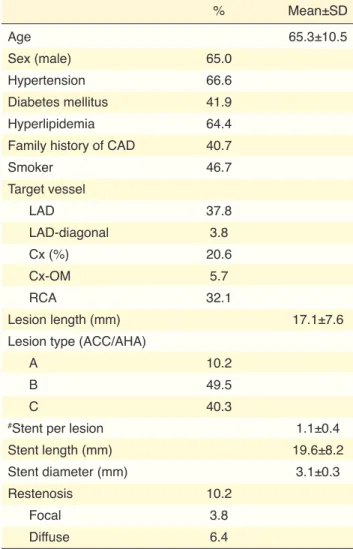
Table 1 shows basal characteristics of the study popu-lation. Mean age was 65.3±10.5, and 65% of the pa-tients were male. Rate of diabetics was 41.9%. Ac-cording to ACC/AHA lesion classification, 49.5% of the lesions were of type B, whereas 40.3% were of type C. Restenosis rate was 10.2%.
Table 1. Baseline characteristics of the study population % Mean±SD Age 65.3±10.5 Sex (male) 65.0 Hypertension 66.6 Diabetes mellitus 41.9 Hyperlipidemia 64.4
Family history of CAD 40.7
Smoker 46.7 Target vessel LAD 37.8 LAD-diagonal 3.8 Cx (%) 20.6 Cx-OM 5.7 RCA 32.1 Lesion length (mm) 17.1±7.6
Lesion type (ACC/AHA)
A 10.2
B 49.5
C 40.3
#Stent per lesion 1.1±0.4
Stent length (mm) 19.6±8.2
Stent diameter (mm) 3.1±0.3
Restenosis 10.2
Focal 3.8
Diffuse 6.4
#: Number of. CAD: Coronary artery disease; LAD: Left anterior descend-ing; Cx: Circumflex; RCA: Right coronary artery.
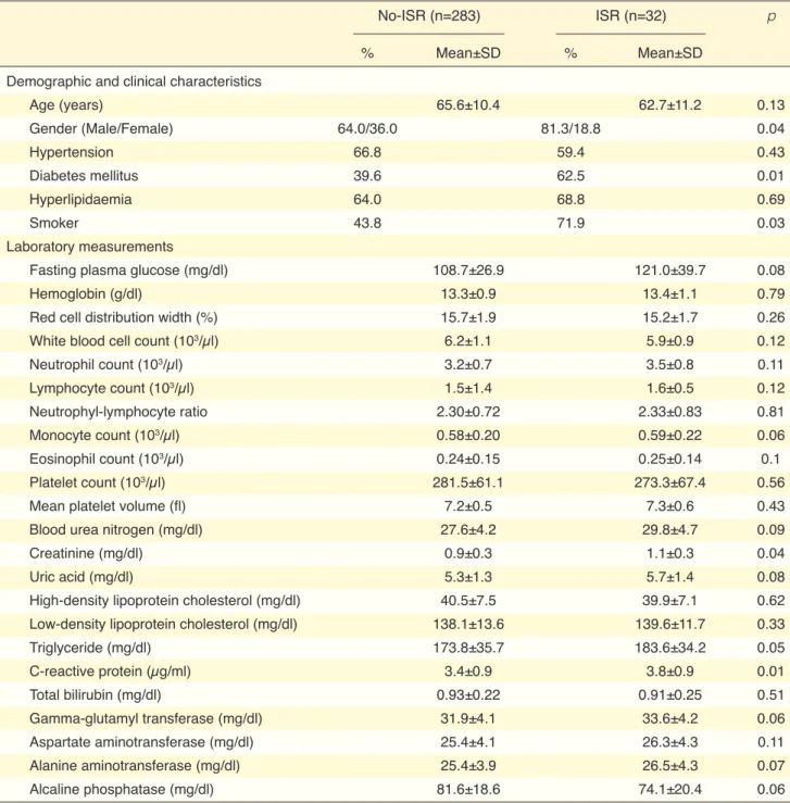
tical significance. There were no significant differenc-es between the two groups with rdifferenc-espect to N/L ratio, monocyte count, eosinophil count, RDW, MPV, and total bilirubin levels (Table 2).
Table 3 demonstrates the angiographic parameters obtained by QCA related to the coronary lesion and When analysed with respect to the presence of
ISR, the ISR group contained more patients who were diabetic and smokers than in the non-ISR group. Serum creatinin and CRP levels were significantly higher in the ISR group. Serum uric acid levels were 5.7±1.4 and 5.3±1.3 in the ISR and non-ISR groups, respectively; however, this result did not reach
statis-Table 2. Demographic, clinical characteristics and laboratory measurements with respect to the presence of in-stent restenosis
No-ISR (n=283) ISR (n=32) p
% Mean±SD % Mean±SD
Demographic and clinical characteristics
Age (years) 65.6±10.4 62.7±11.2 0.13 Gender (Male/Female) 64.0/36.0 81.3/18.8 0.04 Hypertension 66.8 59.4 0.43 Diabetes mellitus 39.6 62.5 0.01 Hyperlipidaemia 64.0 68.8 0.69 Smoker 43.8 71.9 0.03 Laboratory measurements
Fasting plasma glucose (mg/dl) 108.7±26.9 121.0±39.7 0.08
Hemoglobin (g/dl) 13.3±0.9 13.4±1.1 0.79
Red cell distribution width (%) 15.7±1.9 15.2±1.7 0.26
White blood cell count (103/µl) 6.2±1.1 5.9±0.9 0.12
Neutrophil count (103/µl) 3.2±0.7 3.5±0.8 0.11 Lymphocyte count (103/µl) 1.5±1.4 1.6±0.5 0.12 Neutrophyl-lymphocyte ratio 2.30±0.72 2.33±0.83 0.81 Monocyte count (103/µl) 0.58±0.20 0.59±0.22 0.06 Eosinophil count (103/µl) 0.24±0.15 0.25±0.14 0.1 Platelet count (103/µl) 281.5±61.1 273.3±67.4 0.56
Mean platelet volume (fl) 7.2±0.5 7.3±0.6 0.43
Blood urea nitrogen (mg/dl) 27.6±4.2 29.8±4.7 0.09
Creatinine (mg/dl) 0.9±0.3 1.1±0.3 0.04
Uric acid (mg/dl) 5.3±1.3 5.7±1.4 0.08
High-density lipoprotein cholesterol (mg/dl) 40.5±7.5 39.9±7.1 0.62
Low-density lipoprotein cholesterol (mg/dl) 138.1±13.6 139.6±11.7 0.33
Triglyceride (mg/dl) 173.8±35.7 183.6±34.2 0.05 C-reactive protein (µg/ml) 3.4±0.9 3.8±0.9 0.01 Total bilirubin (mg/dl) 0.93±0.22 0.91±0.25 0.51 Gamma-glutamyl transferase (mg/dl) 31.9±4.1 33.6±4.2 0.06 Aspartate aminotransferase (mg/dl) 25.4±4.1 26.3±4.3 0.11 Alanine aminotransferase (mg/dl) 25.4±3.9 26.5±4.3 0.07 Alcaline phosphatase (mg/dl) 81.6±18.6 74.1±20.4 0.06
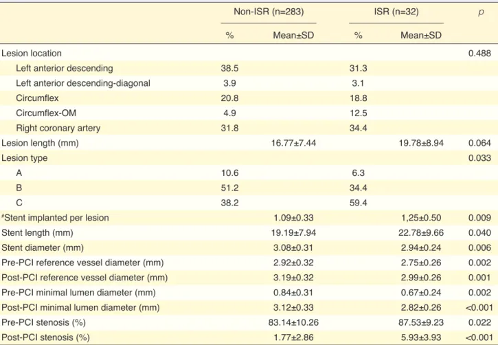
stent implanted. The percentage of type C lesions was higher in the ISR group (59.4% vs. 38.2%, p=0.033). Mean stent length and number of stents implanted per lesion were higher in the ISR group, whereas mean stent diameter was lower. When QCA data were com-pared, pre-procedural and post-procedural mean RVD and MLD were lower in the ISR group, while pre-pro-cedural and post-propre-pro-cedural percentages of diameter stenosis were higher in the ISR group.
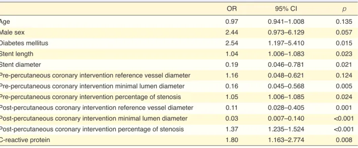
Table 4 shows the results of logistic regression analysis. In the univariate regression analysis (Table 4), DM, serum CRP levels, stent length, stent diam-eter, pre-PCI diameter stenosis, pre-PCI MLD, post-PCI diameter stenosis (residual stenosis), post-post-PCI RVD, and post-PCI MLD were predictors of ISR. Nonetheless, multivariate regression analysis identi-fied only DM and post-PCI diameter stenosis (residual stenosis) as independent predictors of ISR (Table 5).
DISCUSSION
We analysed whether any routinely measured hema-tologic or biochemical parameter might be used as a predictor of ISR in stable CAD in the era of 2nd gen-eration DES. Serum CRP level was identified as an ISR predictor in the univariate regression analysis. However, multivariate regression analysis revealed that only the presence of DM and post-PCI residual stenosis were significant predictors of ISR using 2nd generation DES in stable patients.
In this study, we detected an overall restenosis rate of 10.2%. This is a slightly high rate considering the use of DES; nevertheless, had we restricted coronary angiography procedures only to symptomatic pa-tients, a lower restenosis rate may have been reported. Routine coronary angiography after 6–8 months of stent restenosis remains controversial. In the 2012 ap-propriate use criteria for diagnostic coronary angiog-Table 3. Angiographic parameters with respect to the presence of in-stent restenosis
Non-ISR (n=283) ISR (n=32) p
% Mean±SD % Mean±SD
Lesion location 0.488
Left anterior descending 38.5 31.3
Left anterior descending-diagonal 3.9 3.1
Circumflex 20.8 18.8
Circumflex-OM 4.9 12.5
Right coronary artery 31.8 34.4
Lesion length (mm) 16.77±7.44 19.78±8.94 0.064
Lesion type 0.033
A 10.6 6.3
B 51.2 34.4
C 38.2 59.4
#Stent implanted per lesion 1.09±0.33 1,25±0.50 0.009
Stent length (mm) 19.19±7.94 22.78±9.66 0.040
Stent diameter (mm) 3.08±0.31 2.94±0.24 0.006
Pre-PCI reference vessel diameter (mm) 2.92±0.32 2.75±0.26 0.002
Post-PCI reference vessel diameter (mm) 3.19±0.32 2.99±0.26 0.001
Pre-PCI minimal lumen diameter (mm) 0.84±0.31 0.67±0.24 0.002
Post-PCI minimal lumen diameter (mm) 3.12±0.33 2.82±0.26 <0.001
Pre-PCI stenosis (%) 83.14±10.26 87.53±9.23 0.022
Post-PCI stenosis (%) 1.77±2.86 5.93±3.93 <0.001
Th1 lymphocytes primarily result from classical in-flammatory response; however, eosinophils and Th2 lymphocytes result from allergic inflammation, which is a pathogenetic mechanism in DES but not BMS.[1] Hypersensitivity reactions to the polymer employed in DES underlie allergic inflammation. Although blood eosinophil or monocyte count does not reflect the amount of inflammatory infiltrate in the stented segment, we nonetheless compared their levels be-tween ISR and non-ISR patients. No significant dif-ferences were found.
In BMS restenosis, the main process is neointimal proliferation. This is characterized by smooth muscle cell proliferation and extracellular matrix synthesis, which are driven by arterial inflammation caused by the medial damage and/or penetration of the stent struts into the lipid core of atherosclerotic plaques. [16] Intimal hyperplasia after BMS usually peaks be-tween 6 months–1 year, after which a quiescent pe-riod resumes.[17,18] The mechanism for DES restenosis includes different aspects. Chronic inflammation and impaired endothelial function cause late de novo neo-raphy, routine control angiography after stenting was
considered unnecessary, unless there were symptoms or signs of ischemia.[12] However, in a recently pub-lished study conducted on a large cohort of 10,004 patients, presence of restenosis at follow-up angiogra-phy predicted 4-year mortality, and prognostic value was maintained in asymptomatic patients as well as symptomatic patients.[13]
In a retrospective study of 624 patients who had undergone PCI with BMS due to stable or unstable angina pectoris, pre-procedural N/L ratio was a strong and independent predictor of restenosis.[8] Serum UA, which is associated with atherosclerosis as a result of proinflammatory properties, was also found to be a strong predictor of ISR in BMS in patients with stable and unstable pectoris.[10] CRP is a well-studied marker of systemic inflammation and was found to be related to atherosclerotic events.[14] It was shown to be a sig-nificant predictor of angiographic BMS restenosis in a meta-analysis including 2,747 patients.[15] Inflamma-tion is undoubtedly an important pathogenetic mecha-nism of stent restenosis. Monocytes, neutrophils, and
Table 4. Univariate logistic regression analysis to determine the predictors of in-stent restenosis
OR 95% CI p Age 0.97 0.941–1.008 0.135 Male sex 2.44 0.973–6.129 0.057 Diabetes mellitus 2.54 1.197–5.410 0.015 Stent length 1.04 1.006–1.083 0.023 Stent diameter 0.19 0.046–0.781 0.021
Pre-percutaneous coronary intervention reference vessel diameter 1.16 0.048–0.621 0.124
Pre-percutaneous coronary intervention minimal lumen diameter 0.16 0.045–0.568 0.005
Pre-percutaneous coronary intervention percentage of stenosis 1.05 1.006–1.085 0.024
Post-percutaneous coronary intervention reference vessel diameter 0.11 0.028–0.405 0.001
Post-percutaneous coronary intervention minimal lumen diameter 0.03 0.007–0.140 <0.001
Post-percutaneous coronary intervention percentage of stenosis 1.37 1.235–1.524 <0.001
C-reactive protein 1.80 1.163–2.774 0.008
CI: Confidence interval; OR: Odds ratio. p<0.05 is considered as statistically significant.
Table 5. Multivariate logistic regression analysis to determine the predictors of in-stent restenosis
OR 95% CI p
Diabetes mellitus 2.76 1.084–7.070 0.033
Post-percutaneous coronary intervention percentage of stenosis 1.43 1.121–1.838 0.004
ity. We were not able to serially measure blood levels of the parameters mentioned. There may have been uncontrolled factors which affected the results. In ad-dition, we did not have IVUS or optical coherence tomography data to illustrate the morphology in the stented segment. Control angiography was performed within 1 year of the stenting procedure. Longer-term angiographic results might be different, considering the possibility of later neointimal proliferation with DES[24] and late de novo neoatherosclerosis.[19]
Long-term prospective studies performing control coronary angiography—preferably with IVUS—and obtaining serial measurements of all hematologic, biochemical and inflammatory markers of interest would be beneficial in determining whether these pa-rameters are significant predictors of ISR in DES.
In conclusion, in stable CAD when 2nd generation DES was used, patient-related factors and procedural factors were of paramount importance. None of the pre-procedural blood parameters could be safely used to predict future ISR.
Conflict-of-interest issues regarding the authorship or article: None declared
REFERENCES
1. Niccoli G, Montone RA, Ferrante G, Crea F. The evolving role of inflammatory biomarkers in risk assessment after stent implantation. J Am Coll Cardiol 2010;56:1783–93. CrossRef 2. Kang WC, Ahn TH, Moon CI, Han SH, Shin EK, Kim JS,
et al. Comparison of inflammatory markers and angiographic outcomes after implantation of sirolimus and paclitaxel-elut-ing stents. Heart 2009;95:970–5. CrossRef
3. Doğan A, Kozan Ö, Tüzün N. The physiopathology and treat-ment of in-stent restenosis. Turk Kard Dern Ars 2005;33:115– 25.
4. Moses JW, Leon MB, Popma JJ, Fitzgerald PJ, Holmes DR, O’Shaughnessy C, et al. Sirolimus-eluting stents versus stan-dard stents in patients with stenosis in a native coronary ar-tery. N Engl J Med 2003;349:1315–23. CrossRef
5. Ertaş G, Van Beusekom H. Drug eluting stents: current status and new developments. Anadolu Kardiyol Derg 2012;12:676– 83. CrossRef
6. Kirtane AJ, Gupta A, Iyengar S, Moses JW, Leon MB, Apple-gate R, et al. Safety and efficacy of drug-eluting and bare met-al stents: comprehensive meta-anmet-alysis of randomized trimet-als and observational studies. Circulation 2009;119:3198–206. 7. Kurtul A, Murat SN, Yarlioglues M, Duran M, Karadeniz M,
Ergun G, et al. The association of red cell distribution width with in-stent restenosis in patients with stable coronary artery
atherosclerosis in both BMS and DES; however, this occurs earlier and with greater frequency in DES.[19] Hypersensitivity reaction to the employed polymer is another pathogenetic mechanism of ISR in DES.
Elevated CRP levels were associated with stent thrombosis, death, myocardial infarction, and stroke in patients receiving DES.[20,21] Although pre-proce-dural CRP was an independent predictor of adverse cardiac events, it was not detected as a predictor of stent restenosis with DES. Park et al. showed that while event-free survival was decreased in patients with higher pre-procedural CRP levels who received DES, restenosis rates were not significantly different in the lowest and highest tertiles of CRP.[22] In addi-tion, pre-procedural CRP was not correlated with the degree of diameter stenosis, MLD, or late lumen loss at follow up. In light of these findings, it can be sup-posed that the association between CRP and adverse events reported in those studies may be attributed to stent thrombosis rather than stent restenosis, consid-ering that higher inflammatory activity is associated with increased platelet and clotting cascade activity. [23] In our study, CRP levels were higher in patients with ISR, and it was a predictor of ISR in univariate but not multivariate regression analysis.
Studies describing associations between serum CRP, N/L ratio, serum UA levels, MPV, and RDW are typically performed with BMS. We have not detected any differences with respect to N/L ratio or serum UA levels in patients with or without ISR. This discrepan-cy is probably due to the difference between the study designs. Our study population consisted of patients only with stable CAD, and we used 2nd generation DES. Anti-proliferative and anti-inflammatory effects of the drugs might have prevented us from detecting any inflammatory marker as a predictor of DES ISR.
Kurtul et al. reported that RDW predicted ISR in BMS stable CAD patients.[7] They concluded that im-maturation of red blood cells from inflammatory cy-tokines which cause heterogeneity in RBC size, and oxidative stress which also causes heterogeneity in RBC size were the factors which raised RDW as a predictor of ISR. However, our results differed from that study, as we found similar RDW levels in both ISR and non-ISR patients.
The main limitation of the study was the retrospec-tive design, which prevented us from inferring
causal-vasc Recausal-vasc Med 2008;9:156–65. CrossRef
16. Buja LM. Vascular responses to percutaneous coronary in-tervention with bare-metal stents and drug-eluting stents: a perspective based on insights from pathological and clinical studies. J Am Coll Cardiol 2011;57:1323–6. CrossRef
17. Kimura T, Yokoi H, Nakagawa Y, Tamura T, Kaburagi S, Sawada Y, et al. Three-year follow-up after implantation of metallic coronary-artery stents. N Engl J Med 1996;334:561– 6. CrossRef
18. Komatsu R, Ueda M, Naruko T, Kojima A, Becker AE. Neo-intimal tissue response at sites of coronary stenting in hu-mans: macroscopic, histological, and immunohistochemical analyses. Circulation 1998;98:224–33. CrossRef
19. Park SJ, Kang SJ, Virmani R, Nakano M, Ueda Y. In-stent neoatherosclerosis: a final common pathway of late stent fail-ure. J Am Coll Cardiol 2012;59:2051–7. CrossRef
20. Park DW, Yun SC, Lee JY, Kim WJ, Kang SJ, Lee SW, et al. C-reactive protein and the risk of stent thrombosis and cardio-vascular events after drug-eluting stent implantation. Circula-tion 2009;120:1987–95. CrossRef
21. Park DW, Lee SW, Yun SC, Song HG, Ahn JM, Lee JY, et al. A point-of-care platelet function assay and C-reactive protein for prediction of major cardiovascular events after drug-elut-ing stent implantation. J Am Coll Cardiol 2011;58:2630–9. 22. Park DW, Lee CW, Yun SC, Kim YH, Hong MK, Kim JJ,
et al. Prognostic impact of preprocedural C reactive protein levels on 6-month angiographic and 1-year clinical outcomes after drug-eluting stent implantation. Heart 2007;93:1087–92. 23. Bisoendial RJ, Kastelein JJ, Levels JH, Zwaginga JJ, van den Bogaard B, Reitsma PH, et al. Activation of inflammation and coagulation after infusion of C-reactive protein in humans. Circ Res 2005;96:714–6. CrossRef
24. Kang SJ, Park DW, Mintz GS, Lee SW, Kim YH, Lee CW, et al. Long-term vascular changes after drug-eluting stent im-plantation assessed by serial volumetric intravascular ultra-sound analysis. Am J Cardiol 2010;105:1402–8. CrossRef disease. Platelets 2015;26:48–52. CrossRef
8. Turak O, Ozcan F, Isleyen A, Tok D, Sokmen E, Buyuk-kaya E, et al. Usefulness of the neutrophil-to-lymphocyte ratio to predict bare-metal stent restenosis. Am J Cardiol 2012;110:1405–10. CrossRef
9. Norgaz T, Hobikoglu G, Aksu H, Bolca O, Uyarel H, Eren M, et al. The relationship between preprocedural platelet size and subsequent in-stent restenosis. Acta Cardiol 2004;59:391–5. 10. Turak O, Canpolat U, Özcan F, Mendi MA, Oksüz F, Işleyen
A, et al. Usefulness of preprocedural serum uric acid level to predict restenosis of bare metal stents. Am J Cardiol 2014;113:197–202. CrossRef
11. Zurakowski A, Wojakowski W, Dzielski T, Milewski K, Gościńska-Bis K, Tendera M, et al. Plasma levels of C-re-active protein and interleuk10 predict late coronary in-stent restenosis 6 months after elective in-stenting. Kardiol Pol 2009;67:623–30.
12. Patel MR, Bailey SR, Bonow RO, Chambers CE, Chan PS, Dehmer GJ, et al. ACCF/SCAI/AATS/AHA/ASE/ASNC/ HFSA/HRS/SCCM/SCCT/SCMR/STS 2012 appropriate use criteria for diagnostic catheterization: a report of the Ameri-can College of Cardiology Foundation Appropriate Use Cri-teria Task Force, Society for Cardiovascular Angiography and Interventions, American Association for Thoracic Surgery, American Heart Association, American Society of Echocar-diography, American Society of Nuclear Cardiology, Heart Failure Society of America, Heart Rhythm Society, Society of Critical Care Medicine, Society of Cardiovascular Computed Tomography, Society for Cardiovascular Magnetic Reso-nance, and Society of Thoracic Surgeons. J Am Coll Cardiol 2012;59:1995–2027. CrossRef
13. Cassese S, Byrne RA, Schulz S, Hoppman P, Kreutzer J, Feuchtenberger A, et al. Prognostic role of restenosis in 10 004 patients undergoing routine control angiography after coronary stenting. Eur Heart J 2015;36:94–9. CrossRef
14. Puri R, Nissen SE, Shao M, Uno K, Kataoka Y, Kapadia SR, et al. Impact of baseline lipoprotein and C-reactive protein levels on coronary atheroma regression following high-inten-sity statin therapy. Am J Cardiol 2014;114:1465–72. CrossRef 15. Ferrante G, Niccoli G, Biasucci LM, Liuzzo G, Burzotta F,
Galiuto L, et al. Association between C-reactive protein and angiographic restenosis after bare metal stents: an updated and comprehensive meta-analysis of 2747 patients.
Cardio-Key words: Drug-eluting stent; in-stent restenosis; predictors of
re-stenosis.
Anahtar sözcükler: İlaç kaplı stent; stent içi yeniden daralma;