Address for Correspondence / Yazışma Adresi: Selin Üstün Bezgin, MD, Istanbul Kanuni Sultan Education and Research Hospital, Otorhinolaryngology Department, Istanbul, Turkey E-mail: [email protected]
©Telif Hakkı 2017 Gazi Üniversitesi Tıp Fakültesi - Makale metnine http://medicaljournal.gazi.edu.tr/ web adresinden ulaşılabilir. ©Copyright 2017 by Gazi University Medical Faculty - Available on-line at web site http://medicaljournal.gazi.edu.tr/
doi:http://dx.doi.org/10.12996/gmj.2017.16
Pilomatricoma of the Retroauricular Area
Retroaurikuler Bölgede Pilomatrikoma
Selin Üstün Bezgin
1, Raşit Cevizci
2, Ahmet Keleş
2, Elif Çalış
3, Yıldırım Ahmet Bayazıt
21 Istanbul Kanuni Sultan Education and Research Hospital, Otorhinolaryngology Department, Istanbul, Turkey 2 Istanbul Medipol University, Faculty of Medicine, Department of Otorhinolaryngology, Istanbul, Turkey 2 Istanbul Medipol University, Faculty of Medicine, Department of Pathology, Istanbul, Turkey
ABSTRACT
Pilomatricoma is a benign skin tumor of hair matrix cells. The head and neck is the most commonly affected site of the body. This article describes 41 year-old man who referred to our clinic with an asymptomatic, slow-growing mass on the right retroauricular, suboccipital region. The tumor was excised and histopathologically diagnosed as pilomatricoma. Because of its frequency and predilection of head and neck region, otolaryngologists should be familiar of this tumor.
Key Words: Pilomatricoma, skin neoplasm, calcifying epitelioma.
Received: 09.05.2016 Accepted: 09.28.2016
ÖZET
Pilomatrikoma, kıl folliküllerinin matriks hücrelerinden köken alan benign deri tümörleridir. Bu tümörün en sık görüldüğü yerleşim yeri baş ve boyun bölgesidir. Bu yazıda, 41 yaşında sağ retroauriküler, suboccipital bölgede semptomsuz, yavaş büyüyen kitle ile başvuran bir hasta tarif edilmektedir. Bu tümör çıkarılarak histopatolojik incelemeye gönderilmiş ve pilomatrikoma tanısı almıştır. Baş ve boyun bölgesinde sık görülme eğilimi nedeniyle kulak burun boğaz hekimlerinin bu tümörün klinik sunumunun bilincinde olması ve baş boyun kitlelerinin ayırıcı tanısında bu tümörleri akla getirmesi önerilmektedir.
Anahtar Sözcükler: Pilomatrikoma, deri tümörü, kalsifiye epitelyoma Geliş Tarihi: 05.09.2016 Kabul Tarihi: 28.09.2016
INTRODUCTION
Pilomatricoma, also described as calcifying epitelioma of Malherbe is a skin neoplasm, which originates from hair matrix cells (1-3). This tumor is benign and the neck is the one of the most commonly affected sites of the body. This tumor is actually not uncommon neoplasia that pathologist confirmed this diagnosis for one of every 500 specimens (4). Despite the frequency of pilomatricoma, it is rarely come to mind as prediagnosis by clinicians (5). Its varying presentations may be a cause of misdiagnosis. Thus, it should be important to be awere of this tumor. This article describes a case of pilomatricoma in the retro-auricular, sub-occipital region and discuss this neoplasia.
CASE REPORT
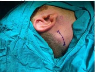
A 41 year-old male patient referred to our clinic with a a right retro-auricular, sub-occipital mass. The tumor has grown in 8 months. No pain, discharge or discoloration occured in this area. There were no other masses or lymphadenopathy or significant medical history of the patient. The superficial, solid, mobile mass measured aproximately 3 cm x 1,5 cm in the right retro-auricular, sub-occipital region (Figure 1). The other parts of ENT examination and systemic examination was normal. Superficial ultrasound examination revealed a 3 cmx 1,5 cm subcutaneous cystic lesion with containing solid areas. Excision surgery was performed under general anesthesia.
Skin incision was made in the sub-occipital region and then by proceeding with blunt dissection of the subcutaneous tissue, the mass was achieved. A firm calcified mass, which is non-adherent to the surrounding tissues was excised (Figure 2). The pathologist explained the diagnosis as pilomatricoma (Figure 3).
Figure 1: Photograph of a mass in the retroauricular, suboccipital region.
Figure 2: Intraoperative photograph of a firm, calcified mass
Figure 3: Photomicrograph of pilomatricoma showing the characteristic basaloid (black arrow) and (blue arrow) shadow cells. Among them, multinucleated giant cells with chronic inflammatory infiltrates are also seen (H&E, ×40)
DISCUSSION
The formation mechanism of pilomatricoma, which is the tumor of hair matrix cell, had explained by the failure of the hair follicle cycle that cause the breakdown in the differentiation of pilar keratinocytes into mature hair follicules. (6,7). Furthermore, the association of pilomatricoma with the genes for myotonic dystrophy, polyoma virüs have been demostrated (8,9). β-catenin gene mutations have also been shown to be associated with pilomatricoma (10).
Clinically, pilomatricoma presents as a superficial, firm, freely mobile, cystic or solid mass. Discoloration as reddish-blue or ulceration of the skin may be seen. These tumors generally are painless, occasionally they present with pain and tenderness (5, 6, 11,12). Pilomatricoma commonly occurs at 5-15 years and at 50-65 years (13) and it has female predominance (5). Although these tumors are solitary lesions, multiple lesions reported in patients with myotonic dystrophy, Turner syndrome, Gardner syndrome, Steinert disease (5, 14). Pilomatricoma can be usually seen in haired parts of the body. Thus, they occur most frequently in the cervicofasial region and the limbs are the second common area. In the head and neck region, because of the high density of hair follicles, the cheek, neck, peri-auricular, and peri-orbital are the common areas fo this tumor. (6,11,15). To avoid misdiagnosis, benign and malignant tumors such as sebaceous and epidermoid cysts, basal cells and scuamous cell carsinoma, vascular lesion, lymph node, lipoma, branchial remnants, preauricular sinuses, chondroma and parotid region tumors should be considered before intervention to the mass (5,12,16,17).
For diagnostic investigation, ultrasonography can be performed for the determination of the position and the calsification of the mass, especially for young patients (6). Computed tomography or magnetic resonance imaging are mainly requested for certain cases such as preauricular and extensive tumors (5, 12). As a part of diagnostic investigation fine needle aspiration has been described (18).
Histopathologically, pilomatricoma is markedly limited nodules with a fibrous capsule. The islands of well-organized malpighian cells are the most frequent microscopic features. The tumor contains basaloid cells with nucleus settled in the peripheral of the island and eozinofilic shadow cells (ghost cells) without nucleus settled in the center (5,6,12). Calsification in the ghost cell region is common feature that ranges from 69% to 85% (18). Malign forms have atypical, undifferentiated cells, a large epithelial cell component and may infiltrate blood vessels and fibrous capsule (12,20).
The target of treatment is the total removal of the tumor (5). This tumor has low recurrence and malignant transformation rate (6). Malignant transformation may occur with advancing ages in males (20). Also death due to the malignant metastatic pilomatricoma was reported (21).
CONCLUSION
Pilomatricoma is one of the most common benign skin neoplasm. Otolaryngologists should consider this tumor in a patient who presented with mobile, superficial, slowly progressive mass in the head and neck region. Cilical presentation is usually suggestive for the diagnosis. Radiologic assesment is important in certain cases. Complete resection of the mass is essential for the treatment. Recurrence and malignant transformation rates are low.
Conflict of interest
No conflict of interest was declared by the authors.
REFERENCES
1. Malherbe A, Chenantais J. Note sur l'epitheliome calcifie des glandes sebacees, Prog. Med. 1880; 8: 826–37.
2. Forbis R, Helwig EB. Pilomatrixoma (calcifying epithelioma), Arch. Dematol. 1961; 83: 606–7.
3. Arnold HL. Pilomatricoma, Arch. Dermatol. 1977; 113.
4. Marrogi, A. J., Wick, M. R., and Dehner, L. P. Pilomatrical neoplasms in children and young adults.Am. J.Dermatopathol. 1992; 14: 87.
5. Lan MY, Lan MC, Ho CY, Li WY, Lin CZ. Pilomatricoma of the head and neck: a retrospective review of 179 cases.Arch Otolaryngol Head Neck Surg 2003; 129: 1327–30.
6. Ashkan Pirouzmanesh, B.S., John F. Reinisch, M.D., Ignacio Gonzalez-Gomez, M.D.,Ebonie M. Smith, B.A., and John G. Meara, M.D., D.M.D. Pilomatrixoma: A Review of 346 Cases.Plast Reconst Surg. 2003;112: 1784-9. 7. Goufman LTDB, Murrell CDRGL, Watkins CDRDV. Quiz case 2: diagnosis: pilomatricoma (calcifying epitelioma of Malherbe). Arch Otolaryngol Head Neck Surg. 2001: 127: 218-20.
8. Harper, P. S. Calcifying epithelioma of Malherbe: Association with myotonic muscular dystrophy.Arch. Dermatol. 1972; 106: 52.
9. Hashimoto, K., Magré, L. A., and Lever, W. F. Electron microscopic identification of viral particles in calcifying epithelioma induced by polyoma virus. J. Natl. Cancer Inst. 1967; 39: 977.
10. Kajino, Y., Yamaguchi, A., Hashimoto, N., Matsuura, A., Sato, N., and Kikuchi, K. -Catenin gene mutation in human hair follicle-related tumors. Pathol. Int. 2001; 51: 543.
Bezgin et al.
Pilomatricoma
GMJ
11. Kwon D, Grekov K, Krishnan M, Dyleski R. Characteristic of pilomatrixoma in children : A review of 137 patients. International Journal of Pediatric Otorhinolaryngology 2014; 78: 1337–41.
12. Duflo S, Nicollas R, Roman S, Magalon G, Triglia JM. Pilomatrixoma of the head and neck in children: a study of 38 cases and a review of the literature, Arch. Otol. 1998; 124: 1239–42.
13. Gupta V, Marwah N, Jain P, Dua S, Gupta S, Sen R. Diagnostic pitfalls of pilomatricoma on fine needle aspiration cytology, Iran J. Dermatol. 2012; 15: 59–61.
14. Trufant J, Kurz W, Frankel A, Muthusamy V, McKinnon W, Greenblatt M, Lazar A, Cook D, Bosenberg MJ. Familial multiple pilomatrixomas as a presentation of attenuated adenomatosis polyposis coli. J. Cutan. Pathol. 2012; 39: 440-3.
15. Schwarz Y, Pitaro J, Waissbluth S, Daniel SJ. Review of head and neck pilomatrixoma. Int Ped Otorhinolaryngol. 2016; 39:573.
16. O’Connor N, Patel M, Umar T, Marcpherson DW, Ethunandan M. Head and neck pilomatricoma: an analysis of 201 cases. British Journal of Oral and Maxillofacial Surgery 2011; 49: 354–8.
17. Aydın S, Bilmez ZE, Erdoğdu S, Altintoprak N, Kayıpmaz Ş. Complicated Giant Pilomatrixoma of the Parotid Region. J Maxillofac Oral Surg. 2016;15111-5.
18. Wang J, Cobb CJ, Martin SE, Venegas R, Wu N, Greaves TS. Pilomatrixoma: clinicopathologic study of 51 cases with emphasis on cytologic features.Diagn Cytopathol 2002; 27: 167–72.
19. Boyd AS, Martin RW III. Pilomatricoma (calcified epitelioma of Malherbe) with secondary ossification. Arch Otolaryngol Head Neck Surg. 1992; 118: 212-5.
20. Hardisson D, Linares MD, Cuevas-Santos J, Contreras F. Pilomatrix carcinoma: a clinicopathologic study of six cases and review of the literature.Am J Dermatopathol 2001; 23: 394–401.
21. Walker DM, Dowthwaite S, Cronin D, Molden-Hauer T, McMonagle B. Metastatic pilomatrix carcinoma: Not so rare after all? A case report and review of the literature. Ear Nose Throat J. 2016;95:117-20.