GENETICS AND MECHANISMS OF ESSENTIAL
TREMOR AND RELATED DISORDERS
A THESIS SUBMITTED TO
THE GRADUATE SCHOOL OF ENGINEERING AND SCIENCE OF BILKENT UNIVERSITY
IN PARTIAL FULFILLMENT OF THE REQUIREMENTS FOR THE DEGREE OF
DOCTOR OF PHILOSOPHY IN
MATERIALS SCIENCE AND NANOTECHNOLOGY
By
Hilal Ünal Gülsüner January, 2015
ii
GENETICS AND MECHANISMS OF ESSENTIAL TREMOR AND RELATED DISORDERS
By Hilal Ünal Gülsüner January, 2015
We certify that we have read this thesis and that in our opinion it is fully adequate, in scope and in quality, as a thesis for the degree of Doctor of Philosophy.
______________________________ Assist. Prof. Dr. Ayşe Begüm Tekinay (Advisor)
______________________________ Prof. Dr. Tayfun Özçelik (Co-advisor)
______________________________ Assoc. Prof. Dr. Mustafa Özgür Güler
______________________________ Assist. Prof. Dr. Özlen Konu
______________________________ Assist. Prof. Dr. Urartu Özgür Şafak Şeker
______________________________ Prof. Dr. Cenk Akbostancı
Approved for the Graduate School of Engineering and Science:
______________________________ Prof. Dr. Levent Onural
iii
ABSTRACT
GENETICS AND MECHANISMS OF ESSENTIAL
TREMOR AND RELATED DISORDERS
Hilal Ünal Gülsüner
Ph.D. in Materials Science and Nanotechnology Advisor: Assist. Prof. Dr. Ayşe Begüm Tekinay
January, 2015
Neurodegenerative disorders are characterized by progressive nervous system dysfunction, and they remain as one of the most challenging disorders known to humankind. These disorders have devastating effects on patients and currently there are no effective therapeutic approaches. Approved medicines provide only symptomatic relief, and inadequacy of information about the molecular mechanisms underlying these conditions restricts the development of new effective therapies. In this thesis, I presented the genetic analysis of two different neurodegenerative disorders and investigate the molecular mechanisms underlying these two disorders: essential tremor and Troyer syndrome. Essential tremor is one of the most prevalent movement disorders; however, its genetic cause and molecular mechanisms remain unknown because of its clinical heterogeneity, age-dependent penetrance, variable expressivity, and relation to other neurodegenerative disorders. In a six-generation consanguineous Turkish family with both essential tremor and Parkinson’s disease, we identified a rare missense mutation of HTRA2 as the causative allele. Family members homozygous for this allele were more severely affected than those heterozygous for this allele. Troyer
iv
syndrome is a very rare autosomal recessive neurodegenerative disorder with only several described cases. Only two reports with truncating mutations have been described. In a consanguineous Turkish kindred with two affected siblings presenting tremor of the hands as well as clinical features similar to that of Troyer syndrome, we identified a novel missense mutation in SPG20. We presented a genotype-phenotype correlation in this family, and the missense SPG20 p.G580R mutation was found to result in a milder form of Troyer syndrome without skeletal abnormalities. Overall, this study provides solutions to complexities of neurodegenerative disorders by suggesting a novel and unifying molecular mechanism underlying essential tremor and Parkinson’s disease. Furthermore, correlation of genotypes and phenotypic differences in patients with Troyer syndrome explains the clinical heterogeneity and variable expressivity of neurodegenerative disorders.
Keywords: Disease gene identification, Essential tremor, hereditary movement disorders, mutation, next-generation sequencing, Troyer syndrome.
v
ÖZET
ESANSİYEL TREMOR VE BENZERİ
HASTALIKLARIN GENETİĞİ VE MEKANİZMASI
Hilal Ünal Gülsüner
Malzeme Bilimi ve Nanoteknoloji Programı, Doktora Tez Danışmanı: Yard. Doç. Dr. Ayşe Begüm Tekinay
Ocak, 2015
Nörodejeneratif hastalıklar ilerleyici sinir sistemi bozuklukları olarak tanımlanırlar ve insanlık tarihinde karşılaşılan en zorlu hastalıklardır. Hastalar üzerindeki etkileri çok tahrip edici boyutlara ulaşabilen nörodejeneratif hastalıkların etkili bir tedavi yöntemleri bulunmamaktadır. Günümüzde kullanılan tedavi yaklaşımları sadece bu hastalıkların belirtilerini azaltmaya yöneliktir. Günümüzde bu tür hastalıkların altında yatan moleküler mekanizmalar ile ilgili yeterli bilgiye sahip olunmaması, yeni ve etkili tedavi yöntemlerinin geliştirilebilmesi konusunda çok büyük engel teşkil etmektedir. Bu çalışmada iki farklı nörodejeneratif hastalık olan esansiyel tremor ve Troyer sendromunun genetik incelemeleri sunulmuştur. Esansiyel tremor en sık görülen hareket bozukluklarından biri olmasına rağmen, bu hastalığın klinik heterojenliği, yaşa bağlı penetransı, değişken ifade edilmesi ve diğer nörodejeneratif hastalıklar ile olan benzerlikleri nedeniyle genetik ve moleküler kökenleri henüz tam olarak tespit edilememiştir. Bu çalışmada, Türkiye’de yaşayan, esansiyel tremor ve Parkinson hastalarının görüldüğü ve akraba evliliği olan 6 kuşaklık bir aile tanımlanmıştır. Bu ailedeki 3 hastadan alınan DNA ile gerçekleştirilen tüm ekzom dizilemesi sonucunda
vi
HTRA2 geninde hastalık yapıcı bir yanlış anlamlı mutasyon tespit edilmiştir. Bu mutasyonu homozigot olarak taşıyan kişilerde hastalık belirtileri heterozigot bireylere göre çok daha şiddetlidir ve bu bireylerde orta yaştan itibaren Parkinson hastalığı gelişmiştir. Troyer sendromu ise çok nadir görülen otozomal çekinik bir hastalıktır. Bu hastalığın nedeni olarak şu anda kadar sadece SPG20 geninde protein ifadesini yok eden iki farklı çerçeve kayması mutasyonu tanımlanmıştır. Bu çalışmada, el titremesi ile birlikte Troyer sendromu belirtilerini taşıyan ve akraba evliliği sonucu dünyaya gelmiş iki kardeş tanımlanmıştır. Bu iki hasta bireyden izole edilen DNA ile gerçekleştirilen tüm ekzom dizileme çalışmaları sonucunda SPG20 geninde yeni bir yanlış anlamlı mutasyon keşfedilmiştir. Bu ailede saptanan SPG20 p.G399S mutasyonu, tanımlanan diğer Troyer vakalarından farklı olarak, iskelet anomalilerine neden olmamaktadır. Bu çalışmalar esansiyel tremor ve Parkinson hastalıklarında yeni ve birleştirici bir mekanizma sunarak, nörodejeneratif hastalıkların karmaşık yapılarına bir çözüm getirmektedir. Ayrıca, Troyer sendromlu ailede tanımladığımız fenotip şiddeti ile genotip arasında saptanan ilişki, nörodejeneratif hastalıkların klinik heterojenliği ve değişken ifadelerini aydınlatmaktadır.
Anahtar sözcükler: Esansiyel tremor, hastalık gen tanımlanması, kalıtsal hareket bozuklukları, Troyer sendromu, yeni nesil dizileme.
vii
viii
Acknowledgement
I would like to express my gratitude to my advisor Dr. Ayşe Begüm Tekinay for her continuous guidance, supervision and encouragement. She always provided me all the support, motivation, and scientific and personal advice which enriched my graduate experience and this thesis.
It is a pleasure to express my gratitude to Dr. Tayfun Özçelik for his valuable discussions, support and encouragement throughout my research.
I would like to express my gratitude to Dr. Mary-Claire King for providing me the opportunity to be a visiting student in her lab, and leading me to work on human genetics, for her insightful comments and motivation. It was an honor to work in your lab.
I would like to express my gratitude to Dr. Mustafa Özgür Güler for his advice, feedback, conversations and critical contribution to my research.
I would like to acknowledge the financial assistance of The Scientific and Technological Research Council of Turkey (TÜBİTAK)-BİDEB 2214/A PhD scholarship.
I am thankful to my colleagues in the NBT and BML groups for their friendship and providing me such a comfortable and warm working environment. I would like to express my special thanks to Hakan Ceylan not only for his friendship, but also for his collaboration, advice and support. I am very grateful to Seher Yaylacı and Samet Kocabey for their friendship and support. I would like to thank to Zeynep Ergül Ülger for her friendship, and technical assistance in the lab.
ix
I would like to thank to everyone in the King lab for providing me a welcoming, warm and fruitful working environment; especially Tom Walsh, Silvia Casadei, Sarah Pierce and Ming K. Lee for their assistance in my experiments.
I am very grateful to my very special friends Gamze Akman, Betül Kayar and Elif İnci Hoşoğlu who had always been there for me, even when we were thousands of miles away. I am very lucky to have great friends like you.
I am indebted to Süleyman Gülsüner for his support, his unconditional love and great patience for all times. He was not only a loving husband but also a mentor all the time; this thesis would have never been possible without you. Words would never express how grateful I am. I am also really grateful to Gönül Gülsüner, Hüseyin Gülsüner, Eda Gülsüner and Gülnur Gülsüner for their support and encouragement.
Finally, I would like to express my deepest gratitude to my parents and my lovely sister, Elif. Without their love, care, encouragement and assistance I would not have finished this thesis. Words do not suffice to thank you. You have most of the credit for what I have achieved and what I will achieve in the future.
HİLAL ÜNAL GÜLSÜNER January, 2015
x
Contents
1 Introduction 1
1.1 Neurodegenerative Disorders 1
1.2 Genetics and Mechanisms of Neurodegenerative Disorders 4
1.2.1 Protein misfolding and aggregation 4
1.2.2 Mitochondrial dysfunction and oxidative stress 5
1.2.3 Endoplasmic reticulum stress 7
1.2.4 Axonal transport deficits 7
1.2.5 Environmental factors 8
1.3 Disease Gene Identification Strategies 9
1.3.1 Traditional Methods 9
1.3.2 Whole Exome Sequencing 11
1.4 Consanguinity 14
1.5 Outline of the Thesis 16
2 Mitochondrial serine protease HTRA2 p.G399S in a kindred with essential tremor
and Parkinson’s disease 18
2.1 Outline 18
2.2 Introduction to Essential Tremor 19
2.2.1 Tremor in human history 19
2.2.2 Clinical features 19
2.2.3 Genetic heterogeneity 20
2.2.4 Etiologic and pathologic heterogeneity 22
2.2.5 Treatments 25
xi
2.3 Materials and Methods 25
2.3.1 Subjects 25
2.3.2 DNA isolation 26
2.3.3 SNP genotyping, haplotype analysis and identity by descent analysis 27 2.3.4 Library construction and whole exome sequencing 29
2.3.5 Bioinformatics 30 2.3.6 Sanger sequencing 31 2.3.7 Population screening 31 2.3.8 Linkage analysis 33 2.3.9 Statistical analysis 33 2.4 Results 33
2.4.1 Clinical features of the ET-1 family 33
2.4.2 Gene discovery 35
2.5 Discussion 42
3 A missense mutation in SPG20 cause Troyer Syndrome 50
3.1 Outline 50
3.2 Introduction to Hereditary Spastic Paraplegias 50
3.2.1 Clinical and genetic features of Hereditary Spastic Paraplegias 50
3.2.2 Troyer Syndrome 52
3.2.3 Aim of this study 54
3.3 Materials and Methods 54
3.3.1 Subjects 54
3.3.2 DNA isolation 55
3.3.3 Library construction and whole exome sequencing 55
xii
3.3.5 Sanger sequencing and population screening 57
3.3.6 Population screening 57
3.4 Results 59
3.4.1 Clinical features of the family 59
3.4.2 Gene discovery 60
3.5 Discussion 61
4 Conclusion and Future Perspectives 71
4.1 Research Summary 71
4.2 Unraveling the genetic bases of complex neurodegenerative disorders 72
4.3 Application to clinical practice 72
Bibliography 74
Appendices 85
xiii
List of Figures
Figure 1.1 Workflow of exome sequencing 12
Figure 1.2 Global prevalence of consanguinity 15
Figure 2.1 Genetic heterogeneity in familial essential tremor 22
Figure 2.2 Pathological examination of the brains of patients with essential tremor 24 Figure 2.3 Quality control of post captured amplified whole exome library 32 Figure 2.4 Pedigree of family ET-1 segregating essential tremor, with genotypes
at HTRA2 G399S 34
Figure 2.5 Archimedes spiral tests of individuals of various ages and genotypes
at HTRA2 p.G399S 37
Figure 2.6 Sanger traces of HTRA2 p.G399S in ET-1 family and its conservation 44 Figure 2.7 Haplotype structure at the HTRA2 locus for the ET-1 family 45 Figure 2.8 Relationship between HTRA2 genotype and age at onset and severity
of tremors of essential tremor 46
Figure 2.9 Schematic representation of HTRA2 protein and its activation against
mitochondrial stress 48
Figure 3.1 Quality control of post captured amplified whole exome libraries of ET-4
Family 56
Figure 3.2 Morphological and radiological analysis of extremities of subject IV:1 60
Figure 3.3 Brain MRI scans of affected subjects 62
Figure 3.4 Pedigree of family ET-4 and segregation of the SPG20 mutation 66 Figure 3.5 Spartin protein and its sequence homology among species 67
xiv
List of Tables
Table 1.1 Clinical and pathological features of hereditary neurodegenerative disorders
and the list of mutated genes 3
Table 2.1 Clinical characteristics of tremor in the affected individuals of family ET-1 28 Table 2.2 Concentrations and qualities of DNA isolated for whole exome sequencing
from ET-1 family 29
Table 2.3 Results of whole exome sequencing of DNA from three severely affected
relatives from ET-1 39
Table 2.4 Homozygous regions shared by three affected relatives 40 Table 2.5 Damaging coding sequence variants shared by exome sequences of three
severely affected relatives of family ET-1 41
Table 3.1 Concentration and quality of DNA extracted from patients for whole exome
sequencing from ET-4 family 55
Table 3.2 Clinical characterization of ET-4 family and comparison with previously
published studies 58
Table 3.3 Results of whole exome sequencing of DNA from two affected relatives from
family ET-4 63
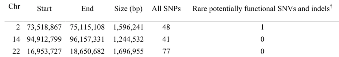
Table 3.4 Homozygous rare variants shared by IV:1 and IV:2 64
xv
ABBREVIATIONS
AD Alzheimer’s disease
ALS Amyotrophic lateral sclerosis
CDK5 Cyclin dependent kinase 5
ET Essential Tremor
HSP Hereditary spastic paraplegia
HTRA2 High temperature requirement protein A2
IBD Identity by descent
NGS Next generation sequencing
PD Parkinson’s disease
PINK1 PTEN induced putative kinase
ROS Reactive oxygen species
SPG20 Spastic paraplegia 20
TS Troyer syndrome
URLs
1000 genomes project 1000genomes.org
GATK broadinstitute.org/gatk/
Mutation Assessor mutationassessor.org
NHLBI exome sequencing project evs.gs.washington.edu/EVS/
Polyphen-2 genetics.bwh.harvard.edu/pph2/
1
Chapter 1
1 Introduction
1.1 Neurodegenerative Disorders
Neurodegeneration is the loss of the structure and death of the neurons in motor, sensory or cognitive systems. Neurodegenerative disorders are characterized by
progressive nervous system dysfunction which begins insidiously and follow a slow but progressive course. The level of neurodegeneration can range from molecular to cellular levels. Individuals first have mild symptoms like problems in coordination or memory. However, as cells start to deteriorate, symptoms progressively get worse, such as patients might lose their ability to walk, move, or think clearly.
There are more than a few hundreds of neurodegenerative disorders. Most of them overlap with each other clinically and pathologically. Neurodegenerative disorders can be classified depending on the pattern of cell loss in different regions of the brain. They are grouped into the basal ganglia, brainstem and cerebellum, cerebral cortex, or spinal cord disorders (1) (Table 1). Basal ganglia disorders are characterized by abnormal movements or defects in the initiation of motor function (2). Parkinson’s disease, which is characterized by loss of dopaminergic neurons and presence of Lewy bodies, is a hypokinetic basal ganglia disorder; that is, higher than normal basal ganglia output causes inhibition of thalamocortical neurons and reduces motor function (2). Huntington’s disease is characterized by chorea and is a hyperkinetic basal ganglia disorder. It is caused by reduced basal ganglia output, which increases thalamocortical
2
function and uncontrollable motor function (2). Essential tremor, Friedreich’s ataxia, multiple systems atrophy, and spinocerebellar ataxia are examples of brainstem and cerebellum disorders. Classification of diseases that predominantly affect brainstem and cerebellum is especially challenging because of the overlap among pathological
symptoms. Cerebral cortex is cerebrum’s outer layer and plays a key role in memory, language, attention, perceptual awareness, and consciousness (3). One of the disorders that occur due to damage in cerebral cortex is Alzheimer’s disease which is
characterized with neuropathological features of neuronal loss, senile plaques, neurofibrillary tangles and acetylcholine deficiency. Diffuse Lewy body disease and Pick’s disease are other examples of cerebral cortex disorders. Spinal cord supports nerve cells that extend from medulla oblongata to brainstem. Defects in spinal cord destroy motor neurons which function in voluntary muscle activities such as speaking, walking, breathing and swallowing (3). The neurodegenerative diseases with defective spinal cord are hereditary spastic paraplegia, amyotrophic lateral sclerosis, and spinal muscular atrophy.
Central nervous system diseases remain one of the most challenging disorders known to humankind. Neurological disorders have devastating affects to patients and there is no effective therapeutic approach. Currently, medications can only provide symptomatic relief, and the development of new effective therapies is prevented by the limited knowledge of the molecular mechanisms underlying these conditions.
3
Table 1.1 Clinical and pathological features of hereditary neurodegenerative disorders and the list of mutated genes
Disease and Predominantly
Affected Site Neuropathological Features Mutated Genes*
Basal Ganglia Huntington’s disease neuronal loss in neurostriatum and cerebral cortex HTT Parkinson’s disease neuronal loss in substantia nigra, Lewy bodies in pigmented neurons
EIF4G1, LRRK2, PARKIN, PARK7, PINK1, SNCA, VPS35
Brain stem and Cerebellum
Essential tremor
large Bergmann glial cells and torpedoes in cerebellum
FUS, DNAJC13 Friedreich’s ataxia neuronal loss in motor
and sensory systems FXN Multiple system atrophy neuronal and glial
inclusions SNCA Spinocerebellar ataxia
neuronal loss in cerebellum and brainstem
Multiple genes (e.g. ATXN1, ATXN2, ATXN3, C10ORF2)
Cerebral Cortex
Alzheimer’s disease
senile plaques,
neurofibrillary tangles, neuronal and synaptic loss
APP, PSEN1, PSEN2
Pick’s disease frontotemporal atrophy,
pick bodies NPC1, NPC2, SMPD1
Spinal cord
Amyotrophic lateral sclerosis
loss of upper and lower motor neurons, Bonina bodies and axonal spheroids
ANG, ALS2, C9ORF72, FIG4, FUS, OPTN, SETX, SOD1, SPG11, TARDBP, UBQLN2, VAPB, VCP Hereditary spastic paraplegia loss of lower motor
neurons
Multiple genes (e.g. ATL1, KIAA0196, PLP1, SPG7, SPG11, SPG20, SPAST, ZFYVE26)
Spinal muscular atrophy loss of lower motor neurons DYNC1H1, SMN1, SMN2, UBA1, VAPB
4
1.2 Genetics and Mechanisms of Neurodegenerative
Disorders
In addition to their extensive clinical diversity, most of the neurodegenerative disorders are genetically heterogeneous. However, the insights gained through genetic studies reveal some overlapping mechanisms (4). These include protein aggregation, oxidative stress, excessive reactive oxygen species formation, mitochondrial
dysfunction, energy depletion and autophagy disruption, endoplasmic reticulum stress, neuroinflammation, and several environmental factors.
1.2.1 Protein misfolding and aggregation
Proteins form networks based on their physical interactions and these networks determine every aspect of life to ensure that cells and organisms function properly. Proteins must maintain their three dimensional structures to stay functionally active in such networks. Chaperone proteins organize protein folding in order to prevent mistakes and malfunctioning proteins. However, partial protein folding or misfolding,
aggregation and deposition have been observed in the brain tissues of neurodegenerative disorders. These disorders are classified as protein conformational diseases or
proteinopathies (5). Some examples of proteinopathies are CAG trinucleotide repeat expansion diseases such as Huntington’s disease and spinocerebellar ataxias (6). Parkinson’s disease, amyotrophic lateral sclerosis, prion disease, and Alzheimer’s disease are also classified as proteinopathies (6). The most important feature of these proteinopathies is that a certain protein folds into an alternative stable conformation which results in its aggregation in the tissues (6). Examples of the aggregated proteins are α-synuclein (encoded by SNCA) in Parkinson’s disease, amyloid-β (cleaved from
5
amyloid-β precursor protein (APP) with the help of enzymes encoded by presenilin1 (PSEN1) and presenilin 2 (PSEN2)) in Alzhemier disease, huntingtin (encoded by HTT) in Huntington’s disease and superoxide dismutase (encoded by SOD1) in amyotrophic lateral sclerosis (6).
The link between protein aggregation and neurodegeneration is not well understood. The hydrophobic patches of these aggregated proteins might interact with other components of the cell and cause damage. In addition, these proteins are part of essential cellular networks, including chromatin organization, vesicular transportation, transcription and translation, cell architecture and protein quality control (7). Therefore, their misfolding and aggregation results in disintegration of these crucial cellular functions (7).
There are several systems used by eukaryotes to remove the malfunctioning proteins and to prevent their aggregation. Cells have protein quality control pathways in order to avoid defects in protein homeostasis. The protein homeostasis is regulated by molecular chaperones, autophagy and ubiquitin proteosome system (UPS) (PARKIN, which is mutated in AD, involves in UPS) to clear proteins expressed in abundance. However, these protein control mechanisms become overwhelmed with age by several genetic and environmental factors. Therefore misfolded proteins can contribute to neurodegenerative disorders by loss-of-function of the protein aggregates, gain-of-toxic-activity, or by causing brain inflammations (6).
1.2.2 Mitochondrial dysfunction and oxidative stress
Mitochondria are present in all cells of the body except erythrocytes and range from hundreds to thousands in number. They provide an organism’s housekeeping
6
functions, including production of the adenosine triphosphate (ATP), biosynthesis of aminoacids, beta-oxidation of fatty acids, and apoptosis (4). Due to their high need of bioenergetics, mitochondria are enriched at presynaptic and postsynaptic terminals of neurons (4).
One of the most common mitochondrial defects that lead to neurodegeneration is oxidative stress. Mitochondria produce ATP through the oxidation of metabolic
intermediates generated during Krebs cycle (8). ATP is generated by electron transport chain (ETC) via redox reactions across the inner mitochondrial membrane. Cells can defend themselves against those reactive oxygen species (ROS), produced during redox reactions, by several antioxidant enzymatic activities including superoxide dismutase and glutathione peroxidase; or scavengers including ascorbic acid (vitamin C),
β-carotene, ubiquinone, lipoic acid and tocopherol (vitamin E) (9). However, disturbances in the regular redox state of the cells might cause increased ROS. The resulting
increased ROS production and insufficiency of antioxidant defense mechanism is called oxidative stress (10). Neuronal tissue is particularly more vulnerable to oxidative stress and damage; moreover, brain contains relatively low levels of antioxidants which lead to neurodegeneration (10). For example, PINK1 encodes a serine/threonine kinase located in mitochondria. Mutations in this gene causes early onset Parkinson’s disease due to stress induced degeneration of dopaminergic neurons (11).
Beside oxidative stress, other major defects in mitochondria that lead to neurodegeneration are abnormal mitochondrial trafficking, distorted inter-organellar communication, and damaged mitochondrial quality control (4). SOD1 mutations induce mitochondrial membrane damage, discharging mitochondrial membrane potential and leading to ALS (11). Several other neurodegenerative diseases including
7
Parkinson’s disease, Alzheimer’s disease and Huntington’s disease are known to be caused by alterations or dysfunction in mitochondrial dynamics (12).
1.2.3 Endoplasmic reticulum stress
Endoplasmic reticulum (ER) is an important organelle for protein synthesis, folding, post-translational modification and transport (13). Accumulation of misfolded proteins and changes in calcium homeostasis lead to ER stress due to the loss of ER integrity or normal function. In order to restore the effects of ER stress, unfolded protein response (UPR) is activated by changing the expression of ER chaperones and enhancing degradation of misfolded or aggregated proteins. Sustained ER stress can lead to the activation of cell death pathways (13).
The defects in the cellular processes involving ER cause different degrees of ER stress contributing to neurodegenerative disorders such as Alzheimer’s disease,
amyotrophic lateral sclerosis, Parkinson’s disease, PolyQ diseases such as Huntington’s disease, and transmissible spongiform encephalophaties (TSE), or prion disease (13-18).
1.2.4 Axonal transport deficits
Motor neurons are large polarized cells. Their function is dependent on the intracellular transport of molecules along their axons. Anterograde axonal transport (towards the axon tip) carries proteins and lipids to the distal synapse and mitochondria (19). Retrograde transport (away from the synapse towards the soma) is essential for the clearance of defective proteins from the axon (19). Kinesin and dynein complexes are responsible for the axonal transport of cargoes in the anterograde and retrograde direction, respectively.
8
Defects in axonal transport are common factors in many neurodegenerative disorders. In Alzheimer’s disease, mutant presenilin 1 and amyloid-β impairs cargo attachment to kinesins and damages axonal transport (20, 21). Mutations in heat shock factor binding protein 1 (HSBP1) cause microtubule deacetylation and disruption of neurofilament assembly resulting in Charcot-Marie-Tooth disease (22). More than 40% of pure hereditary spastic paraplegias (HSPs) occur due to mutations in spastin (SPG4) which results in a progressive axonopathy affecting the cortiospinal tract (23). In ALS, mutations in dynactin 1 (DCTN1) protein harm microtubule-based axonal transport (24). The most common cause of ALS occurs due to the mutations in superoxide dismutase (SOD1). Mutant SOD1 together with glutamate inhibits cargo from binding to the motor protein which slows axonal transport (25).
1.2.5 Environmental factors
Acute or chronic exposure to environmental toxins can also cause
neurodegeneration. Lead poisoning is one of the most common examples which cause myelin loss and axonal degeneration. Other metals (mercury, aluminum, zinc),
pesticides, electromagnetic fields, brain injuries, inflammation, lifestyle and dietary factors are also associated with the increased risk of neurodegenerative disorders (26).
Currently, there is no cure for neurodegenerative diseases, but the medications aim to ease the symptoms. The advances in technology allow the identification of new disease causing genes every day, which results in the exploration of common pathways that lead to certain neurological disorders. This will allow the development of new targeted therapeutic approaches for these pathways.
9
1.3 Disease Gene Identification Strategies
As discussed in section 1.2., neurological disorders are influenced by both defective genes and environment. Identification of genes responsible for diseases is the first step to understand the human genome, to understand the pathophysiology of the disease, and the role of underlying proteins and pathways (27).
1.3.1 Traditional Methods
The most commonly used technique for the identification of Mendelian disease genes have been positional mapping through linkage analysis (28). Prior biological or medical knowledge is not needed to perform linkage analysis; therefore it is unbiased. The theory behind linkage analysis was developed by Thomas Morgan in 1910 that traits that are co-inherited are likely to be due to “factors” that are “linked” (29). Genes located in close proximity on a chromosome are less likely to have recombination events between them, and they tend to co-segregate together. Based on this, the first genetic map to estimate the distance between trait-causing genes based on
recombination was created. If the two traits are co-segregated in a family, then they are assumed to be linked (30). Logarithm of odds (LOD) analysis compares the probability of obtaining the observations if the two loci are linked, to the probability of observing the same data by chance (30). For Mendelian disorders, a LOD score greater than 3 suggests linkage. In 1977 spinocerebellar atrophy (SCA1) became the first
neurolodegenerative disorder linked to a chromosome (HLA region of chromosome 6) (31). Later, in 1983, Huntington’s disease became the first neurologic disorder mapped to a genomic locus using polymorphic DNA markers (32). Although this technology allowed the identification of ~3,000 Mendelian disease loci (28), this approach yields only regions of linkage and not the causative gene.
10
The discovery of DNA sequencing through chain termination of
dideoxynucleotides (ddNTPs) by Frederic Sanger in 1977 was a breakthrough in genomic technologies. This approach allowed the sequencing of candidate genes identified through positional mapping, and had been the most widely used sequencing technique for approximately 30 years (33). Nevertheless, identification of the genes present in the mapped region required several labor-intensive steps, such as cloning the region, and then determining the disease causing gene. For example, although
Huntington’s disease was the first neurodegenerative disorder mapped to a certain locus, it took 10 years of research to clone the region and identify the disease causing gene (34). Medium-throughput DNA sequencing through automated capillary electrophoresis allowed the identification of human reference genome during the Human Genome Project from 1990 to 2001. This development eased the identification of disease causing mutations by eliminating several steps of cloning that required several years of research. However, even with the human reference sequence, most of the disease causing genes remained elusive due to large linkage regions containing hundreds of genes, which increased the costs of follow-up sequencing projects (35).
In addition to linkage analysis, homozygosity mapping is another powerful method to identify recessive disease genes by identifying homozygous regions shared by the affected family members when the family is either consanguineous or practice endogamy (36). This technique relies on genome wide single nucleotide polymorphism (SNP) arrays of affected and unaffected family members.
Despite these advances in technology, a particular category of diseases remained largely unsolved: rare genetic disorders due to the limited number of family members,
11
or diseases with de novo events, locus heterogeneity, reduced penetrance or variable expressivity.
1.3.2 Whole Exome Sequencing
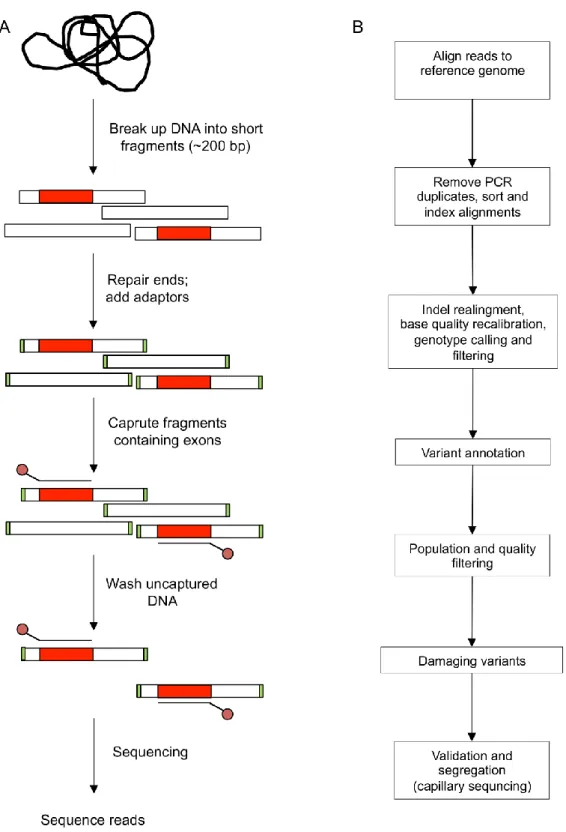
Since 2005, next generation sequencing (NGS) platforms become available and reduce the cost and time of whole genome sequencing. NGS platforms are also applied to target the protein coding regions of the genome (known as the “exome”) through sequence capture by hybridization and massively parallel sequencing (Figure 1.1A). This allows the sequencing of the best known ~1% of the human genome which is estimated to hold ~85% of the mutations associated with disease phenotypes (28). Currently, the cost of whole exome sequencing technology is reduced to ~$600. Discovery of Mendelian disease genes through exome sequencing has been growing exponentially since it was first demonstrated in 2009 (37). Ongoing gene identification reveals genetic causes underlying neurological disorders and illuminates new
biochemical pathways.
Whole exome sequencing has successfully been applied for the identification of disease causing mutations in rare Mendelian diseases such as brain malformations, many forms of cerebellar ataxia and spastic paraplegia (38-40). Along with rare Mendelian diseases, whole exome sequencing has also been applied to common and complex disorders to identify highly penetrant variants in familial forms of Parkinson’s disease, Alzheimer’s disease, essential tremor, and multiple sclerosis (41-44).
The main steps required for whole exome sequencing are shown in Figure 1.1A. Genomic DNA is randomly sheared into 200-300 bp by sonication. The ends of the fragments are converted into blunt ends by removing 3’ overhangs and filling 5’
12
Figure 1.1 Workflow of exome sequencing. (A) Isolated genomic DNA is randomly sheared
into 200-300 bp followed by end repair, A-tailing and indexed paired-end adapter ligation. Exomes are captured and hybridized to biotinylated capture probes, and finally libraries are sequenced. (Copyright 2012 Nature Publishing Group. From Bras et al (35) with permission)
13
overhangs. Then the 3’ ends of the fragments are adenylated (A-tailing). Corresponding thymine nucleotide on the 3’ end of the adapters is ligated to the complementary
fragment. DNA fragments with adapter on both ends are amplified by PCR. The resulting exome library is hybridized to biotinylated DNA beads. The captured DNA is amplified by PCR and is sequenced (Figure 1.1A).
Whole exome sequencing generates millions of short sequence reads per run and the main challenge of whole exome sequencing is to make sense of the large amount of data. Identification of disease genes requires several steps of analytical and functional filtering and analyzing the data to sort through thousands of variants that differ from the human reference sequence (Figure 1.1B). The first analytical step after receiving whole exome sequence data is to map the millions of short sequences to a reference human genome. This is obtained from the reference assembly formed by the Human Genome Project. In order to detect the regions differ from the reference human genome, overlapping reads are required with a minimum coverage of 8 reads for heterozygous genotype calls and 4 reads for homozygous calls. A minimum of 30 reads per base is generally needed throughout the genome to reach the ideal depth of sequencing. Since sequencing platforms have error rates, it is important to differentiate the true variants from sequencing artifacts (45). In order to detect the variants that are rare or private to the subject, variants present in public databases (dbSNP, 1000genomes, EVS, or a set of unaffected individuals) are filtered out. During this filtration, the threshold for minor allele frequency (MAF) should be set accordingly. The next step is the functional filtering where detection of the functional variants among neutral coding variants is needed. Nonsense mutations and frameshift insertions and deletions resulting in a premature stop codon generally lead to truncated and usually nonfunctional protein products. In addition, several computational tools are used to predict the impact of
14
missense variants on protein function, such as SIFT, PolyPhen2, and MutationAssessor, in order to distinguish pathogenic variants from neutrals (46-48). These tools use
biochemical properties of amino acid changes and multiple sequence alignments to obtain the degree of conservation among aminoacid residues in closely related sequences. Finally the candidate variants are verified by genotyping or capillary sequencing, and segregation status is analyzed if the family members are available.
Now exome sequencing becomes the de facto approach to identify disease causing mutations. Although whole exome sequencing has increased the disease gene identification rate, there are still some major reasons why one can not find a variation associated with the disease. These are the lack of or poor sequence coverage of the coding regions, false negative calls in regions with poor quality sequence data, mutations in non-coding regions, and misinterpretation of missense variants (49). Whole genome sequencing and new prediction methods have been used to increase the success rate of disease gene identification.
1.4 Consanguinity
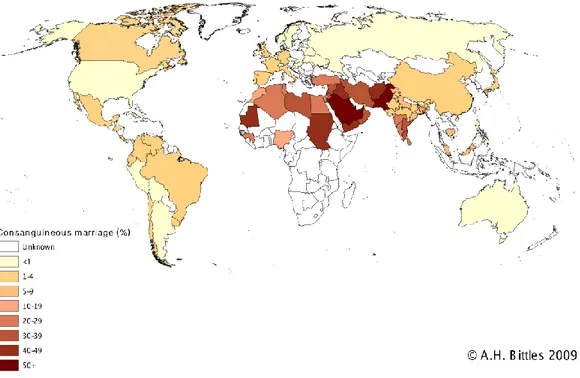
Individuals related as second cousins or closer are defined as consanguineous (50). The most common consanguineous marriages occur between first cousins. Since spouses share 1/8 of their genome, their progeny are homozygous for 1/16 of all loci. Consanguineous marriages are culturally favored in some countries, such as North Africa, West Asia and South India, and constitute around 20-50% of all marriages (51) (Figure 1.2). Consanguineous marriage rate in Turkey is around 20%.
15
Figure 1.2 Global prevalence of consanguinity. (http://www.consang.net with permission from
A.H. Bittles)
Consanguineous marriages have detrimental health effects as it increases the probability of disease causing mutations to reside in homozygous regions (52). Consanguineous populations have limited gene pool by founder effect; moreover, the risk of observing recessive phenotypes due to two copies of the same mutation is increased. A genomic region is classified as identical by descent (IBD) if contiguous SNPs share the same genotype in the family members. The IBD regions shared by all affected individuals that differ from the unaffected individuals are considered as disease causing region. Availability of high-density genome-wide SNP genotyping panels ease to trace IBD regions by analyzing the shared homozygous regions in consanguineous families with recessive disorders. (53). This can lead to the identification of founder mutations in isolated populations. In addition, consanguineous marriages may also introduce individuals with common disorders with a much severe phenotype due to
16
homozygosity; therefore, allows the identification of causative dominant mutations with dosage effect (54).
1.5 Outline of the Thesis
In this dissertation, my work on the genetic analysis of neurodegenerative disorders is divided into four chapters, including this introductory section.
The second chapter addresses my work on the analysis and characterization of a family segregating essential tremor and Parkinson’s disease. Essential tremor is one of the most frequent movement disorders of humans, but its causes remain largely
unknown. In a six-generation consanguineous Turkish family with both essential tremor and Parkinson’s disease, we identified a rare missense mutation of HTRA2 as the
causative allele. Family members homozygous for this allele were more severely
affected than those heterozygous for this allele. The same mutation had previously been associated with Parkinson’s characteristics in mouse mutants and with Parkinson’s disease in some, but not all, epidemiologic studies. Our results suggest that HTRA2 might be responsible for essential tremor in some families and that homozygosity for damaging alleles of HTRA2 may be responsible for Parkinson’s disease.
The third chapter covers the genetic analysis of a family segregating Troyer syndrome. We identified a consanguineous Turkish kindred with two affected siblings presenting clinical features similar to that of Troyer syndrome. Affected siblings have a novel missense mutation in SPG20. Frameshift mutations in SPG20 resulting into loss of function of spartin protein had previously been identified in Troyer syndrome patients. The clinical examinations showed that affected individuals in the Turkish family lack skeletal abnormalities observed in Amish and Omani populations with loss
17
of function mutations in SPG20. These findings suggest that truncated mutations in SPG20 cause a much severe phenotype than that of missense mutations, which indicates a genotype phenotype correlation.
The final chapter summarizes my research and offers future perspectives on the genetic and molecular analysis of neurodegenerative disorders, and applications of these findings to clinical medicine.
18
Chapter 2
2 Mitochondrial serine protease HTRA2
p.G399S in a kindred with essential
tremor and Parkinson’s disease
The majority of the work described here was published in: Unal Gulsuner H., Gulsuner S., Mercan F.N., Onat O.E., Walsh T., Shahin H., Lee M. K., Dogu O., Kansu T.,
Topaloglu H., Elibol B., Akbostanci C., King M.-C., Ozcelik T., Tekinay A.B.
“Mitochondrial serine protease HTRA2 p.G399S in a kindred with essential tremor and Parkinson disease”, Proc Natl Acad Sci USA, 2014, doi: 10.1073/pnas.1419581111
2.1 Outline
Essential tremor is one of the most frequent movement disorders of humans and can be associated with substantial disability. Some but not all persons with essential tremor develop signs of Parkinson’s disease, and the relationship between the conditions has not been clear. In a six-generation consanguineous Turkish kindred with both
essential tremor and Parkinson’s disease, we carried out whole exome sequencing and pedigree analysis, identifying HTRA2 p.G399S as the allele likely responsible for both conditions. Essential tremor was present in persons either heterozygous or homozygous for this allele. Homozygosity was associated with earlier age at onset of tremor (P < 0.0001), more severe postural tremor (P < 0.0001), and more severe kinetic tremor (P =
19
0.0019). Homozygotes, but not heterozygotes, developed Parkinson signs in the middle age. Among population controls from the same Anatolian region as the family,
frequency of HTRA2 p.G399S was 0.0027, slightly lower than other European
populations. HTRA2 encodes a mitochondrial serine protease. Loss of function of HtrA2 was previously shown to lead to parkinsonian features in motor neuron degeneration (mnd2) mice. HTRA2 p.G399S was previously shown to lead to mitochondrial dysfunction, altered mitochondrial morphology, and decreased protease activity; but epidemiologic studies of an association between HTRA2 and Parkinson’s disease yielded conflicting results. Our results suggest that in some families, HTRA2 p.G399S is responsible for hereditary essential tremor and that homozygotes for this allele develop Parkinson’s disease. This hypothesis has implications for understanding the pathogenesis of essential tremor and its relationship to Parkinson’s disease.
2.2 Introduction to Essential Tremor
2.2.1 Tremor in human history
Tremor is a rhythmic and oscillatory movement (55). This phenomenon was first recognized in ancient India (5000-3000 BC), Egypt (700 BC), and Greece (~400 BC) (56). There are also records of patients with kinetic and rest tremors in the writings of Galen of Pergamon (130-200 AD), Franciscus Sylvius (1680) and Gerard van Swieten (1745) (56). However, the first complete study was reported in 1887 by Dr. Charles Dana, with the presence of tremor in several large families (56).
2.2.2 Clinical features
Essential tremor (ET) is a progressive central nervous system disorder and is one of the most frequent movement disorders in humans (57). It is characterized by postural
20
or kinetic tremor of upper extremities, but head, legs, voice and other regions of the body may also be affected (58). Kinetic tremor occurs during voluntary movements such as drinking, eating or writing. The intentional tremor can be identified during a finger-to-nose movement if the amplitude of tremor increases when approaching to the target. Postural tremor might also occur in patients with essential tremor when they hold their arms horizontally against gravity. The familial forms of essential tremor show heterogeneity in terms of age at onset, rate of progression of tremor, and tremor
distribution (59). Rest tremor, which occurs while a body part is relaxed and supported against gravity, has also been observed in patients with essential tremor (59). It is suggested that motor system abnormalities, such as basal ganglia, might cause the basis of rest tremor in essential tremor (58).
Essential tremor is a slowly progressive disorder. Over the years, tremor might spread from the arms to the other unaffected body regions (58). Moreover, some essential tremor patients might develop Parkinson’s disease later in their lives (60).
The worldwide prevalence of ET is 0.9%, increasing up to 4.6% in elderly populations (57). According to a population based study in Turkey, the prevalence of essential tremor is 4.0% among individuals older than 40 (61).
2.2.3 Genetic heterogeneity
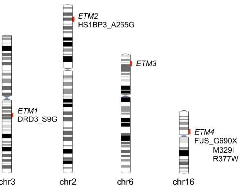
ET is genetically heterogeneous (Figure 2.1). The first chromosomal region was mapped at chromosome 3q13 (ETM1; OMIM 190300) (62) in a genome-wide study including 16 Icelandic families with 75 affected individuals. ET was segregating in an autosomal dominant pattern. The logarithm of odds (LOD) score was 3.71; however, the highest single-family LOD score was 1.29, below the significant threshold to map a monogenic disorder to a marker. Common variant DRD3 p.S9G (OMIM 126451)
21
located in the ETM1 region has been proposed as a risk factor for ET (63) in 23 of 30 French families, but this could not be replicated in other populations (64, 65).
Therefore, these results should be taken cautiously.
ETM2 (OMIM 602134) locus was mapped at chromosome 2p22-24 in a large American-Czech family segregating essential tremor in an autosomal dominant pattern with a significant LOD score of 5.92 (66). The same group confirmed the linkage to ETM2 lous in four independent American families (67). HS1BP3 p.A265G located in the ETM2 region was found in two families with ET (68). However, subsequent association and linkage studies were not able to identify any association or linkage to this region (69, 70). Thus the causality of ETM2 remains unknown.
Linkage to chromosome 6p23 (ETM3; OMIM 611456) was identified in a genome-wide linkage study including seven American families with 325 affected individuals segregating ET in an autosomal dominant pattern (71). However, sequencing the 15 candidate genes in ETM3 locus did not identify any pathogenic variants. Two different linkage studies in Italian families with essential tremor excluded any linkage to ETM3 locus (70, 72). Genome wide association studies reported common intronic variants in LINGO1 (73) and in SLC1A2 (74) to be associated with ET, but meta-analysis indicated that it is unlikely that any of these variants are causative (75).
Recently, a nonsense mutation p.Q290X in the RNA-binding protein FUS was identified by whole exome sequencing in a large family with ET (ETM4; OMIM 614782) (43). Further screening of ET cases for FUS revealed two rare missense variants, (76, 77) suggesting that mutations in FUS explain a subset of cases of ET.
22
Figure 2.1 Genetic heterogeneity in familial essential tremor. Linkage studies revealed three
genomic regions segregating with the condition. No clearly casual mutations have been identified in these regions, except two common variants associated with the disese. Recently a nonsense mutation in the RNA-binding protein FUS was identified by whole exome sequencing.
Another study showed that DNAJC13 p.N855S, which had previously been identified in Parkinson’s disease patients, was also present in two unrelated patients with essential tremor (78).
2.2.4 Etiologic and pathologic heterogeneity
Disease etiology refers to the study of initial or primary causes and origin of the disease. The etiological studies showed that both genetic and environmental factors might be operating in combination for the cause of essential tremor (79). Environmental factors play a role in late-onset neurodegenerative disorders like essential tremor, Alzheimer’s disease and Parkinson’s disease. Case studies show that there are both familial and sporadic forms of essential tremor (58). The causes of these sporadic forms
23
of essential tremor are not fully known but β-carboline alkaloids and lead are some of the putative environmental factors that have been examined with several case-control studies (80, 81). β-carboline alkaloids are present in human diet, such as in animal proteins, and are found to be elevated in the blood concentration of essential tremor patients than in controls (80). Lead, a neurotoxin which causes cerebellar damage, has also been found to be higher in blood from essential tremor patients than controls (81). However, these studies need to be replicated with additional case-control studies. It is possible to suggest the “presence of a gene-environment or two-hit (increased genetic susceptibility followed by exposure to environmental factors) model” for essential tremor (59).
Disease pathophysiology refers to the study of mechanisms operating within an organism and its changes after the disease occurrence. The pathophysiological basis of essential tremor is relatively unknown. The histopathological examinations of brains of indicated that essential tremor cases can be clustered into two categories: those with cerebellar degenerative changes and those with brainstem Lewy bodies (82). The majority of essential tremor cases have cerebellum with larger amounts of Bergmann glial cells and higher Purkinje cell axons, as well as other microscopic changes in the cerebellum (torpedoes) and have been referred as “cerebellar essential tremor” (Figure 2.2A). Bergmann glial cells are nonspecific pathological response to injury, and
torpedoes are swelling of the Purkinje cell axons which results in Purkinje cell loss (83). These findings are not unexpected since the tremor is believed to be mediated by
cerebello-thalamo-cortical pathway (58). Moreover, the intentional component of essential tremor further supports an abnormality of cerebellar function (58). A smaller portion of essential tremor cases show Lewy bodies mainly in the locus coeruleus which is distinctive from brainstem
24
Figure 2.2 Pathological examination of the brains of patients with essential tremor. (A)
Fusiformal axonal swelling (torpedoe) is observed on a modified silver stain of cerebellar Purkinje cell. Torpedoes are pathological accumulation of phosphorylated neurofilaments. (B) Haematoxylin-eosin staining of the locus coeruleus shows multiple Lewy bodies, with as many as four per neuron. (Copyright 2005 Elsevier. From Louis et al. (84) with permission)
Lewy bodies where the patterns are observed prominently in substantia nigra pars compacta (84) (Figure 2.2B). This pattern is different from that of healthy and aged controls in whom Lewy body formation is not observed or very rare (82). It might be suggested that essential tremor patients who develop Parkinson’s disease in later ages go on to develop more widespread (such as nigral) Lewy bodies; however, further evaluation is needed.
Therefore, it might be suggested that essential tremor is “a family of diseases rather than a single entity” (85) due to the presence of multiple genes and environmental factors as the cause for the disease, the evidence of variable pathologic patterns, and the broad variety in the clinical features.
25
2.2.5 Treatments
There is no cure for essential tremor; however, some treatments can ease the symptoms for some patients (86). The cornerstone therapeutic drugs are anticonvulsant primidone and β-adrenoceptor antagonist propranolol (87). Deep brain stimulation (thalamic stimulation) is required to the patients who failed to benefit from the
medications (88). Ethanol is also known to release the symptoms of tremor by binding to the GABAA receptors and facilitate GABAergic neurotransmission; however, there are also serious drawbacks of chronic alcohol usage for treatment (58).
Identification of new causative genes for essential tremor will highlight new biochemical pathways to allow the design of new effective treatments.
2.2.6 Aim of this study
In this study, we examined a 6-generation family (ET-1) segregating ET or ET as a predominant feature of PD. We carried out whole exome sequencing of DNA from three severely affected individuals in the family in order to identify the causative mutation.
2.3 Materials and Methods
2.3.1 Subjects
Family ET-1 is of Turkish origin. The proband was first evaluated at Ankara University Medical School. He and his informative relatives were followed at Ankara University Medical School and Hacettepe University Medical School. This project was approved by the ethics committees of all participating universities and informed consent was obtained from all individuals. Each participant was examined for essential tremor
26
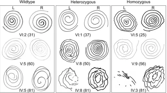
using the criteria of both the Washington Heights-Inwood Genetic Study of essential tremor (WHIGET) and the Consensus Statement of the Movement Disorder Society (MDS) on Tremor (Table 2.1) (89, 90). Each participant was rated for rest and postural tremors and was asked to perform four different tasks (pouring water, drinking water from a cup, finger-to-nose movement, and drawing spirals) to elicit kinetic tremor. During the examination, severity of tremor was rated during each task (89). Participants were evaluated for features of Parkinson’s disease using the diagnostic criteria of the UK Parkinson’s Disease Society Brain Bank. Diagnosis of Parkinson’s disease required presence of bradykinesia plus at least one of muscular rigidity, resting tremor, or
postural instability (91). We collected histories about distribution and severity of tremor and change of these parameters over time. Participants were interviewed about
concurrent use of drugs that might cause action or resting tremor and for symptoms of hyperthyroidism, which was ruled out by TSH tests as needed. Clinical assessments were carried out by at least two neurologists without knowledge of participants’ genotypes. An additional 25 families with hereditary essential tremor, 59 patients with Parkinson’s disease, and 364 healthy controls, ages 20-30 and from the same central Anatolian region as family ET-1, were recruited for genetic analysis from Ankara University Medical School, Hacettepe University Medical School, and Bilkent University. Unaffected individuals showed no signs of disease at the time of
examination. Because controls were young adults, they serve as population controls.
2.3.2 DNA isolation
DNAs of all family members were extracted from blood using Nucleospin Blood Kit (Macherey-Nagel) according to manufacturer’s protocol.
27
2.3.3 SNP genotyping, haplotype analysis and identity by descent analysis
DNA samples from five affected subjects (IV:3, IV:4, IV:8, V:8 and VI:5) and one unaffected subject (IV:5) were genotyped using GeneChip mapping 250K NspI SNP arrays and analyzed using GTYPE software (Affymetrix).
Identity by descent (IBD) regions shared by all genotyped affected family members were determined using a pipeline developed by King Lab (Figure B; Table B). SNP genotype data of a tio (unaffected mother (IV:5), affected father (IV:4), and
affected daughter (V:8)) was used to determine the hypothetical disease alleles for each informative marker. Each marker was then compared in the additional three affected relatives (IV:3, IV:8, and VI:5). If genotypes of one of the three subjects is opposite homozygous at one marker (e.g. disease allele is A, opposite homozygous is BB) then this marker was called IBD0. Then, IBD0 SNPs were merged into segments to define IBD0 regions. A region was assigned as IBD0 if it contains at least 2 informative IBD0 SNPs in a minimum 500 Kb segment. All IBD0 segments, centromeric and telomeric regions, and sequence gaps were filtered out from the whole genome. Remaining regions with at least 3 Mb in size were considered potential IBD segments.
In order to determine whether essential tremor in family ET-1 is linked to DNAJC13 or to genomic regions previously reported to be associated with essential tremor (43, 62, 66, 71, 78), haplotype analysis were performed. Haplotypes were generated in a similar method used for IBD analysis to evaluate the possibility of a disease-causing mutation in any of these regions.
28
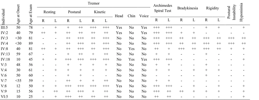
Table 2.1 Clinical characteristics of tremor in the affected individuals of family ET-1
Indi vi du al A ge a t O nse t A ge a t Ex am Tremor Archimedes
Spiral Test Bradykinesia Rigidity
Post ur al Inst abi li ty H ypom im ia
Resting Postural Kinetic
Head Chin Voice
R L R L R L R L R L R L
III:5 50 78 - + + ++ +++ +++ Yes No Yes +++ +++ - - + + - + IV:2 40 79 ++ + ++ ++ ++ ++ Yes No Yes +++ +++ + + - - - -IV:3 <30 81 - - ++ +++ ++ +++ No No No +++ +++ ++ ++ +++ ++ +++ ++ IV:4 <30 89 - - ++ +++ ++ +++ No No No +++ +++ ++ ++ +++ ++ +++ ++ IV:8 40 81 ++ + ++ +++ ++ +++ No Yes No ++ + +++ ++ +++ ++ + + IV:13 59 67 - - + ++ + ++ No No No + + - - + - - -IV:18 10 45 + + +++ +++ +++ +++ No Yes Yes +++ +++ - - - + V:3 48 56 - - + + + + No No No + + - - + - - -V:4 30 61 - + + + + + No No No + + - + + + - -V:6 50 60 - - + + - - No No No - - - - + - - -V:7 <55 59 - - ++ + + ++ No No No + + - - - -V:8 12 50 + + +++ +++ +++ +++ Yes No No +++ +++ - + - + - + V:9 15 56 + ++ ++ +++ + ++ No No No +++ ++ ++ + + + - + VI:5 10 25 - + +++ ++ ++ ++ No No No ++ ++ - - - +
Abbreviations: For tremor +: low amplitude or barely perceivable tremor, ++: moderate amplitude tremor (1-2 cm), +++: large amplitude, severe tremor (>2 cm).(89) For Archimedes spiral test, bradykinesia, rigidity, postural instability and hypomimia +: mild, ++: moderate, +++: severe. R: right hand, L: left hand. None of the individuals had intentional tremor. Subject IV:9 was deceased and is not included in the table. Subject VI:5 was assessed based on his clinical history and is not included in the table.
29
2.3.4 Library construction and whole exome sequencing
Three severely affected individuals (IV:3, IV:4 and VI:5) were selected for whole exome sequencing and their high quality DNA’s were isolated (Table 2.2).
Table 2.2 Concentrations and qualities of DNA isolated for whole exome sequencing from ET-1 family
Sample ID Concentration (ng/µl) A260/280 A260/230
IV:3 215.7 1.9 2.0
IV:4 252.1 1.9 2.2
VI:5 131.5 1.8 1.7
Library construction for whole exome sequencing was carried out according to manufacturer’s protocol (Illumina TruSeq DNA Sample Preparation Guide). For whole exome sequencing, DNA samples were diluted in suspension buffer to a final amount of 2.5 μg in 55 μl. DNA was sheared into 200-300 bp for 120 s in sonicator at 6ºC
(Covaris) in order to generate dsDNA fragments with 3’ or 5’ overhangs. The sheared DNA was run on a gel for quality check; the final amount of the sheared DNA should be 1 μg. End repair was performed in order to convert the overhangs resulting from fragmentation into blunt ends by incubating the fragmented DNA with End Repair Mix for 30 min at 30ºC. The fragmented DNA was cleaned up using AMPure XP Beads. 160 μl AMPure XP Beads were added to each sample and this mixture was placed to
magnetic stand. The DNA samples bound to beads were washed with 80% ethanol and dried. DNA was resuspended in Resuspension Buffer. This was followed by A-tailing by incubating the DNA fragments with A-tailing mix for 30 min at 30ºC. This reaction allows single ‘A’ nucleotides to be added to the 3’ ends of the blunt fragments to prevent them from ligating to one another during adapter ligation. Multiple paired-end
30
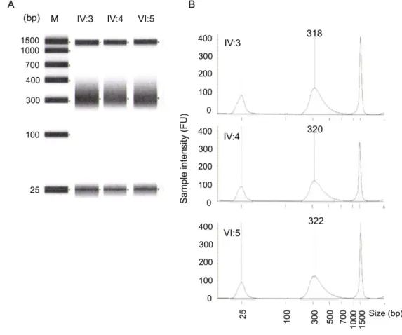
indexing adapters with corresponding T nucleotides on their 3’ ends were ligated to the complementary dsDNA overhangs by incubating at 30ºC for 10 min. The ligation reaction was inactivated by the addition of Stop Ligation Buffer. This step provided DNA fragments for hybridization onto a flow cell. The DNA fragments were cleaned up with AMPure XP Magnetic Beads. After this, DNA fragments with adapters on both ends are amplified with PCR. DNA quality was checked at this step by running on Agilent Technologies 2220 TapeStation instrument. Exomes were captured by SeqCap EZ Exome v2 (Roche) and hybridized to biotinylated capture probes at 47 ºC for 72h. The post-captured exomes were amplified with LM-PCR. The amplified captured exomes were cleaned up using Qiagen QIAquick PCR Purification Kit, and the final concentration and quality were calculated by running on Agilent D1K ScreenTape and analyzing with Agilent Technologies 2220 TapeStation software (Figure 2.3). Libraries were sequenced on an Illumina HiSeq2500.
2.3.5 Bioinformatics
Paired-end sequence reads were aligned to the human reference genome (hg19) using Burrows-Wheeler Aligner (BWA, v0.6.1-r104) (92). Removal of PCR duplicates, sorting and indexing were done using SAMtools v0.1.18 (93). Indel realignments and base quality score recalibration (BQSR) were done with Genome Analysis Tool Kit (GATK, v3.0-0-g6bad1c6) using recommended parameters (94). Genotypes were called and filtered using GATK Unified Genotyper and Variant Filtration tools. Variants were annotated using our in-house pipeline. Common SNPs and artifacts were excluded using dbSNP v138, the NHLBI Exome Sequencing Project, the 1000 Genomes Project, and 700 exomes previously sequenced in our lab. Variants were defined as potentially damaging if they led to premature stop codon or were mutations with scores on in silico
31
prediction tools SIFT P≤ 0.05, PolyPhen2 score ≥ 0.8, and MutationAssessor score ≥ 1.95 (Table S2) (46-48).
2.3.6 Sanger sequencing
Genotypes for candidate variants for 24 informative relatives of family ET-1 were determined by capillary sequencing (ABI 3130xl Genetic Analyzer). All coding regions, potential regulatory regions and miRNA binding sites of HTRA2 were
sequenced in probands from 25 unrelated families with multiple relatives with essential tremor. Primers were designed using Primer3 (Table C1) (95). Products were analyzed via gel electrophoresis and Sanger sequenced. Sanger traces were analyzed with CLCBio Main Workbench software package (CLCBio Inc.). Family ET-1 was genotyped with FAM- and HEX- labeled primers for polymorphic markers on chromosome 2 flanking HTRA2 (ABI 3130xl Genetic Analyzer). Data was analyzed with GeneMapper v4.0 software package (Applied Biosystems).
2.3.7 Population screening
TaqMan genotyping assay (Life Technologies) was used for screening HTRA2 p.G399S in Parkinson’s disease patients and controls. Custom TaqMAn SNP
genotyping assay was designed to amplify and detect both alleleles with probes labeled with FAM or VIC dyes. One homozygous, one heterozygous, two no template controls and 364 Turkish control DNA samples (10 ng each) were added into each well of 384 optical plate. TaqMan Universal PCR Master Mix (No AmpErase UNG) (2X) and SNP Genotyping Assay (20X) were mixed into the required volume and added into the each well of 384-DNA reaction plate. The reaction was performed on ABI 7900HT Fast
32
Figure 2.3 Quality control of post captured amplified whole exome library. (A) Gel image of
post-captured DNA library. 1500 bp and 25 bp are intrinsic upper and lower markers,
33
Real-Time PCR System with optimized conditions. After PCR amplification, an allelic discrimination plate read was set and performed, and the automatic allele calls were converted into genotypes (Figure D).
2.3.8 Linkage analysis
A LOD score for linkage of HTRA2 to essential tremor in the ET-1 family was calculated using LINKAGE v6.0 (96) under an autosomal dominant mode of
inheritance with penetrance for homozygous or heterozygous genotypes of 1.0 at age 40 and older and 0.6 before age 40, no phenocopies, and a mutant allele frequency of 0.01 in the general population.
2.3.9 Statistical analysis
Statistical significance was evaluated by one-way analysis of variance (ANOVA), or analysis of covariance (ANCOVA), as appropriate.
2.4 Results
2.4.1 Clinical features of the ET-1 family
The family is from central Anatolia, where consanguineous and endogamous marriages are common practice. Ancestors of the extended family have lived in the same area for more than 400 years. Essential tremor is known to have segregated in the family for generations. Twenty-four individuals from the family were clinically
assessed (Figure 2.4). Sixteen family members showed varying degrees of tremor (Table 2.1; Figure 2.5), eleven of whom were diagnosed with ET and five of whom (IV:2, IV:3, IV:4, IV:8 and V:9) were diagnosed with ET co-existing with PD.
34
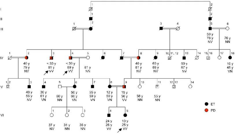
Figure 2.4 Pedigree of family ET-1 segregating essential tremor, with genotypes at HTRA2 G399S. Pedigree of family ET-1.Arrow indicates the proband.
Individuals with ET are shown with filled black symbols, and with PD are shown with filled red symbols. Age at onset for affected individuals, and current ages and genotypes are indicated under the symbols. Individuals who underwent exome sequencing (IV:3, IV:4, VI:5) are represented with asterisks. V indicates the variant allele (Ser) at HTRA2 G399S; N indicates the wildtype allele (Gly). V:11: declined clinical examination.
35
Diagnoses of ET co-existing with PD were made as follows. IV:3 (age 81), IV:4 (age 89), and V:9 (age 56) developed mild to moderate postural and action tremor before age 30 and bradykinesia, rigidity, resting tremor, and postural instability with increasing age. IV:2 (age 79) developed postural and action tremor at middle age and subsequently bradykinesia and resting tremor. IV:8 (age 81) developed postural and action tremor at middle age and subsequently resting tremor, bradykinesia, rigidity, and postural instability. For all five of these patients, all the indicated signs were apparent upon physical examination for this study. Subject V:8, whose clinical features were
particularly severe, was diagnosed with ET at age 12 and with Hashimoto’s thyroiditis at age 49. Subject IV:13 was using propranolol at the time of examination. Subjects V:3, V:4, V:6 and V:7 had mild tremor of at least 4 years’ duration. VI:4 has mild tremor with an age at onset of 24 according to his medical history, but his clinical scores could not be obtained by a face-to-face physical exam. Subject IV:9 provided a DNA sample but died before clinical evaluation; according to her clinical history she had mild hand and head tremor. Subject V:11 agreed to provide a DNA sample but declined subsequent clinical evaluation. Ages of onset of tremor ranged from ~10 years to 60 years. Some family members with tremor were not able to determine the exact age of onset as their tremor developed over many years. ET appeared to be inherited as an autosomal dominant trait spanning six generations.
2.4.2 Gene discovery
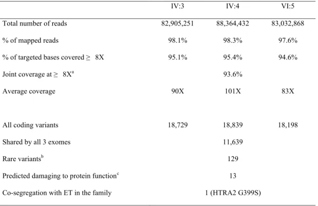
Results of exome sequencing of three severely affected relatives, IV:3, IV:4 and VI:5, are summarized in Table 2.3. The proportion of targeted genomic regions covered at least 8-fold was 95.1% for IV:3, 95.4% for IV:4, 94.6% for VI:5, and 93.6% jointly (Figure E1). Given that the kindred includes multiple consanguineous marriages, we