Yazışma Adresi/Address for Correspondence: Dr. Emine Kılıç Bağır, Cukurova University Faculty of Medicine, Department of Pathology, Adana,Turkey E-mail: [email protected],
ARAŞTIRMA / RESEARCH
Histopathologic subtypes of pediatric lymphomas and relation to
Ebstein-Barr virus: an immunohistochemical and in-situ hybridızation
study
Pediatrik lenfomalarda histopatolojik subtip ve Ebstein-Barr virus ilişkisi:
immünohistokimyasal ve in situ hibridizasyon çalışması
Emine Kılıç Bağır
1, Arbil Açıkalın
1, Melek Ergin
1, Gülay Sezgin
2, Serhan Küpeli
2,
Gülşah Seydaoğlu
31Cukurova University Faculty of Medicine, Department of Pathology, 2Department of Pediatric Oncology, 3Department
of Biostatistics, Adana, Turkey
Cukurova Medical Journal 2018;43(4):868-875
Abstract Öz
Purpose: The aim of this study was toexplore the
histopathologic subtypes of pediatric lymphomas and their relation with Ebstein-Barr virus infection in our region.
Materials and Methods: In this retrospective study, 87
children including 36 cases with Hodgkin lymphoma and 51 cases with non-Hodgkin lymphoma were included in the study. The pathologic slides were used to investigate immunohistochemical staining with ebstein-barr virüs latent membrane protein-1 and in-situ hybridization.
Results: The most common histopathological subtype in
hodgkin lymphoma cases was mixed cellular classical hodgkin lymphoma (55.6%). The most common histopathological subtype in Non-Hodgkin lymphoma cases was Burkitt lymphoma (51.0%). Ebstein-Barr virüs latent membrane protein-1 was positive in 29 Hodgkin lymphoma. All non-Hodgkin lymphoma cases were stained negative with ımmunohistochemical. The positive staining rate of Hodgkin lymphoma cases with in-situ hybridization was 83.3% and this rate was 27.5% in non-Hodgkin lymphoma cases. The differences between groups for both staining methods were significant.
Conclusion: The results of this study show that the
distribution of histopatholojik subtypes of pediatric lymphomas similar to in developing countries. And we have observed that in-situ hybridization is more specific than immunohistochemistry because Ebstein-Barr virus positivity is more detected in in-situ hybridization.
Amaç: Bu çalışmanın amacı bölgemizdeki pediatrik
lenfomaların histopatolojik alt tiplerinin oranını ve bunların Ebstein-Barr virüs enfeksiyonu ile olan ilişkisini incelemektir.
Gereç ve Yöntem: Hodgkin lenfoma tanısı alan 36 olgu
ve non-hodgkin lenfoma tanısı alan 51 olgu retrospektif arşiv taraması yapılarak çalışmaya dahil edildi. Olgulara immünohistokimyasal olarak Ebstein-Barr virüs latent membran proteini-1 ve in-situ hibridizasyon uygulandı.
Bulgular: Hodgkin lenfoma olgularında; en sık rastlanan
histopatolojik alt tipler sırası ile mikst sellüler klasik hodgkin lenfoma (%55,6) idi. Non-Hodgkin lenfoma olgularında en sık rastlanan histopatolojik alt tip Burkitt lenfoma (%51) idi. Ebstein-Barr virüs latent membran proteini-1 ile 29 hodgkin lenfoma olgusunda pozitif iken non-hodgkin lenfoma olgularında immünohistokimya ile pozitif boyanma saptanmadı. Hodgkin lenfoma olgularında in situ hibridizasyon ile boyanma oranı % 83.,3 iken non-Hodgkin lenfoma olgularında bu oran % 27.5 idi.. Her iki boyama yöntemi için gruplar arasındaki fark istatistiksel olarak anlamlıydı.
Sonuç: Bu çalışmada, olgularımızın histopatolojik
dağılımlarının gelişmekte olan ülkelerdeki pediatrik lenfoma olguları ile benzer olduğunu göstermektedir. Aynı zamanda in situ hibridizasyon’da Ebstein-Barr virüs pozitifliği daha fazla olması nedeniyle in situ hibridizasyonun ebstein barr virüs tespitinde immünohistokimyadan daha spesifik olduğunu düşünmekteyiz.
Key words: Ebstein-Barr virüs, hodgkin lymphoma;
INTRODUCTION
Lymphoma includes a group of heterogenous diseases with different epidemiologic, histologic, immunologic and prognostic characteristics that originates from lymphoreticular cells. The first symptom is generally tumoral enlargement of lymph nodes. Lymphomas constitutes approximately 10% of pediatric malignancies in the developed countries. Hodgkin lymphoma (HL) and non-hodgkin lymphomas (NHL) constitutes 40% and 60% of lymphomas, respectively1. The role of Ebstein-Barr
Virüs (EBV) in the development of childhood lymphomas is known. The prevalence of lymphoma is high in malaria-endemic areas. This suggests the role of Plasmodium Falciparum in the pathogenesis. Aside from burkitt lymphoma (BL), EBV was also shown to have a role in the pathogenesis of HL and nasopharynx carcinoma1,2.
EBV can be demonstrated by various methods such as immunohistochemistry, PCR, Southern-Blot, in situ hybridization. In situ hybridization and immunohistochemistry allows the infected cell to be viewed morphologically. In situ hybridization (ISH) is a technique that investigates the specific DNA or RNA in tissue sections bu using complementary nucleic acid sequence or probes. The aim of ISH is generally to investigate the viral infections such as human papilloma virus (HPV), EBV and human immunodeficiency virus (HIV). The viral genome is explored by ISH technique. The ability to recognize latent infections is the superiority of ISH over immunohistochemical (IHC) staining. Latent EBV infections may be shown by ISH technique. This study aimed to explore the distibution of the histologic subtypes of HL and NHL in pediatric age group and the role of EBV infection in this group, compared with the literature.
MATERIALS AND METHODS
This study was performed on 106 pediatric patients with Hodgkin or non-Hodgkin lymphoma diagnosis, who were diagnosed at Cukurova University Faculty of Medicine Pathology Department between 2001 and 2008. We excluded the 19 patients because of the lack of parafine blocks in pathology archive. Demographic data (age, gender), histopathologic subtype and tumor localizations of the rest 87 patients were documented from the hospital file
archive.
The formaline- fixed, parafine- embedded tissues of the 87 patients stained with Hematoxyline eosine (HE) were selected and five µm thick sections were taken for IHC and ISH studies. The study was approved by the ethics committee of Cukurova University. (Ethics committee approval no:7.3.2006/3/3)
Immunohistochemistry method
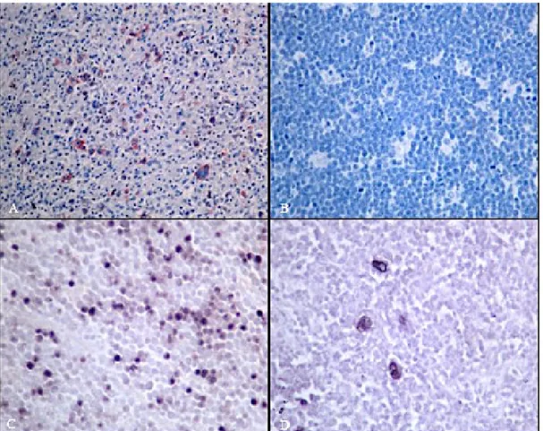
The samples on Poly-L-lysine slides were deparaffinised and washed with distilled water containing hydrogen peroxide (3%). The slides were subjected to antigen retrieval procedure in microwave-oven in the citrate buffer solution (ph.6). Then, the slides were washed 3 to 5 minutes with 0.01 M Phosphate Buffer Saline solution (ph: 7.2-7.4). EBV latent membrane protein (LMP1) (1:50, DAKO, Denmark) as the primary antibody was instilled and incubated for 2 hours at the room temperature. The slides were washed, secondary antibody (LSAB+system-HRP, DAKO) was instilled and incubated for 30 minutes. The slides were washed again, AEC chromogen (3 amino 9 ethyl carbazole) (DAKO, ABD) was instilled, colored with Mayer's Hematoxylin and closed with the water-based material. Detection of Reed-Steinberg cell staining pattern and cytoplasmic granular red-colored staining in the atypical cells were considered positive (Figure 1A, B).
In-situ hybridization (ISH) method
Tissue sections were deparaffinized, incubated in xylene and alcohol (99%, 95%), washed, and incubated for 30 minutes at 37 ᵒC after instilling proteinase K. The slides were washed 2 times with distilled water and dried after dehydration in alcohol solutions (%99,%95). For hybridization, epstein-barr Virus probe ISH Kit (Novacastra, UK) was instilled. It was washed 3 times with Tris-buffered saline (TBS) after incubation for 2 hours at the room temperature. The slides were incubated 10-min with blocking solution, washed again with TBS solution and incubated for 20-min in anti-FITC/AP (vial A). The slides were washed three times with TBS and washed again for 5-min in alkaline phosphatase substrate buffer. In order to show alkaline phosphatase activity, vial B and levamisole/vial C were added and incubated at the room temperature for a night. The slides were counterstained with
mayer's hematoxylin, closed with the water-based material. Detection of reed-steinberg cell staining pattern and dark brown-colored nuclear staining in
the atypical cells were considered positive (Figure 1C, D).
Figure 1. Immunohistochemical and In-situ hybridization staining examples of cases
Positive staining in Reed-sternberg cells (IHC LMP-1x400)(A), negative staining in a burkitt lymphoma case (IHC LMP-1x200) (B), positive result with in-situ hybridization in a burkitt lymphoma case (ISH-EBERx200) (C) and nodular sclerosis classical HL case (ISH-EBERx 400) (D)
Statistical analysis
Histopathological findings, demographic data, IHC and ISH results were evaluated statistically by chi-square test between both groups of patients.
RESULTS
Eighty-seven cases included in the study; 51 were Non-Hodgkin's lymphoma (NHL) (58.6%) and 36 were Hodgkin's lymphoma (HL) (41.4%). Age and gender were not statistically significant in HL and
NHL cases. (Table 1).
Table 1. Demographic characteristics of the groups Parameter Hodgkin Lymhoma Non-hodgkin
Lymphoma p
Age [median
(min-max)] 10.80 (4-15) 10.1 (3-15) 0.125 Gender
(male/female) 27/9 32/19 0.426
The most common histopathological subtype was mixed cellular classical HL (MCCHL) (55.6%) followed by nodular sclerosis classical HL (NSCHL)
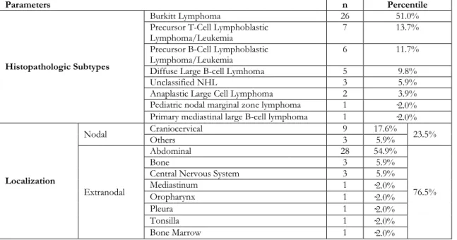
(16.6%). The most frequent involvement area was craniocervical region. nodal involvement was eight times more frequent than extranodal involvement. The histopathological subtypes and involvement areas of HL are presented in Table 2. The most common histopathological subtype was BL (51.0%) followed by precursor T-cell lymphoblastic leukemia/lymphoma (PTCLL) (13.7%). The most frequent involvement area was abdominal (54.9%) followed by craniocervical area (17.6%). The
histopathological subtypes and involvement areas of NHL are presented in Table 3.
EBC LMP1 was positive in 29 cases and all of them had HL. All of the NHL cases were stained negative with IHC. The difference in staining ratios between HL and NHL cases was statistically significant (p<0.001). In terms of ISH, most of the cases with HL had positive staining (83.3%), most of the non-Hodgkin lymphoma cases had negative staining (72.5%).
Table 2. Histopathologic subtypes and localization of Hodgkin lymphoma
Parameters n Percentile
Histopathologic Subtypes Mixed cellular classical HL 20 55.5% Nodular sclerosis classical HL 6 16.6% Nodular lymphocyte predominant
classical HL 1 2.7%
Lymphocyte rich classical HL 0 0%
Unclassified 9 25.0%
Involvement area Nodal Craniocervical 25 69.4% 88.8%
Others 7 19.4% Extranodal Abdominal 1 2.8% 11.2% Bone Marrow 1 2.8% Mediastinum 1 2.8% Lung 1 2.8% HL: Hodgkin lymphoma
Table 3. Histopathologic subtypes and localization of Non-hodgkin Lymphoma.
Parameters n Percentile
Histopathologic Subtypes
Burkitt Lymphoma 26 51.0%
Precursor T-Cell Lymphoblastic
Lymphoma/Leukemia 7 13.7%
Precursor B-Cell Lymphoblastic
Lymphoma/Leukemia 6 11.7%
Diffuse Large B-cell Lymhoma 5 9.8%
Unclassified NHL 3 5.9%
Anaplastic Large Cell Lymphoma 2 3.9%
Pediatric nodal marginal zone lymphoma 1 ̴2.0% Primary mediastinal large B-cell lymphoma 1 ̴2.0%
Localization
Nodal Craniocervical Others 9 3 17.6% 5.9% 23.5%
Extranodal
Abdominal 28 54.9%
76.5%
Bone 3 5.9%
Central Nervous System 3 5.9%
Mediastinum 1 ̴2.0% Oropharynx 1 ̴2.0% Pleura 1 ̴2.0% Tonsilla 1 ̴2.0% Bone Marrow 1 ̴2.0% NHL. Non-hodgkin lymphoma
Tablo 4. Immunohistochemical staining and in-situ hybridization results in Hodgkin and Non-hodgkin lymphoma cases. Hodgkin Lymphoma n (%) Non-hodgkin Lymphoma n (%) Total n
LMP1-IHC staining Negative Positive 29 (80.6%) 7 (19.4%) 51 (100.0%) 0 58 29
Total 36 51 87
EBER-ISH staining- Negative Positive 30 (83.3%) 6 (16.7%) 14 (27.5) % 37 (72.5%) 43 44
Total 36 51 87
EBER: Ebstein Barr-encoding region, ISH. In-situ hybridization, IHC. Immunohystochemistry, LMP-1. Latent membrane protein-1
The difference was also statistically significant (p<0.001). Thirty HL cases (83.3%) and 14 NHL cases (27.4%) had positive results with either IHC or ISH staining methods. All NHL cases had negative results with IHC staining, some of them had positive results with ISH staining. All IHC-positive cases also had IHC-positive results with ISH staining. Fifteen cases with negative IHC staining
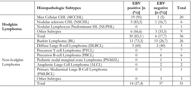
had positive results with ISH staining. IHC staining and ISH staining results are shown in Table 4.Cases with positive test results for EBV with both IHC and ISH methods were evaluated according to histopathologic subtypes of HL and NHL. The results are shown in Table 5. Nineteen over 20 cases with MCCHL, 5 over 6 cases with NSCHL had positive staining for EBV with both methods.
Table 5. Ebstein Barr virus positivity in subtypes of Hodgkin and Non-hodgkin Lymphoma. Histopathologic Subtypes positive [n EBV
(%)] EBV negative [n (%)] Total Hodgkin Lymphoma Mixt Cellular CHL (MCCHL) 19 (95) 1 (5) 20 Nodular sclerosis CHL (NSCHL) 5 (83,3) 1 (16,7) 6
Nodular Lymphocyte Predominant HL (NLPHL) 0 1 1
Other Subtypes 6 (66,6) 3 (33,3) 9
Total 30 (83,3 ) 6 (17,7) 36
Non-hodgkin Lymphoma
Burkitt Lymphoma (BL) 11 (73,3) 15 (26,7) 26
Diffuse Large B-cell Lymphoma (DLBCL) 3 (60) 2 (40) 5
Precursor T-cell Lymphoma (PTCL) 0 7 7
Precursor B-cell Lymphoma (PBCL) 0 6 6
Pediatric nodal marginal zone Lymphoma (PNMZL) 0 1 1
Anaplastic Large Cell Lymphoma (ALCL) 0 2 2
Primary Mediastinal Large B-Cell Lymphoma
(PMLBCL) 0 1 1
Other Subtypes 0 3 3
Total 14 (27,4) 37 51
CHL: Classic hodgkin lymphoma, HL: Hodgkin lymphoma
DISCUSSION
Expression of BCL-2, p53 suppressor gene mutation and damaged immune response are accused in the pathogenesis of HL3. The most
important findingthat shows the role of EBV in the pathogenesis of HL is the high incidence of HL in patients with infectious mononucleosis2,4.
In this study, we performed EBER-ISH and LMP1-IHC to show the presence of EBV in tissue sections. These two methods were selected because of their superiority to Southern-blot and PCR tests in detecting the pathology morphologically at the cell level. We think that EBER-ISH method is more sensitive than LMP1-IHC staining. It should be emphasized that the negative IHC staining despite
positive ISH may be secondary to tissue fixation procedures.
There are serologic, IHC, ISH and PCR-based studies in Turkey related to lymphoma and EBV relation5-16. In these studies, EBV presence in HL
was reported as 43-55%. Yılmaz et al. detected LMP1 positivity by 61.5% through IHC staining in pediatric HL series of 52 cases covering the Southeast Region in Turkey5. In these series,
MCCHL was the most frequent subtype. In the study of Zeytinoğlu et al., which was conducted with a few number of cases and without grouping per age, EBV presence was 50% in HL with EBER-ISH while it was detected 23.8% in NHL16. In most of
the studies, it was shown that the relation of MCCHL with EBV was stronger than NSCHL. In a series of 63 cases, Aktaş et al. found EBER-ISH positivity by 82.5% in all HL cases and by 93.9% in MCCHL cases14. In our study, in compliance with
the studies in the literature, EBV positivity was 83.3% when both methods were evaluated together in cases with HL and 95% of them were MCCHL cases.
Weinred and Amstrong highlighted that EBV incidence in MCCHL in Brazil and Saudi Arabia was higher than the cases in England17,18. They
concluded that 100% EBV positivity in all HL cases in Kenya was probably associated with high BL incidence.
In the distribution of NHL sub-groups, the significance of geographical localization is striking. While in Japan NHL is rare, B-cell lymphomas constitute the half of childhood cancer in equator line countries. Likewise, the excess of B-cell lymphoma in Brazil attracts attention. The studies conducted show the role of EBV19-21. The majority
of the studies conducted with EBV in NHL intended to analyze the EBV prevalence in BL and to present the impact of the virus in this lymphoma type. The association of EBV and HL was first asserted during the researches regarding the etiology of BL in 196421. In the later years, it was determined
that EBV played a role in the etiology of many malignant diseases like BL, nasopharynx carcinoma, HL and B-cell lymphoma in immunodeficiency disorders22,23. Significant differences were detected
between the association of EBV with endemic (African type) and sporadic BL. While higher antibody titers against EBV was found in 80-90% of endemic BL patients, this ratio was around 15-20% in sporadic BL patients18-24. In some non-BL NHL
patients, there are high anti-EBV-VCA titers. In addition, unlike the endemic cases, EBV genome could only be shown in tumor tissues of patients who developed ataxia telangiectasia, X-linked lymphoproliferative syndrome and B-cell lymphoma following organ transplantation20,21. Ebstein barr
vırus positivity in childhood group for BL differs depending on the country, however, the ratios reported were between 34-80%.(25-28) In our study, EBV positive cases detected with EBER-ISH are 42.3%.
In the literature, EBV positivity was also reported in DLBCL patients19,20. In this study, EBV positivity
was detected only in three of five cases (60%) in DLBCL patients. Although this result is statistically significant when compared to other groups, we consider that further studies are required on this matter because of our small number of cases. Çavdar et al. reported that there are more cases with an abdominal location in a pediatric age group in Turkey and that the average age was 5.5 years. The authors found serum EBV-VCA-IgG positivity in 94.8% of BL patients6. Ertem et al. found serum
EBV-VCA-IgG positivity in all 63 pediatric BL patients included in the study10. Due to high
incidence of EBV positivity in BL cases in Turkey and due to abdominal localization of some cases, these cases were suggested to be neither African nor American type, but an "intermediate form”9-12,14.
Denis Burkitt asserted that BL develops as a result of the interaction of reticuloendothelial system exhausted with chronic and severe infection attacks of malaria or other parasites with virus(es). It was shown that EBV-specific immunity is impaired during Plasmodium falciparum attacks, number and function of T-helper and T-suppressor cells are decreased, and in return, the number of B lymphocytes and immunoglobulin synthesis are increased. There is also limited evidence that the precursor merozoite antigen of plasmodium falciparum may activate EBV-infected lymphocytes29,30. Futher studies are warrented to
explore the relationship between EBV a malaria infection. Although our region is an endemic malaria region we could not demostrate malaria in our cases. The HL and EBV relation is as strong as endemic BL and nasopharynx carcinoma. The NHL and EBV relation is weaker than the HL relation. However, the virus plays an active role in the pathogenesis even in some types of NHL. In this
study, the positive rates of EBV, especially MSHL, BL and DBBHL, indicate that EBV has a disease-predisposing role in a significant proportion of lymphoma patients; but it is noteworthy that socioeconomic conditions, genetic characteristics and immunosuppressive factors are important in the development of the disease as a whole.
The limitation of this study was the imbalance between the histopathological groups in terms of number of patients.
Although the average age of patients in this study was similar to those in developed countries. However, when the histopathologic, IHC and ISH results of our cases evaluated together, it was seen that they have the same pathologic characteristics with the cases in developing countries. We believe that more extensive studies should be conducted in which epidemiological, serological and immunological studies are conducted together to better clarify the etiology of pediatric malign lymphomas.
REFERENCES
1. Ozoya OO, Sokol L, Dalia S. EBV-related malignancies, outcomes and novel prevention strategies. Infect Disord Drug Targets. 2016;16:4-21. 2. Anagnostopoulos I, Hummel M. Epstein-Barr virus
in tumours. Histopathology. 1996;29:297-315. 3. Pasman PC, Tiebosch A, Erdkamp FL, Vrints LW,
Breed WP, Schouten HC. P53 as a marker of the malignant cell in Hodgkin's disease. Ann Oncol. 1994;5:89-91.
4. Jarrett RF, MacKenzie J. Epstein-Barr virus and other candidate viruses in the pathogenesis of Hodgkin's disease. Semin Hematol. 1999;36:260-9. 5. Yilmaz F, Uzunlar AK, Sogutcu N, Ozaydin M.
Hodgkin's disease and association with Epstein-Barr virus in children in Southeast Turkey. Saudi Med. J. 2005;26:571-5.
6. Cavdar AO, Tacoy A, Babacan E, Gozdasoglu S, Arcasoy A, Topuz U et al. Hodgkin's disease in Turkish children: a clinical and histopathologic analysis. J Natl Cancer Inst. 1977;58:479-481. 7. Tinguely M, Brundler MA, Gogos S, Kerl K, Borisch
B. Epstein-Barr virus association in pediatric abdominal non-Hodgkin-lymphomas from Turkey. Arch Immunol Ther Exp (Warsz). 2000;48:317-22. 8. Durmaz R, Aydin A, Koroglu M, Aker H, Ozercan
IH, Atik E et al. Detection and genotyping of Epstein-Barr virus by polymerase chain reaction in tissues obtained from cases with Hodgkin's disease in Turkey. Acta Virol. 1998;42:375-81.
9. Tacyildiz N, Cavdar AO, Ertem U, Oksal A, Kutluay L, Uluoglu O et al. Unusually high frequency of a 69-bp deletion within the carboxy terminus of the LMP-1 oncogene of Epstein-Barr virus detected in Burkitt's lymphoma of Turkish children. Leukemia. 1998;12:1796-1805.
10. Ertem U, Duru F, Pamir A, Tacyildiz N, Dagdemir A, Akcayoz A et al. Burkitt's lymphoma in 63 Turkish children diagnosed over a 10 year period. Pediatr Hematol Oncol. 1996;13:123-34.
11. Cavdar AO, Yavuz G, Babacan E, Gozdasoglu S, Unal E, Ertem U et al. Burkitt's lymphoma in Turkish children: Clinical, viral [EBV] and molecular studies. Leuk Lymphoma 1994;14:323-30.
12. Cavdar AO, Gozdasoglu S, Yavuz G, Babacan E, Unal E, Uluoglu O et al. Burkitt's lymphoma between African and American types in Turkish children: Clinical, viral (EBV), and molecular studies. Med Pediatr Oncol. 1993;21:36-42.
13. Ataoglu O, Fen T, Suer O, Daglı M, Akı Z, Yamac K. Epstein-Barr Virus latent membrane protein 1 (LMP-1) in Hodgkin's lymphoma patients in Turkey. Turk J Haematol. 2001;18:123-6.
14. Aktas S, Kargi A, Olgun N, Diniz G, Erbay A, Vergin C. Prognostic significance of cell proliferation and apoptosis-regulating proteins in Epstein-Barr virus positive and negative pediatric Hodgkin lymphoma. Lymphat Res Biol. 2007;5:175-82. 15. Tanyildiz HG, Yildiz I, Bassullu N, Tuzuner N,
Ozkan A, Celkan T et al. The role of Epstein-Barr virus LMP-1 immunohistochemical staining in childhood Hodgkin lymphoma. Iran J Pediatr. 2015;25:e2359.
16. Zeytinoglu A, Hekimgil M, Erensoy S, Aydemir S, Berber S, Cagirgan S et al. Investigation of Epstein-Barr virus DNA and RNA in tissues of patients with lymphoma. Mikrobiyol Bul. 2005;39:473-81. 17. Weinreb M, Day PJ, Niggli F, Powell JE, Raafat F,
Hesseling PB et al. The role of Epstein-Barr virus in Hodgkin's disease from different geographical areas. Arch Dis. Chil. 1996;74:27-31.
18. Armstrong AA, Alexander FE, Paes RP, Morad NA, Gallagher A, Krajewski AS et al. Association of Epstein-Barr virus with pediatric Hodgkin's disease. Am J Pathol. 1993;142:1683-8.
19. Araujo I, Foss HD, Bittencourt A, Hummel M, Demel G, Mendonca N et al. Expression of Epstein-Barr virus-gene products in Burkitt's lymphoma in Northeast Brazil. Blood. 1996;87:5279-86.
20. Lanzkowsky P, Lipton JM, Fish JD. Manual Pediatric Hematology and Oncology, 6th edition. London,
Academic Press, 2016.
21. Reiter A. Non-Hodgkin lymphoma in children and adolescents. Klin Pediatr. 2013;225:87-93.
22. Capone G, Fasano C, Lucchese G, Calabro M, Kanduc D. EBV-associated cancer and autoimmunity: searching for therapies. Vaccines (Basel). 2015;3:74-89.
23. Pollock BH, Jenson HB, Leach CT, McClain KL, Hutchison RE, Garzarella L et al. Risk factors for pediatric human immunodeficiency virus-related malignancy. JAMA. 2003;289:2393-9.
24. Epstein MA, Achong BG, Barr YM. Virus particles in cultered lymphoblasts from Burkitt's lymphoma. Lancet. 1964;1:702-3.
25. Anwar N, Kingma D, Boch AR, Mourad M, Raffeld M, Franklin J et al. The investigation of Ebstein-Barr viral sequences in 41 cases of Burkitt's lymphoma from Egypt. Cancer. 1995;76:1245-52.
26. Peylan-Ramu N, Diment J, Krichevsky S, Ben-Yehuda D, Bhatia K, Magrath IT. Expression of EBV encoded nuclear small non-polyadenylated RNA (EBER) molecules in 32 cases of childhood Burkitt's lymphoma from Israel. Leuk Lymphoma. 2001;40:405-11.
27. Klumb CE, Hassan R, De Oliveira DE, De Resende LM, Carrico MK, De Almeida Dobbin J et al. Geographic variation in Epstein-Barr
virus-associated Burkitt's lymphoma in children from Brazil. Int J Cancer. 2004;108:66-70.
28. Mansoor A, Stevenson MS, Li RZ, Frekko K, Weiss W, Ahmad M et al. Prevalence of Epstein-Barr viral sequences and EBV LMP1 oncogene deletions in Burkitt's lymphoma from Pakistan: Epidemiological correlations. Hum Pathol. 1997;28:283-8.
29. Whittle HC, Brown J, Marsh K, Blackman M, Jobe O, Shenton F. The effects of Plasmodium falciparum malaria on immune control of B lymphocytes in Gambian children. Clin Exp Immunol. 1990;80:213-8.
30. Pinkus GS, Said JW. Hodgkin's disease, lymphocyte predominance type, nodular--a distinct entity? Unique staining profile for L&H variants of Reed-Sternberg cells defined by monoclonal antibodies to leukocyte common antigen, granulocyte-specific antigen, and B-cell-specific antigen. Am J Pathol. 1985;118:1-6.