1 Department of Urology, Diyarbakır Gazi Yasargil Training and Research Hospital, Diyarbakir, Turkey 2 Department of Radiology, Diyarbakır Gazi Yasargil Training and Research Hospital, Diyarbakir, Turkey
3 Department of Urology, Silvan Dr Yusuf Azizoglu State Hospital, Diyarbakir, Turkey
Yazışma Adresi /Correspondence: Selçuk Altın,
Department of Urology, Gazi Yasargil Training and Research Hospital, Diyarbakir, Turkey Email: [email protected]
ORIGINAL ARTICLE / ÖZGÜN ARAŞTIRMA
Our Initial Experiences with Laparoscopic Urologic Surgery
Laparoskopik Ürolojik Cerrahi Başlangıç Deneyimlerimiz
Selçuk Altın1, Ramazan Topaktaş1, Ali Akkoç1, Cemil Aydın1, Reha Girgin1, Zeynep Banu Aydın2, Kadir Yıldırım3
ÖZET
Amaç: Kliniğimizde gerçekleştirilen ürolojik laparoskopik
cerrahi olguların sonuçları ve komplikasyonlarını geriye dönük olarak değerlendirmek.
Yöntemler: Ocak 2012 - Ocak 2015 tarihleri arasında
la-paroskopik operasyon geçiren toplam 115 hasta geriye dönük incelendi. Hastaların demografik özellikleri, preo-peratif tanıları, laparoskopik yaklaşım şekli, ameliyat ve yatış süreleri, cerrahi işlem esnasında ve sonrasında ge-lişen komplikasyonlar ve açık cerrahiye geçilen hastalar açısından incelendi.
Bulgular: Hastaların 61’i kadın, 54’ü erkek ve ortalama
yaşları 52,4±11,7 yıl idi. Altmış sekiz olguda transperito-neal, 47 olguda ise retroperitoneal girişim uygulandı. Top-lam 29 hastaya basit böbrek kisti eksizyonu, 25 hastaya basit nefrektomi, 22 hastaya üreterolitotomi, 19 hastaya radikal nefrektomi, 15 hastaya piyelolitotomi ve 5 has-taya da piyeloplasti yapıldı. Toplam 115 vakanın 4’ünde (%3,4) açık operasyona geçildi. Bu hastalar dışında ma-jör komplikasyon ve mortalite görülmedi. En sık yapılan ameliyatların ortalama süreleri ise; böbrek kist eksizyonu 62 (50-110) dk, basit nefrektomi 125 (95-140) dk, üretero-litotomi 108 (90-130) dk, radikal nefrektomi 141 (105-175) dk, piyelolitotomi 116 (95-140) dk ve pyeloplasti 166 (150-190) dk idi. Ortalama yatış süresi 3,7±2,8 gün (2-11) idi.
Sonuç: Uyguladığımız laparoskopik cerrahilerin
sonuç-ları, başarı ve komplikasyon oranları literatürle uyumlu bulundu. Laparoskopik cerrahi, teknolojinin gelişmesi, de-neyimlerin artmasıyla ayrıca hastalar tarafından daha iyi tolere edilmesi nedeniyle açık cerrahiye alternatif, güvenli ve minimal invaziv bir tedavi yöntemi olduğunu düşün-mekteyiz.
Anahtar kelimeler: Laparoskopi, başlangıç deneyimleri,
retrospektif
ABSTRACT
Objective: Retrospectively, to evaluate outcomes and
complications of urological laparoscopic surgery cases performed in our clinic.
Methods: A total of 115 patients who received
laparo-scopic surgery between January 2012 and January 2015 were retrospectively evaluated. Included patients were assessed in terms of demographic characteristics, pre-operative diagnosis, type of laparoscopic approach, dura-tion of surgery and hospitalizadura-tion, complicadura-tions before and after surgery, and postoperative requirement for open surgery.
Results: 61 of included patients were women, 54 were
male, and the mean age was 52.4±11.7 years. Sixty-eight patients underwent transperitoneal and 47 patients ceived retroperitoneal procedures. While 29 patients re-ceived renal cyst excision, 25 had simple nephrectomy, 22 had ureterolithotomy, 19 had radical nephrectomy, 15 had pyelolithotomy and 5 had pyeloplasty. Four (3,4%) of the 115 patients required converting to open surgery. Except these patients, no major complication or mortal-ity was encountered. The mean duration of surgery for the most commonly applied procedures were as follows: renal cyst excision 62 (50-110) min, simple nephrectomy 125 (95-140) min, ureterolithotomy 108 (90-130) min, rad-ical nephrectomy 141 (105-175) min, pyelolithotomy 116 (95-140) min, and pyeloplasty 166 (150-190) min. The mean hospital stay was 3.7±2.8 (2-11) days.
Conclusion: The success and complication rates of the
laparoscopic surgeries performed in our clinic were con-sistent with those reported in the literature. In the light of technological advances and increasing experience, as well as based on the higher tolerance exhibited by pa-tients, we believe that laparoscopic surgery is a minimally invasive method that is a safe alternative to open surgery.
Key words: Laparoscopy, initial experiences,
INTRODUCTION
Recently, laparoscopic surgery has been increas-ingly employed in urology due to improvements in technical capabilities and experience. Laparoscopic surgery is recognized as a minimally invasive sur-gery that has become increasingly popular world-wide during the 90s which has also been started to be used since the beginning of 2000 in Turkey. Laparoscopic surgery comes with many advantages compared to open surgery such as lesser degree of pain and hemorrhage, shorter hospital stay, and bet-ter cosmetic results [1]. Laparoscopy was first ap-plied for pelvic lymphadenectomy in urology [2], which was followed by its successful application for laparoscopic nephrectomy in children and adults [3]. In the following years, it has been started to be used for various indications such as pyeloplasty, ureteral stone, nonpalpable testes, and adrenal sur-gery. In this study, we retrospectively evaluated our case series of 115 patients in terms of outcomes and complications of laparoscopic surgery.
METHODS
The medical records of 115 patients who received laparoscopic surgery at the Urology Department of
Diyarbakir Gazi Yasargil Teaching and Research Hospital between January 2012 and January 2015 were retrospectively evaluated. The laparoscopic procedures were performed, in a single center by 4 surgeons using transabdominal and retroperitoneal methods, with no manual manipulation. Patients were evaluated with regard to age, gender, preop-erative diagnosis, laparoscopic approach, duration of surgery and hospital stay, analgesic requirement, complications during or after surgery, pre-operative and post-operative laboratory results, blood transfu-sion, and converting to open surgery. In our clinic, as part of a routine clinical procedure, patients with a urinary infection were treated with oral or paren-teral antibiotics depending on the urinary culture re-sults. Prior to the operation, surgical informed con-sent was obtained from each patient, while patients were put on magnesium citrate starting from one night before surgery and were also given prophylac-tic antibioprophylac-tics (cefuroxime axetil 50 mg/kg) 1 hour before the procedure. All patients underwent gen-eral anesthesia. Transperitoneal laparoscopic meth-od was performed by making the patients assume a 70° lateral decubitus position, while retroperitoneal method was carried out with the patients at com-plete lumbar (lateral decubitus) position (Figure 1). Figure 1. Patient was
posi-tioned in flank position and trocar positioning for right transperitoneal approach.
The duration of surgery was described as the time interval between placement of the first trocar and closure of the skin. A febrile status was de-scribed as a temperature above 38°C. Postopera-tively, at day 1, all patients were evaluated via blood count and biochemical profile after which they were discharged. Follow-up assessment was scheduled for 1 week later. All patients were scheduled to visit for follow-up assessment at postoperative 1 and 6 months. The values were expressed as mean ± stan-dard deviation.
Operative technique
Transperitoneal access was achieved using a Verres needle and retroperitoneal access was achieved by
a 1.5 cm incision through the Petit triangle. Sub-sequently, the retroperitoneal area received bal-loon dilatation, guided by a finger. Pneumoperito-neum was induced to achieve a CO2 pressure of 20 mmHg. The first trocar placement was performed at umbilical level, lateral to the rectus in transperito-neal approach, and through the Petit triangle in ret-roperitoneal approach. In both methods, 3 standard ports (2 10mm and 1 5mm) were used. Depending on the difficulty encountered during liver retraction and dissection, the port number was increased. Af-ter the insertion of the trocars, intraabdominal pres-sure was reduced to 12 mmHg. During the dissec-tion, both ultrasonic (Harmonic Scalpel-Ethicon ®)
and thermal (LigaSure-Covidien ®) energy sources were employed. The transperitoneal approaches in-cluded the routine severance of triangular hepatic ligament and the white line of Toldt on the right. The posterior hepatic ligament and the adrenal extension of the ascending colon were medialized. Ureter was found and suspended. On the left, unlike procedures on the right, splenecolic ligament was also severed and the colon was completely medialized.
After this stage at simple and radical nephrec-tomy, in both approaches, nonabsorbable and lock-ing polymeric clips of large and X-large size (Hem-O-Lok™, Research Triangle Park, NC) were placed first on the artery and then on the vein. The insignif-icant (<7 mm) vascular structures were treated with metal clips or ligatures. Ureter was severed by clos-ing the metal clip. The hemorrhage was controlled under low pressure (6 mmHg). The excised renal tissue was put into a laparoscopic bag (Endocatch II, Covidien-ABD) and removed through a cutane-ous incision as small as possible. No morcellation was employed to the tissues.
Prior to the cyst excision, cases suspected of having a problem in the renal calyceal system were evaluated by retrograde pyelography. Cyst fluid was removed via aspiration and the excised cyst wall specimen was sent for pathologic interpretation.
In ureterolithotomy and pyelolithotomy cases, the stones were removed using a grasper through a longitudinal incision made by scissors in some cases and scalpel in others, without applying any energy. Following the procedure, antegrade double J stent insertion was applied in each patient. The in-cision site was closed using a water-proof 4-0 Vicryl suture.
In cases of dismembered pyeloplasty, antegrade insertion of the ureteral catheter was performed be-fore the operation. Subsequent to the ureteral
spatu-lation, antegrade insertion of a 4.7 F double J stent was achieved. Anastomosis was performed using a 4-0 Vicryl. While the insertion sites of the 10 mm trocars were closed with double suture layers in-cluding the fascia and skin, the insertion sites of the 5 mm trocars were closed using only single-layer sutures through the skin. All patients received peri-operative ureteral catheter insertion and a drain was placed at the end of the procedure.
RESULTS
Of 115 patients, 61 were female and 54 were male. The mean age was 52.4±11.7 years (range: 21-72 years). The demographic data of the study popu-lation are shown in Table 1. As 68% of the cases (59.1%) underwent transperitoneal approach, 47 (40.8%) received retroperitoneal approach. While 29 (25.2%) had simple renal cyst excision, 25 (21.7%) had simple nephrectomy, 22 (19.1%) had ureterolithotomy, 19 (16.5%) had radical nephrec-tomy, 15 (13%) had pyelolithotomy and 5 (4.3%) had pyeloplasty.
Table 1. Demographic characteristics of the patients
(n=108)
Gender female/male (%) 53 / 47
Mean age, year* 52.4 ±11,7 (21-72)
Mean duration of hospital stay (day)* 3.7±2.8 Preoperative hemoglobin (mg/dl)* 13.9±1.5 Postoperative hemoglobin (mg/dl)* 12.8±1.4 * mean ± standard deviation
The 68 transperitoneal cases included 20 renal cyst excisions, 14 simple nephrectomy, 13 radical nephrectomy, 10 ureterolithotomy, 7 pyelolithoto-my, and 4 pyeloplasty. The 47 retroperitoneal cases included 12 ureterolithotomy, 11 simple nephrecto-my, 9 renal cyst excisions, 8 pyelolithotonephrecto-my, 6 radi-cal nephrectomy, and 1 pyeloplasty (Table 2).
Diagnosis Transperitoneal Retroperitoneal Total
Simple nephrectomy Nonfunctional kidney 14 11 25
Radical nephrectomy Renal tumor 13 6 19
Cyst excision Simple cyst 20 9 29
Ureterolithotomy Ureteral stone 10 12 22
Pyelolithotomy Pelvic stone 7 8 15
Pyeloplasty Ureteropelvic stenosis 4 1 5
Table 2. Type and
num-ber of laparoscopic ap-proaches
In both approaches, the mean duration of tro-car insertion was 17±10.8 (10-30) minutes. As 22 patients required the insertion of a 4th trocar, no such need was observed in others. Four of the cases (3.4%) required converting to open surgery. The un-derlying cause for converting to open surgery in 3 of those cases (2.6%) (2 renal masses and 1 non-functional kidney) was occurrence of renal vein in-jury during placement of a vascular clip, inability to reach the pedicle due to adhesions, and
uncorrect-able technical imaging error (during retroperitoneal pyeloplasty). The patients who converted to open surgery due to hemorrhage, received one unit of perioperative erythrocyte suspension, . In these pa-tients, vital signs were stable during postoperative period. Other than those, no major complication or mortality associated with anesthesia or surgery was observed. Perioperative and postoperative results are shown in Table 3.
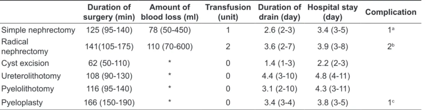
Table 3. Comparison of operative data relative to types of laparoscopic surgery Duration of
surgery (min) blood loss (ml)Amount of Transfusion(unit) Duration ofdrain (day) Hospital stay(day) Complication
Simple nephrectomy 125 (95-140) 78 (50-450) 1 2.6 (2-3) 3.4 (3-5) 1a Radical nephrectomy 141(105-175) 110 (70-600) 2 3.6 (2-7) 3.9 (3-8) 2b Cyst excision 62 (50-110) * 0 1.4 (1-3) 2.2 (2-3) Ureterolithotomy 108 (90-130) * 0 4.4 (3-10) 4.8 (4-11) Pyelolithotomy 116 (95-140) * 0 3.1 (2-10) 4.3 (3-11) Pyeloplasty 166 (150-190) * 0 3.4 (3-4) 3.8 (3-5) 1c * Insignificant hemorrhage
a. Converted to open surgery because of inability to reach the pedicle due to adhesions in a nonfunctional kidney with
a history of surgery
b. Converted to open surgery because of hemorrhage associated with a hem-o-lock injury during hilar vascular control c. Converted to open surgery because of a monitoring error.
All the patients were mobilized within 24 hours. The ureteral catheters were removed after a mean period of 1.3±0.8 (1-8) days and the drain was removed after a mean period of 3.1±1.1 (2-10) days. The mean hospital stay was 3.7±2,8 (2-11) days. The drainage was continued for 7 days in a radical nephrectomy case, 10 days in 3 pyelolithotomy and ureterolithotomy cases, and 8 days in 2 cases. The cases with higher duration of drainage had a serious drainage fluid and they exhibited regression in their follow up, requiring no additional intervention. The ureteral catheters were removed after a mean period of 18.9±4.5 (15-27) days.
DISCUSSION
Recently, as a result of the technological advances, laparoscopic surgery, a minimally invasive sur-gical method, has been replacing open surgery in urologic surgery. Laparoscopic surgery presents many advantages over open surgery to the patients with lesser degree of pain and hemorrhage, shorter
hospital stay, and better cosmetic outcomes, while providing a better view and safer procedure to the surgeons [1].
Laparoscopic interventions were applied as transperitoneal and retroperitoneal approaches. In the literature, the transperitoneal approach is often preferred, however, in our clinic, we use both of them [4,5]. The most important factor in choosing between these two approaches is the experience and preference of the surgeon, however, both methods have their own ups and downs. Transperitoneal ap-proach provides a better range of movement due to larger surgical site and higher distances between the ports, however, in order to reach the retroperitoneal located kidney via transperitoneal approach, the mobilization of intraabdominal organs is required. In retroperitoneal approach, in addition to the dif-ficulty of controlling renal hilus, it is hard to reach the upper pole of kidney and the distal ureter, while there is a need to learn to work in this area. How-ever, it also presents some advantages such as faster kidney reach, absence of any risk for intraperitoneal
organ injury and postoperative adhesion associated with shorter hospital stay, and its easy-to-apply na-ture in patients with a history of abdominal surgery [6,7].
Studies have not shown any significant differ-ence between transperitoneal and retroperitoneal approaches with regard to complications, need for postoperative analgesia, and duration of hospital stay [7]. Since the surgeons in our clinic have a wide spectrum of experience, 68 patients (59%) received transperitoneal and 47 patients (41%) received ret-roperitoneal approaches.
Following the first recognition of laparoscopic nephrectomy by the authorities, it has been increas-ingly used in the treatment of upper urinary system diseases. Laparoscopic surgery is most commonly applied in simple and radical nephrectomy proce-dures, with comparable safety and lower complica-tion rates as compared to the open surgery [8]. In the literature, the most common complications of laparoscopic nephrectomy cases are hemorrhage, spleen, liver and intestinal injury, unsuccessful or-gan removal, abdominal wall hematoma, and in-traperitoneal abscess [9,10]. Rasweiler et al. con-ducted a study reporting their first 100 laparoscopic
experiences and noted converting to open surgery in 17 cases which was explained by them with the difficulty of the procedure and the individual learn-ing curve, however, in our case series, only 4 cases necessitated converting to open surgery due to hem-orrhage, technical errors and adhesions [11]. Con-verting to open surgery is necessary in the presence of large vessel injury, impaired hemodynamics, or-gan injury, and in cases where laparoscopic experi-ence is not enough. In our nephrectomy series of 25 cases, the mean surgery time was 125 (95-140), mean blood loss was 78 (50-450) ml, while in our radical nephrectomy series of 19 cases same values were 141 (105-175) min and 110 (70-600) ml, re-spectively. In the literature, the mean duration of simple nephrectomy is 200 (120-300) min and the mean amount of blood loss is 210 (50-1500) ml [12] which were consistent with our results.
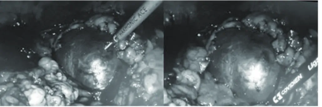
In our clinic, our first laparoscopic experience was with simple renal cyst excision (Figure 2). While the mean duration of surgery for simple renal cyst decortication was 45 minutes in our country, it was 62 minutes in our series [13,14]. We believe that the longer duration in our study may be associ-ated with those being our first laparoscopic cases and the natural consequence of a learning curve.
Figure 2. Renal cyst excision
through the transperitoneal approach
Laparoscopy has been first practiced for stone surgery in urology by Wickkam in 1979 [15]. Lapa-roscopic ureterolithotomy has been reported to be safe and beneficial in cases of large ureteral stones impacted in the mucosa or in patients with solitary kidney where ESWL (Extracorporeal Shock Wave Lithotripsy) and endourological technology fail to succeed [16]. Of 134 laparoscopic ureterolithotomy performed by Simforoosh, 114 were transperitoneal and 20 were retroperitoneal [17]. The mean duration of surgery was 143 min., stone-free rate was 100%, and side effect rate was 10%. In our series, 45.4%
of the laparoscopic ureteral stone cases were treated with transperitoneal and 54.5% were treated with retroperitoneal approaches, while the mean duration of surgery was 108 min and the stone-free rate was 100%. Postoperatively, 3 of our patients demon-strated prolonged urinary leakage and the drain was removed without any need for intervention at post-operative 10 days. Transperitoneal approach has a downside that infected urine may contact with the peritoneal space or organs. However, Janetschek et al. reported no side effects in their series of transab-dominal surgeries [18]. Laparoscopic kidney stone
treatment is indicated in cases where methods such as ESWL, PNL (percutaneous nephrolithotomy), and flexible ureterorenoscopy (F-URS) fail; or in the presence of large or complex stones in ectopic or rotated kidneys or when the patient does not want to undergo open surgery or other invasive methods [19].
In the present study, the kidney stones were single and all were larger than 2 cm with no risk of push back to the calyceal system. Some of our pa-tients had rotation anomaly and had received ESWL therapy with no success. Some had underwent suc-cessful treatments in other clinics but their stones had recurred. Thus, we performed laparoscopic pyelolithotomy in 15 patients. In a study compar-ing the open and laparoscopic pyelolithotomy, the mean duration of surgery was 94.43 min, the mean hospital stay was 3.8 days, and the mean duration of drainage was 2.7 days in the laparoscopic group [20]. In the present study, the mean duration of sur-gery was 116 minutes, while the mean duration of drainage and hospital stay were 3.1 and 4.3 days, re-spectively, which were consistent with the literature. Two of our pyelolithotomy patients demonstrated prolonged urinary leakage and the drains were re-moved without any need for additional intervention at 8 and 10 days. Considering that we do not have PNL and F-URS devices in our hospital, we can
say that laparoscopic procedures are not an alterna-tive to the modern techniques routinely performed in kidney stones. We believe that laparoscopic ap-proach should be carried out only in the absence of other more appropriate endoscopic methods among candidates of open surgery.
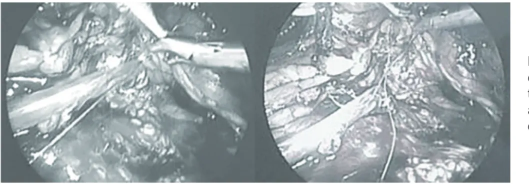
Laparoscopic pyeloplasty has been first started to be used at the beginning of 1990s and today it has become the most preferred minimally invasive method in obstruction of ureteropelvic junction [21]. According to the reports of various institutions performing laparoscopic pyeloplasty, laparoscopic approach yields outcomes similar to those of open surgery and provides a better quality of life [22,23]. Laparoscopic pyeloplasty may be carried out using retroperitoneal or transperitoneal approach depend-ing on the experience and preference of the surgeon (Figure 3). Other factors involved in the decision are history of surgery, morbid obesity, and presence of crossing vessels. Generally, we prefer the transperi-toneal approach because of our higher experience with that. Converting to open surgery during lapa-roscopic pyeloplasty is generally associated with failure to dissect the ureteropelvic region, ureteral stent migration, or failure to approximate ureter and pelvis, however, in our series, converting to open surgery was due to sudden loss of vision because of an imaging system failure in one case [24,25].
Figure 3. Dismembered
py-eloplasty was performed through the transperitoneal approach and anastomosis of the ureteropelvic junction
In conclusion, the search of patients and sur-geons for better functional outcomes and lower mor-bidity along with scar-free surgeries will continue. In this regard, laparoscopy is recognized as a safe and practicable technique acting as an alternative to open surgery in the recent years due to technologi-cal advances. In Turkey, it is an easily practicable technique, as well. Our initial experiences with laparoscopic surgery are promising and consistent
with the literature. We believe that our improving experience in laparoscopic surgery will also make more of our patients prefer this technique which is associated with high patient satisfaction.
Declaration of Conflicting Interests: The
au-thors declare that they have no conflict of interest.
Financial Disclosure: No financial support
REFERENCES
1. Demir Ö, Öztürk B, Eğriboyun S, Esen AA. Initial experi-ence with urologic laparoscopic surgery in our clinic and the learning process. Dokuz Eylül Üniversitesi Tıp Fakül-tesi Derg 2010;24:105-112.
2. Schuessler WW, Vancaillie TG, Reich H, Griffith DP. Trans-peritoneal endosurgical lymphadenectomy in patients with localized prostate cancer. J Urol 1991;145:988-991. 3. Clayman RV, Kavoussi LR, Soper NJ, et al. Laparoscopic
nephrectomy: initial case report. J Urol 1991;146:278-282. 4. Chung JH, Lee SW, Lee KSet al. Safety of en bloc ligation
of the renal hilum during laparoscopic radical nephrectomy for renal cell carcinoma: a randomized controlled trial. J Laparoendosc Adv Surg Tech A 2013;23:489-494.
5. Rassweiler J. Laparoscopic radical prostatectomy is also on-cologically safe and effective! BJU Int 2013;112:158. 6. Leclair MD, Vidal I, Suply E, et al. Retroperitoneal
laparo-scopic heminephrectomy in duplex kidney in infants and children: a 15 -year experience. Eur Urol 2009;56:385-389. 7. Kim C, McKay K, Docimo S. Laparoscopic nephrectomy in
children: systemic review of transperitoneal laparoscopic and retroperitoneal approaches. Urology 2009;73:280-284. 8. Dağgulli M, Utanğaç MM, Bozkurt Y, et al. Our laparo-scopic radical nephrectomy experiences. Dicle Med J 2014;41:732-737.
9. Keeley FX, Tolley DA. A review of our first 100 cases of laparoscopic nephrectomy: Defining risk factors for com-plications. Br J Urol 1998;82:615-618.
10. Siqueira TM, Kuo RL, Gardner TA, et al. Major compli-cations in 213 laparoscopic nephrectomy cases: The Indi-anopolis experience. J Urol 2002;168:1361-1365.
11. Rassweiler JJ, Seemann O, Henkel T, et al. Retroperitoneos-copy. Technique and experiences with the first 100 patients. Urol A 1996; 35: 185-195.
12. Kural AR, Demirkesen O, Akpınar H, et al. Our initial experiences with laparoscopic nephrectomy. Turk J Urol 2004;30:414-421.
13. Hatipoğlu NK, Penbegül N, Söylemez H, et al. Urological laparoscopic surgery: Our experience of first 100 cases in Dicle University. J Clin Exp Invest 2012;3:44-48.
14. Bayraktar AM, Ölçücüoğlu E, Taştemur S, et al. Initial re-sults of our laparoscopic urological surgery: Firs 32. Fırat Medical Journal 2014;19:75-78.
15. Wickham JEA. The surgical treatment of renal lithiasis. In: Urinary Calculous Disease. edn. Edited by JEA W: New York, NY: Churchill-Livingstone; 1979: 145-198.
16. Gaur DD, Trivedi S, Prabhudesai MR, et al. Laparoscopic ureterolithotomy: technical considerations and long-term follow-up. BJU Int 2002;89:339-343.
17. Simforoosh N, Bassiri A, Danesh A, et al. Laparoscopic management of upper urinary tract stones: a report of 134 cases. Urology 2006;68:15.
18. Nambirajan T, Jeschke S, Albqami N, et al. Role of laparos-copy in management of renal stones: single-center experi-ence and review of literature. J Endourol 2005;19:353-359. 19. Türk C, Knoll T, Petric A, et al. Guidelines on urolithiasis.
European Association of Urology, Guidelines 2015. 20. Patloo AM, Sarmast AH, Khan MA, et al. Laparoscopic
retroperitoneal pyelolithotomy and open pyelolithotomy: a comparative study. Turk J Urol 2012;38:195-200.
21. Schuessler WW, Grune MT, Tecuanhuey LV, Preminger GM. Laparoscopic dismembered pyeloplasty. J Urol 1993;150:1795-1799.
22. Eden CG. Minimally invasive treatment of ureteropelvic junction obstruction: a critical analysis of results. Eur Urol. 2007;52:983-989.
23. Tan BJ, Rastinehad AR, Marcovich R, et al. Trends in urt-ereropelvic junction obstruction management among urolo-gists in the United States. Urology 2005;65:260-264. 24. Soulie M, Seguin P. Urological complications of
laparo-scopic surgery: Experience with 350 procedures at a single center. J Urol 2002;165:1960-1963.
25. Rassweiler JJ, Teber D, Frede T. Complications of laparo-scopic pyeloplasty. World J Urol 2008;26:539-547.