STUDIES ON RELEASE OF KETOROLAC TROMETHAMIN AND
INDOMETHACIN FROM OPHTHALMIC HYDROGEL INSERTS
OFTALMİK HİDROJEL İNSERTLERDEN KETOROLAK TROMETAMİN VE
İNDOMETAZİN’İN SALIMI ÜZERİNDE ÇALIŞMALAR
Ayşegül KARATAŞ, Tamer BAYKARA
Ankara University, Faculty of Pharmacy, Department of Pharmaceutical Technology, 06100 Tandoğan-Ankara, TURKEY
ABSTRACT
Hydrogels are hydrophilic, three-dimensional networks which are able to imbibe large amounts of water or biolojical fluids, and thus resemble to a large extent biological tissue. The well-tolerated safety and biocompatability of Poly (2 – hydroxyethly methacrylate) and poly (2 -hydroxypropyl methacrylate ) hydrogels especially are evident in their use as contact lens and drug release system. In this study, inserts of water-soluble Ketorolac tromethamin (KT) and water- insoluble indomethacin (IND) were prepared using hydrogels such as Poly (butyl methacrylate) (pBMA), Poly (2-hydroxyethyl methacrylate) (pHEMA), and poly (2-hydroxypropyl methacrylate) (pHPMA), and a plasticizer such as Polyethylene glycol 300 (PEG) by film casting method. Swelling properties of these inserts was determined and they were irradiated with an absorbed dose of 1.2 Mrad by means of a Co- 60 source. The effects of these parameters on the drug release were examined. The mechanism of drug release was identified by means of the semi-empirical equation developed by Korsmeyer and Peppas. Swelling of the hydrogels and release of drugs from the hydrogels increased with size of side chain, hydroxyl groups on the side chain of the acrylate and using PEG 300 in the formulation, which increase hydrophilicity. Water–soluble KT showed higher release than water–insoluble IND. It was also observed no effect of irradiation dose on the release of drugs from the inserts. Release of KT mainly fit to the Fickian diffusion mechanism, whereas drug release of IND mainly showed the non-Fickian release mechanism according to their n exponent values.
ÖZET
Hidrojeller suyu ya da biyolojik sıviları büyük miktarlarda içen ve böylece büyük çapta biyolojik dokuya benzeyen hidrofilik ve üç boyutlu ağlardır. Özellikle hidroksietil metakrilat) ve poli(2-hidroksipropil metakrilat) hidrojellerinin iyi-tolere edilme emniyeti ve biyogeçimliliği onların kontak lens ve etkin madde salım sistemi olarak kullanımının bir kanıtıdır. Bu çalışmada, suda çözünen ketorolak trometamin’in (KT) ve suda çözünmeyen indometazin’in (İND) poli (bütilmetakrilat) (pBMA), poli(2-hidroksietil metakrilat) (pHEMA) ve poli(2-hidroksipropil metakrilat) (pHPMA) gibi hidrojeller ve plastizer olarak Polietilen glikol 300 (PEG) kullanılarak film hazırlama yöntemiyle insertleri hazırlanmıştır. Bu insertlerin şişme özellikleri tayin edilmiştir ve insetler Co-60 kullanılarak 1.2 Mrad doz ile ışınlanmıştır. Etkin madde salımı üzerinde bu parametrelerin etkisi incelenmiştir. Etkin madde salım mekanizması Korsmeyer ve Peppas tarafından geliştirilen yarı-emprik salım mekanızmasıyla tanımlanmıştır. Hidrojellerin şişmesi ve hidrojellerden etkin maddelerin salımı yan zincir büyüklüğü ile, akrilatın yan zinciri üzerinde hidroksil gruplarının yeralmasıyla ve formüllerde PEG 300’ün kullanılmasıyla hidrofilisitenin artmasından dolayı artmıştır. Suda çözünür KT ile suda çözünmeyen İND’den daha fazla salım görülmüştür. İnsertlerden etkin maddelerin salımı üzerinde sterilizasyon dozunun bir etkisinin olmadığı da gözlenmiştir. KT’nin salımı başlıca Fick difüzyon mekanizmasına uyum gösterirken, İND’in salımı onların n eksponent değerlerine göre başlıca Fick difüzyon mekanizmasına uymayam salım göstermiştir.
Anahtar kelimeler: Ketorolak trometamin, İndometazin, H idrojel, Işınlama, Oftalmik
INTRODUCTION
A variety of carriers have been explored to modify the response to drugs that are delivered topically to the eye. These include gels, nanoparticles, polymer matrices, liposomes, and the Ocusert system (1).
Provision of a matrix to sustain drug release in the eye can be achieved in several ways. One of them is hydrogel insert. It can serve as a drug reservoir. It appears to be extremely effective way of maintaining a therapeutic concentration of drug on the eye surface for considerable periods of time (1) In addition, the ability of hydrogels to release an entrapped drug in to aqueous medium and to regulate the release drug by control of swelling and cross-linking makes them particularly suitable for controlled release applications. Hydrogels can be applied for the release of both hydrophilic and hydrophobic drugs (2).
Hydrogels are three dimensional, hydrophilic crosslinked, polymeric networks that have capacity to swell by absorbing water or biological fluids (3-6). Because of the high amount of water they have the structure that resembles the natural tissue. Hydrogels can be synthesized by
using hydrophilic monomers, polymers or copolymers, depending on the type of the monomers or polymers or the composition of the copolymers. Their hydrophilicity and, their swelling capacity as a result of hydrophilicity may differ. The crosslinking ratio is one of the most important factors that affect the swelling of hydrogels (3-8).
Synthetic hydrogels such as poly (2 -hydroxyethlymethacrylate) and poly (2 -hydroxypropyl methacrylate) have also been used for various biomedical applications ranging from soft contact lenses (9) to drug delivery systems because of their inertness, good biocompatibility and well controlled release of entrapped drug in an aqueous medium (10).
The use of soft contact lenses as drug delivery devices may produce a miracle of therapeutic opportunities. Since the development of poly(hydroxyethyl methacrylate), pHEMA, hydrogels as soft contact lenses, important efforts have been made to use them as drug vehicles for both chronic (e.g. glaucoma) and acute (e.g. infections or inflammatory process) diseases (11, 12)
Indomethacin and Ketorolac tromethamine are non-steroidal anti-inflammatory drugs (NSAIDs) and have been widely used as topical antiinflammatory agents to the eye (13-15)
Any formulation instilled in the eye must be sterile. A variety of sterilization techniques may be used to sterilize ophthalmic medications. These include sterile filling, autoclaving, and sterilization by irradiation. Of all these techniques, sterilization by irradiation is the one best suited to ocular inserts containing drug (16)
The objective of this study was to prepare the insert of water-insoluble indomethacin and water-soluble ketorolac tromethamine using hydrogels such as Poly (butyl methacrylate), Poly (2-hydroxyethly methacrylate) and poly (2-hydroxypropyl methacrylate) and a plasticizer such as polyethylene glycol 300, by film casting method, to investigate the release of both indomethacin and ketorolac tromethamine from hydrated hydrogel inserts and swelling properties of hydrogel inserts, and to evaluate effect of irradiation dose of sterilization on drug release from the hydrogel inserts.
MATERIALS AND METHODS Materials
Indomethacin (IND) (Selectechemie A.G., Switzerland) and Ketorolac tromethamine (KT) (DEVA, Turkey) were used as model drugs. Poly (butyl methacrylate) (pBMA), Poly (2-hydroxyethyl methacrylate) (pHEMA), and poly (2-hydroxypropyl methacrylate) (pHPMA) were
purchased from Aldrich Chemical Com. Inc. (Wisconsin, USA). Polyethylene Glycol (PEG) 300 was supplied by Dolder Ltd. (Switzerland).
Methods
Preparation of the hydrogel inserts
Inserts were prepared by film casting method. Poly (butyl methacrylate) Poly (2-hydroxyethyl methacrylate) and poly (2-hydroxypropyl methacrylate) hydrogels were used as polymers. Polyethylene Glycol 300 was added to inserts as a plasticizer (Table 1).
Plasticizer and drugs were added to the solution. After hydrogels had dissolved in methanol. The polymer mixture containing dissolved drugs was poured into petri dish and evaporated in a desiccator by an aspirator for 3 h. The mixture was allowed to stand overnight in a desiccator (10). A thin film of polymer containing drug was cut out as a disc (6 mm in diameter and 10 mg in weight). The discs used as inserts contained 1% Indomethacin and 0.5 % Ketorolac tromethamine individually. The inserts were stored in a desiccator at 4 oC until use for 3 weeks.
Table 1. Formulations of the inserts; amounts of drugs, hydrogels and plasticizer as a percentage per insert
Formulation IB1 IE1 IE2 IP1 IP2 KB1 KE1 KE2 KP1 KP2
Indomethacin 1 1 1 1 1 Ketorolac tromethamine 0.5 0.5 0.5 0.5 0.5 Poly (butyl methacrylate) 99 99.5 Poly (2-hydroxyethyl methacrylate) 99 84 99.5 84.5 Poly (2-hydroxypropyl methacrylate) 99 84 99.5 84.5 Polyethylene Glycol 300 15 15 15 15 Swelling studies
Dynamic swelling studies (17) were performed by placing previously weighed and dried inserts in to 5 ml of the buffer solution having pH 7.4 at 35±0.5 0C. After removal of surface liquid
30, 60, 90, 120, 180, 240, 300, 360 min which are also selected for release studies. The percentage of swelling was calculated by the following equation:
% Swelling = 100 [(Wt – W0) / W0], (1)
where W0 is the initial weight and Wt the final weight of the gel at time t. Data points are means of
three determinations.
In vitro release studies
The release of KT and IND from inserts was studied in pH 7.4 phosphate buffer at 35±0.5 0C
(18). The insert was carefully pressed onto a glass microscope slide without lubricant. The slide was then immersed in to 5 ml pH 7.4 phosphate buffer at 35±0.5 0C. This was removed from the
buffer solution at the selected time intervals (10, 20, 30, 60, 90, 120, 180, 240, 300, 360 min) and the solution was analyzed spectrophotometrically at 319.5 nm for KT and at 266 nm for IND. The slide was then placed in another beaker containing a fresh solution of buffer at 35 ±0.5 0C. The
procedure was carried out repeatedly. The amount of drugs at each time interval was totaled. The percentage of drugs released from the inserts was obtained (19). All the experiments were performed in triplicate. To prove the analytical validation of release studies linearity and range, accuracy and precision tests were carried out. For linearity and range, Correlation coefficient (r) was found greater than 0.999 for both drugs and the concentration ranges were found to be 1 –14 µg/ml for KT and 1-15 µg/mlfor IND. For accuracy, mean recovery values (n=6) were calculated as 100.17 % with 1.12 RSD % for KT and 100.33 % with 1.49 RSD % for IND. Within-day precisions (n=6) were determined as 0.783 RSD % for KT and 1.77 RSD % for IND. For KT, limit of detection (LOD) and quantification (LOQ) were found to be 0.0552 µg/ml and 0.164 µg/ml
respectively. As for IND, LOD and LOQ were calculated as 0.0603 µg/ml and 0.183 µg/ml
respectively.
Sterilization of the hydrogel inserts
All inserts were irradiated with an absorbed dose of 1.2 Mrad by means of a Co-60 source. Irradiation was conducted at Ankara Nuclear Research Center in Agriculture and Animal Science (ANRCAAS). Irradiation system is constructed in an 800 m2 building, which is formed by
irradiation chamber and warehouse. The irradiation mechanism of the facility is Hungarian made wet storage SVST Co-60-1 type, using computer controlled tote box transport system.
Sterility tests of the hydrogel inserts
The sterility test (20) was carried out using by direct inoculation of the culture media with the product to be examined. Sealed package was opened using aseptic precautions and the inserts were placed in the culture medium. Then the inserts were incubated in soya-bean casein digest medium (pH 7.3) at 35 ±0.5 0C for 14 days. The experiments were performed in duplicate.
RESULTS AND DISCUSSION Swelling behavior
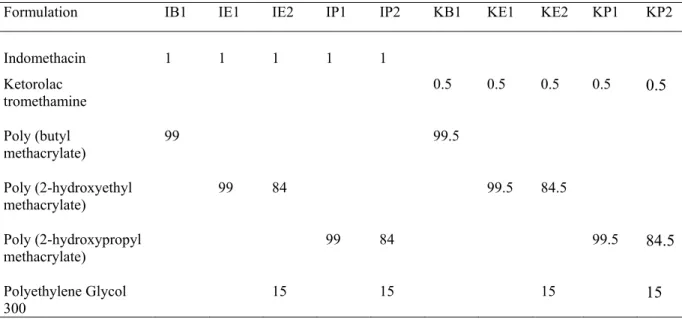
Dynamic swelling behaviors of hydrogels are shown in Figure.1. As it is seen, the dried hydrogels swell due to the absorption of water into the porous structure. pHPMA hydrogel has a much more sensitive response when it is compared with pBMA and pHEMA hydrogels,. pHPMA hydrogel swells rapidly and can achieve a much higher equilibrium-swelling ratio than pBMA and pHEMA hydrogels (21). Generally, swelling increases with size of the side chain and number of hydroxyl groups on chain of the acrylate (22, 23). Water –soluble polymers owe their solubility properties to the presence of functional groups (mainly OH, COOH, NH2) which can be useful for
the formation of hydrogels (24).
0 50 100 150 200 0 100 200 300 400 Time (minute) Swelling, %
Figure 1. Dynamic swelling behavior of the hydrogel inserts. Size of the samples were 6 mm in diameter and 0.3 mm thickness. (∋) pBMA insert (∀) Pure pHEMA insert (4) pHEMA with PEG insert
(∗) Pure pHPMA insert (Η) pHPMA with PEG insert (Each point represents the mean ± SE of three experiments).
PEG has more swelling properties when it is compared to the pure hydrogel. Especially, the swelling capacities of pHPMA were the highest in the hydrogels prepared with PEG. Swelling ratio was increased from 100 % for pHPMA insert without PEG to 175 % for pHPMA insert with PEG in 2 hours. Figure 1 shows that the hydrogel insert absorbed aqueous solution from the environment; meanwhile PEG portion of the insert was released due to its high water solubility. Since groups that constitute the hydrogel network would remain stable, it is thought that the PEG portion of the hydrogel had been leaked out from the insert. The widened space inside the insert would give more free space that causes faster release of the drug from the insert (25).
Release kinetics
A simple semi-empirical equation (26, 27) is introduced to represent the drug release process of swelling polymer:
Mt/M∞ = ktn (2)
The diffusional kinetic constant k defines the characteristics of a polymer network system. The diffusional coefficient n represents the mechanism of the transportation in the equation, n=0.50 is the Fickian release, and n=1.00 is the Case-II release. Between these two limiting cases, the non-Fickian (anomalous or coupled diffusion/relaxation) release behavior is found in the intermediate between Fickian and Case –II. It is defined, as the non-Fickian release with n value is between 0.50 and 1.0 in the semi-empirical equation.
Depending on the rate of polymer relaxation at the glass/rubbery swelling front, the swelling process and the associated drug release may exhibit Fickian or non-Fickian behavior. Typically, for a polymer slab, Fickian diffusion is characterized by a square root time dependence in both the amount diffused and the penetrating diffusion front position. On the other hand, Case-II transport, which is completely governed by the rate of polymer relaxation, exhibits a linear time dependence in both the amount diffused and the penetrating swelling front position. In most cases, the intermediate situation, which is often termed non-Fickian or anomalous diffusion, will exist whenever the rates of Fickian diffusion and polymer relaxation are comparable (28).
Simply, the swelling triggers the release of the loaded drugs. The widened space between the chains brings the release of the loaded drugs. Eq. (2) was made to explain the release profile of the loaded drugs and is being widely used for explaining release phenomena from the swellable hydrogels (25).
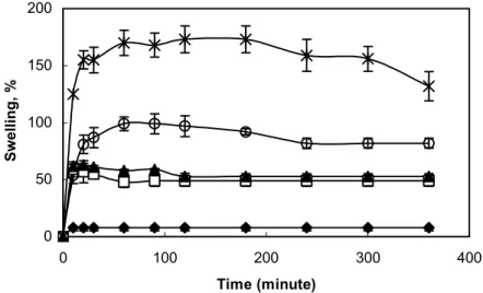
Table 2 Analysis of release data from hydrogel inserts using Eq. (2) Code of the formulation n ± SD k x 10-2 (m-n) r2 IB1 0.633±0.015 0.741 0.965 IE1 0.515±0.018 2.61 0.998 IE2 0.603±0.009 1.86 0.997 IP1 0.566±0.045 3.34 0.990 IP2 0.760±0.020 2.21 0.996 KE1 0.424±0.032 5.89 0.997 KE2 0.489±0.013 5.48 0.994 KP1 0.518±0.040 5.67 0.976 KP2 0.690±0.057 5.99 0.999
The results shown in Table 2 indicate that the release mechanism is non-Fickian (or anomalous) for all cases of IND except for IE1, as it would be expected for swellable matrices (29). The exponent values (n) indicated that inserts (except for KP2) containing KT exhibited mainly the diffusional release mechanism. The Fickian diffusion mechanism of release from the KE1, KE2, IE1 and KP1 formulations was apparent from the n values (0.424, 0.489, 0.515 and 0.518, respectively), which approximated to the value of 0.5 characteristic of the release mechanism. Furthermore, the insert containing PEG produced a change in drug release kinetics ranging from Fickian to non-Fickian. n values of the insert containing PEG, ranging from 0.515 to 0.603; from 0.566 to 0.760, and from 0.518 to 0.690, were indicative of a non- Fickian release mechanism. Lee (28) reported that the sorption of water in hydrogels generally exhibits non-Fickian (or anomalous) behavior ranging from Fickian to Case-II release. As it is seen in Table 2, a significant change between the release kinetic of KE1 and KE2 inserts was not occurred. It could be explained that diffusion rate of KT from the insert was rapid due to its hydrophilicity by comparison with swelling rate of pHEMA with PEG. Thus KT release from PHEMA with PEG hydrogels was mainly controlled by diffusion mechanism according to its n value (0.489).
Drug release
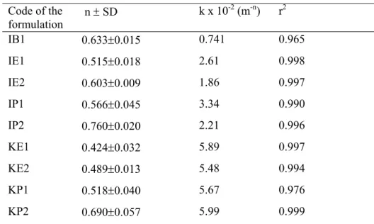
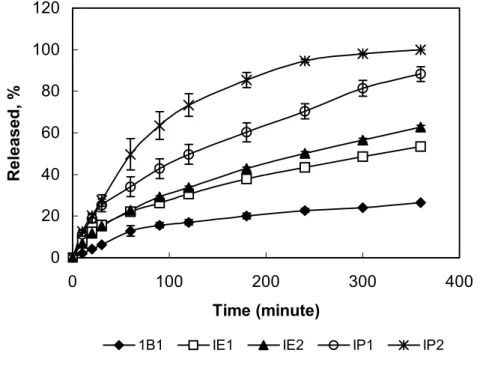
Drug release profiles of the hydrogel inserts containing water soluble KT and water-insoluble IND are shown in Figure 2 and in Figure 3. Hydrophilic KT (except for KB1 formulation) showed higher release than IND did. As shown in Table 2, diffusional release constant, k values were also support these results. The highest k values (5.89, 5.48, 5.67 and 5.99) were obtained with the KT hydrogels. Release was appeared to be correlated with the drug
hydrophilicity. The drug release was affected by the type of drug. Nonetheless, one common observation has been reported the higher drug release associated with the high water content materials and the pHEMA-containing material, regardless of the type of drug investigated.(9).
0 20 40 60 80 100 120 0 100 200 300 400 Time (minute) Released, % KB1 KE1 KE2 KP1 KP2
Figure 2. Release profiles of ketorolac tromethamin from the hydrogel inserts (Each point represents the mean ± SE of three experiments).
A remarkable increase in the drugs release (for both KT and IND) was observed in the pHPMA inserts containing PEG (KP2 and IP2), in comparison of pure pHPMA (KP1 and IP1) (Figure 2,3). This could be explained by the fact that PEG addition to the hydrogel inserts has increased the swelling degree of pHPMA hydrogels up to 175 % from 100 % and hence thesolvent penetration inside the hydrogel is faster which lead to greater drug release than that of pure hydrogels. As water penetrates a glassy hydrogel insert containing drug, the polymer swells and its glass transition temperature is lowered, At the same time, the dissolved drug diffuses through this swollen rubbery region into the external releasing medium (28).It was also found that the release of KT and IND from pHEMA hydrogel (KE2, IE2) was increased by use of PEG in the formulations. But this increase was less than with release from pHPMA hydrogels with PEG (Figure 2 and Figure 3). A small release was seen in KB1 formulation containing KT (Figure 2). It could be a result of KT fraction present at the surface of the insert. As an expected result, the
slowest drug release was obtained from IB1 insert (Figure 3), since pBMA hydrogel had the smallest swelling properties.
0 20 40 60 80 100 120 0 100 200 300 400 Time (minute) Released, %
1B1 IE1 IE2 IP1 IP2
Figure 3. Release profiles of the indomethacin from the hydrogel inserts (Each point represents the mean ± SE of three experiments).
As shown in Figure 3, the couples of IP1, IP2 and IE1, IE2 formulations showed the same release behavior during the first 30 min and first 60 min, respectively. After then the difference in release between the formulations was significant. Furthermore, both of the release profiles were characterized by a biphasic pattern. The first part, from zero to 30 min and 60 min, included one apparent mechanism namely diffusion that govern the drug release out of the hydrogel inserts, since the swelling of the hydrogel was constant. During the second phase from 30 to 360 min and from 60 to 360 min, swelling was not constant and hence, swelling mechanism affected the drug release.
Effect of the sterilization on drug release
Although 2.5 megarads (Mrad) of absorbed radiation was historically selected. It is desirable and acceptable to employ lower doses for some dosage forms such as an inserts (30). Standard sterilization dose of 2.5 Mrad has been proved unsuitable for the inserts. Significant changes in dissolution properties of the inserts occurred post irradiation. Probe studies indicated that irradiation doses of less than 2 Mrad were acceptable so far as produce stability was concerned
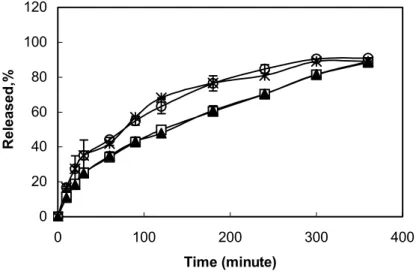
(31). Selection of an irradiation dose of not less than 7 times the D10 value of 0.17 and less than 2 Mrad assured product sterility without product alteration. In our study, we selected 1.2 Mrad irradiation dose 7 times the D10 value of 0.17 and we investigated the effect of selected irradiation dose on release of drugs from the inserts. Figure 4. shows the results of release experiments of IND and KT for two formulations as an example before and after irradiation at 1.2 Mrad. To compare the release profiles of drugs (KT and IND) before and after the irradiation, f1-difference and f2
-similarity factors were used. The values of 0-15 for f1 and the values of 50-100 for f2 have ben
reported acceptable (32). f1(3.1) and f2(79) values for KT and f1(1.2) and f2(96) values for IND
showed the similarity of the release profiles and the irradiation was suggested not to induce any difference in release of the inserts.
0 20 40 60 80 100 120 0 100 200 300 400 Time (minute) Released,%
Figure 4. Release profiles of indomethacin before (∀) after (4) irradiation and Ketorolac tromethamin before (∗) after (Η) irradiation from Poly (2-hydroxypropyl methacrylate) hydrogel (Each
point represents the mean ± SE of three experiments).
After the incubation into the culture media of the hydrogel inserts, no evidence of microbial growth was found. Therefore, the hydrogel insert to be examined complied with the sterility test. Selected dose by irradiation was found suitable for the sterility assurance level of the hydrogel inserts.
CONCLUSION
The use of different types of hydrogels and water-soluble additive such as PEG was found effective on swelling of the hydrogel insert.
Release of KT from the hydrogel inserts was faster than IND. Release of KT from hydrogel inserts was mainly occurred by diffusion controlled mechanism, while IND was released the hydrogel inserts was mainly with diffusion and swelling controlled mechanism together. It was also found that selected irradiation dose did not affect release of drugs from the hydrogel inserts.
ACKNOWLEDGEMENTS
The authors would like to thank Prof. Dr.N. Altanlar (Microbiology Department) for Sterility tests of hydrogel inserts
REFERENCES
1. Hsiue, G-H., Guu, J-A., and Cheng, C-C. “ Poly (2-hydroxyethyl methacrylate) film as a drug delivery system for pilocarpine” Biomaterials, 22, 1763-1769 (2001).
2. Khullar, P., Khar, R.K., and Agarwai, S. P. “Evaluation of guar gum in the preparation of sustained-release matrix tablets” Drug Dev. Ind. Pharm., 24(11), 1095-1099 (1998).
3. Pijls, R.T., Sondercamp, T., Daube, G.W., Krebber,R., Hanssen, H.L., Nuitjts, R. M.M.A., and Koole, L.H. “ Studies on a mew device for drug delivery to the eye” Eur.J. Pharm. Biopharm., 59, 283-288 (2005).
4. Hoffman, A.S. “Hydrogels for biomedical applications” Adv. Drug Del. Rev., 43, 3-12 (2002). 5. Huang, G., Gao, J., Hu, Z., St. John, J.V., Ponder, B.C., and Moro, D. “Controlled drug
release from hydrogel nanoparticle networks” J. Cont. Rel., 94, 303-311 (2004).
6. Gupta, P., Vermani, K., and Garg, S. “Hydrogels: from controlled release to pH-responsive drug delivery” DDT, 7, 10 May, 569-(2002).
7. Park, K., Shalaby, W.S.W. and Park, H. Biodegradable hydrogels for drug delivery, Technomic Publishing Co., Inc, lancester-Basel, 2 (1993).
8. Peppas, N.A., bures, P., Leobandung, W., Ichikawa, H. “Hydrogels in pharmaceutical formulations” Eur. J. Pharm. Biopharm., 50, 27-46 (2000).
9. Karlgard, C.C.S., Wong, N.S., Jones, L.W, and Moresoli, C. “ In vitro uptake and release studies of ocular pharmaceutical agents by silicon-containing and p-HEMA hydrogel contact lens materials” Int. J. Pharm., 257, 141-151 (2003).
10. Sasaki, H., Tei, C., Nishida, k., and Nakamura, J. “Drug release from an ophthalmic insert of a beta-blocker as an ocular drug delivery system” J. Cont. Rel., 27, 127-137 (1993).
11. Garcia, D.M., Escobar, J.L., Noa, Y., bada, N., Hernaez, E., and Katima, I. “Timolol maleat release from pH-sensible poly (2-hydroxyethyl methacrylate-co-methacrylic acid) hydrogels” Eur. Polym. J., 40, 1683-1690 (2004).
12. Alveraz-Lozenzo, C., Yanez, f., Barreiro-Iglesias, R., and Concheiro, A. “ Imprinted soft contact lenses as norfloxacin delivery systems” J. cont. Rel., 113, 236-244 (2006).
13. Flack, A.J. “Nonsteroidal anti-enflammatory drugs in ophthalmology” Int. Ophthalmol. Clin., 33, 1-7 (1993).
14. Jampol, L.M., Jain, S., Pudzisz, B., and Weinreb, R.N. “ Nonsteroidal anti-inflammatory drugs and cataract surgery” Arch. Ophthalmol., 112, 891-894 (1994).
15. Schalnus, R., ”Topical nonsteroidal anti-inflammatory therapy in ophthalmology” Ophthalmologica, 217, 89-98 (2003).
16. Bawa, R.,”Ocular inserts” in Ophthalmic drug delivery systems, Mitra, A.K.(Ed.) Marcel Dekker Inc., New York, 223 (1990).
17. El-Sherbiny, I.M., Lins,R.J., Abdel-Bary, E.M., and Harding, D.R.K., “Preparation, characterization, swelling and in vitro drug release behavior of poly(N-acryloylglycine-chitosan) interpolymeric pH and thermally-responsive hydrogels” Eur. Polymer J., 41, 2584-2591 (2005).
18. Karatas, A., Baykara, T., “Studies on indomethacin insert prepared using water-soluble polymers I. Effect of the preparation method and polymers on drug release” S.T.P. Pharma Sci., 10(2), 187-193 (2000).
19. Hussain, A.A., Higuchi, T., Shell, J.W., “Bioerodible ocular device” United States Patent, 3,960,15, June 1, 1976
20. European Pharmacopoeia, 5th Edition, Council of Europe, Strasbourg, Vol:1, 445-450
(2005).
21. Hongyan, H., Cao, X., and Lee, L.J., “Design of a novel hydrogel-based intelligent system for controlled drug release” J. Cont. Rel., 95, 391-402 (2004).
22. Katima, I., Novoa, R., and Zuluaga, F., “ Swelling kinetics and release studies of teophylline and aminophilline from acrylic acid/n-alkyl methacrylate hydrogels” Eur. Polym. J., 37, 1465-1471 (2001).
23. Gates, G., harmon, J.P., Ors, J., and Benz, P., “Intra and intermolecular relaxations 2,3-dihydroxypropyl methacrylate and 2-hydroxyethyl methacrylate hydrogels” Polymer, 44, 207-214 (2003).
24. Hennink, W.E., Van Nostrum, C.F., “ Novel crosslinking methods to design hydrogels” Adv. Drug Del. Rev., 54, 13-36 (2002)
25. Nam, K., Watanabe, J., and Ishihara, K., “Modelling of swelling and drug release behavior of spontaneously forming hydrogels composed of phospholipid polymers” Int. J. Pharm., 275, 259-269 (2004)
26. Korsmeyer R.W., Gurny, R., Doelker, E., Buri, P., and Peppas, N.A.,” Mechanism of solute release from porous hydrophilic polymers” Int. J. Pharm. 15, 25-35 (1983)
27. Ritger, P.L., Peppas, N.A., “A simple equation for description of solute release II. Fiction and anomalous release from swellable devices” J. Cont Rel., 5, 37-42 (1987)
28 Lee, P.I., “Kinetics of drug release from hydrogel matrices” J. Cont. Rel. 2, 277-288 (1985) 29. Colombo, P., Catellani, N.A., Peppas, N.A., Maggi, L., and Conte, U., “Swelling
characretistics of hydrophilic matrices for controlled release. New dimensionless number to describe the swelling and release behaviour” Int. J Pharm., 88, 99-109 (1992)
30. Jacops, G.P., “Radiation in the sterilization of Pharmaceuticals” in Sterile pharmaceutical manufacturing applications for the 1990’s. Vol.1, Groves, M.J., Olson, W.P., Anisfeld, M.H. (Eds), Interpharm press, London, (1991)
31. Garfinkle, B., Cochran, J., Snyder, J., Thrasher, G., Thompson, B., Orlowski, S., Harwood, R.J., and Cohen, E.M., “Validation of a radiosterilization procedure for sterilization of a solid-state ophthalmic insert system” J.Parenter. Sci. Technol., 39, 246-250 (1985)
32. Moore, J.W., Flanner, H.H., “ Mathematical comparison of curves with an emphasis on in vitro dissolution profiles” Pharm. Tech., 20, 64-74 (1996)
Received: 27.03.2007 Accepted: 06.07.2007