© 2016 Journal of Minimal Access Surgery | Published by Wolters Kluwer ‑ Medknow 148
Laparoscopic spleen‑preserving distal pancreatectomy
for a primary hydatid cyst mimicking a mucinous
cystic neoplasia
Tugan Tezcaner, Yahya Ekici, Onur Huseyin Aydın, Gonca Barit1, Gökhan Moray Departments of General Surgery and 1Pathology, Faculty of Medicine, Baskent University, Ankara, Turkey Address for Correspondence: Dr. Tugan Tezcaner, 5. Sokak No:48 Doktor Ofisleri Bahçelievler, Ankara 06480, Turkey.
E‑mail: [email protected]
Abstract
Pancreatic hydatid cysts are fairly rare. The disease can be encountered concurrently with systemic involvement or as an isolated pancreatic involvement. We report the first case of spleen‑preserving laparoscopic distal pancreatectomy for a pancreatic hydatid cyst. There was no complication or recurrence. A 55‑year‑old woman was admitted to our centre with epigastric and back pain. Upper abdominal magnetic resonance imaging revealed a solitary cystic lesion with septations at the pancreatic tail level measuring 24 mm × 18 mm, which was initially thought to be a pancreatic mucinous cystic neoplasia. She underwent laparoscopic spleen‑preserving distal pancreatectomy and cholecystectomy. Her post‑operative course was uneventful and histopathological examination revealed a hydatid cyst in the pancreatic tail.
Key words: Echinococcus granulosus, minimal invasive
surgery, parasitic pancreatic cyst
fairly rare, with studies reporting an incidence of <1%.[2]
The disease can be encountered concurrently with systemic involvement or as an isolated pancreatic involvement. We report the first case in the international literature of spleen‑preserving laparoscopic distal pancreatectomy for a pancreatic hydatid cyst.
CASE REPORT
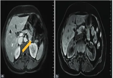
A 55‑year‑old woman was admitted to our centre with epigastric and back pain of gradually increasing severity over the last month. The patient’s medical history indicated no known diseases. Her laboratory values were abnormal. Upper abdominal magnetic resonance imaging (MRI) revealed seven hypodense lesions in the liver parenchyma with regular contours; the lesions were initially thought to be haemangiomas, the largest of which measured 21 mm in diameter and was located in the superior part of the left lobe medial segment of the liver. MRI also revealed a solitary cystic lesion with septations at the pancreatic tail level measuring 24 mm × 18 mm, which was initially thought to be a pancreatic mucinous cystic neoplasia [Figure 1a].
INTRODUCTION
Pancreatic cystic neoplasms, mostly in asymptomatic patients, are being diagnosed in increasing numbers owing to the widespread availability of cross‑sectional imaging techniques.[1] Pancreatic hydatid cysts are
Access this article online Quick Response Code: Website:
www.journalofmas.com
DOI:
10.4103/0972-9941.195578
Unusual Case
Cite this article as: Tezcaner T, Ekici Y, Aydın OH, Barit G, Moray G. Laparoscopic spleen-preserving distal pancreatectomy for a primary hydatid cyst mimicking a mucinous cystic neoplasia. J Min Access Surg 2017;13:148-50. Date of submission: 07/05/2016, Date of acceptance: 10/06/2016 This is an open access article distributed under the terms of the Creative Commons Attribution-NonCommercial-ShareAlike 3.0 License, which allows others to remix, tweak, and build upon the work non-commercially, as long as the author is credited and the new creations are licensed under the identical terms.
For reprints contact: [email protected]
Tezcaner, et al.: Laparoscopic distal pancreatectomy for primary hydatid cyst
Journal of Minimal Access Surgery | Volume 13 | Issue 2 | April-June 2017 149
Echinococcus granulosus is endemic. Diagnosis is rendered even
more difficult in conditions without a simultaneous hydatid cyst lesion in the liver as was the case in our patient. Since the main complaint of our patient was epigastric and lower back pain, and the patient had a cyst of pancreatic origin, surgery was absolutely necessary to rule out malignancy. A single lesion is observed in 90% of pancreatic hydatid cyst cases. The cysts are located in the head, corpus and tail of the pancreas in 50–57%, 24–34% and 16–19% of the cases, respectively.[3]
Treatment options are open or laparoscopic surgery, minimally invasive interventions, biopsy‑aspiration‑injection‑reaspirati on and medical treatment.[4] Open surgical procedures have
been accepted as the gold standard among the treatment options;[4] however, developing surgical techniques and
technologies has allowed laparoscopic operations for hydatid cysts to be performed more safely. Laparoscopic surgical procedures performed by experienced surgeons are at least as efficient and safe as open surgical procedures, especially in patients who are planning to undergo complete resection without opening the cyst.[5] Faraj et al.[6] first reported a
laparoscopic partial cystectomy for an isolated pancreatic hydatid cyst without any recurrence for 6 months. Unless cystic neoplasms are ruled out, resection with clear margins should be performed without opening the cyst.
CONCLUSION
Primary infection of the pancreas with E. granulosus, leading to hydatid cysts, is a fairly rare condition. As in Endosonography‑assisted aspiration failed because of
the dense contents of the cyst. Since a diagnosis of pancreatic mucinous neoplasia could not be ruled out, a decision to perform laparoscopic spleen‑preserving distal pancreatectomy and cholecystectomy was made. Four trocars were placed: One measuring 12 mm, two 10 mm and one 5 mm [Figure 2a]. The omental bursa was opened through the gastrocolic ligament, along the stomach’s greater curvature. The pancreas was freed from the splenic artery and vein at the corpus level. The distal part of the pancreas was then separated from the body with an endoscopic linear stapler. The distal pancreas was dissected from its bottom edge, behind the body; moreover, after reaching the splenic vessels, the small pancreatic veins and arteries were clipped with titanium clips [Figure 2b]. The body of the pancreas was then enforced with polypropylene sutures and the specimen was retrieved via Pfannenstiel suprapubic incision [Figure 2c]. Her post‑operative course was uneventful and the patient was discharged without complications 5 days after the surgery. The post‑operative histopathological report revealed a pancreatic hydatid cyst [Figure 2d]. Thus, she was administered albendazole 10 mg/kg/day for 8 weeks immediately after the operation. Follow‑up abdominal MRIs obtained at the 6th month and the 1st year after the
operation revealed no fluid collection and no recurrence of the disease [Figure 1b]. In addition, she has shown no evidence of serologic recurrence.
DISCUSSION
Pancreatic hydatid cysts should be taken into consideration when a pancreatic cystic lesion is identified in regions where
Figure 2: (a) Port placement. (b) The pancreas is freed from the splenic artery and vein at the corpus level. (c) Completed resection and reinforcement of the body of the pancreas with polypropylene single sutures. (d) Cyst formation within the pancreas parenchyma and inflammatory granulation tissue with foreign body giant cells around the cyst; the cyst wall is formed from an eosinophilic laminated layer (H and E, ×25)
d c
b a
Figure 1: (a) Magnetic resonance imaging at the level of the upper abdomen shows a solitary cystic mass with septations in the region of the pancreatic tail. (b) Follow-up abdominal magnetic resonance imaging obtained during the year after the operation shows no fluid collection and no recurrence of the disease
b a
Tezcaner, et al.: Laparoscopic distal pancreatectomy for primary hydatid cyst
Journal of Minimal Access Surgery | Volume 13 | Issue 2 | April-June 2017 150
this case presented here, the first diagnosis considered radiologically based on the results of imaging studies is a pancreatic mucinous cystadenoma. Laparoscopic resections can be performed safely for cystic lesions of the distal pancreas, including hydatid cysts. With appropriate surgical resection followed by adequate medical treatment, complications and recurrence rates may be kept at low levels.
Financial support and sponsorship
Nil.
Conflicts of interest
There are no conflicts of interest.
REFERENCES
1. Laffan TA, Horton KM, Klein AP, Berlanstein B, Siegelman SS, Kawamoto S,
et al. Prevalence of unsuspected pancreatic cysts on MDCT. AJR Am J
Roentgenol 2008;191:802‑7.
2. Dziri C. Hydatid disease – Continuing serious public health problem: Introduction. World J Surg 2001;25:1‑3.
3. Makni A, Jouini M, Kacem M, Safta ZB. Acute pancreatitis due to pancreatic hydatid cyst: A case report and review of the literature. World J Emerg Surg 2012;7:7.
4. Gundes E, Kucukkartallar T, Cakir M, Aksoy F, Bal A, Kartal A. Primary intra‑abdominal hydatid cyst cases with extra‑hepatic localization. JCEI 2013;4:175‑9.
5. Mehrabi A, Hafezi M, Arvin J, Esmaeilzadeh M, Garoussi C, Emami G, et al. A systematic review and meta‑analysis of laparoscopic versus open distal pancreatectomy for benign and malignant lesions of the pancreas: It’s time to randomize. Surgery 2015;157:45‑55.
6. Faraj W, Selmo F, Khalifeh M, Jamali F. Laparoscopic resection of pancreatic hydatid disease. Surgery 2006;139:438‑41.