Case report
A 50-year-old man presented with a 2-week history of upper abdominal pain, nausea, and abdominal disten-tion. The pain was relieved
tempo-abdominal swelling occupying the upper abdomen. Blood investiga-tion, liver function tests and tumor markers were within normal limits. An ultrasound exam showed a large septated cystic mass that extended rarily with analgesics. There was no
history of alcoholism, jaundice, fe-ver, vomiting, gastrointestinal bleed-ing, or weight loss. The patient did not have any prior history of pancre-atitis. Physical examination revealed JBR–BTR, 2014, 97: 97-99.
PanCreatiC CystiC LymPhangioma: diagnostiC aPProaCh with
mdCt and mr imaging
G. Macin1, K. Hekimoglu1, H. Uner2, C. Tarhan1
Lymphangiomas are rare congenital benign tumors arising from the lymphatic system mostly encountered in the neck and axillary regions of pediatric patients. Pancreatic cystic lymphangiomas very rarely occur in adults. radio-logically, the lesion may mimic pancreatic carcinoma and should be considered in the differential diagnosis of any patient found to have an abdominal cystic mass.
in this article, we present a 50-year-old man who presented with pain in the upper abdomen, nausea, and abdominal swelling. on computed tomography (Ct) and magnetic resonance (mr) imaging, a gross septated cystic lesion was detected in the upper abdomen which extended from the pancreatic corpus to the left liver lobe. the patient under-went complete resection of tumor. Pathology revealed a cystic lymphangioma.
Key-word: Lymphangioma.
From: Department of 1. Radiology, 2. Pathology, Baskent University, School of
Medicine, Ankara, Turkey
Address for correspondence: Dr G. Macin, M.D., Baskent Universitesi Hastanesi,
Radyoloji ABD, Fevzi Cakmak Cad. 10. sok. No: 45, Bahcelievler, Ankara 06490 Turkey. E-mail: [email protected]
✱
✱
✱
✱
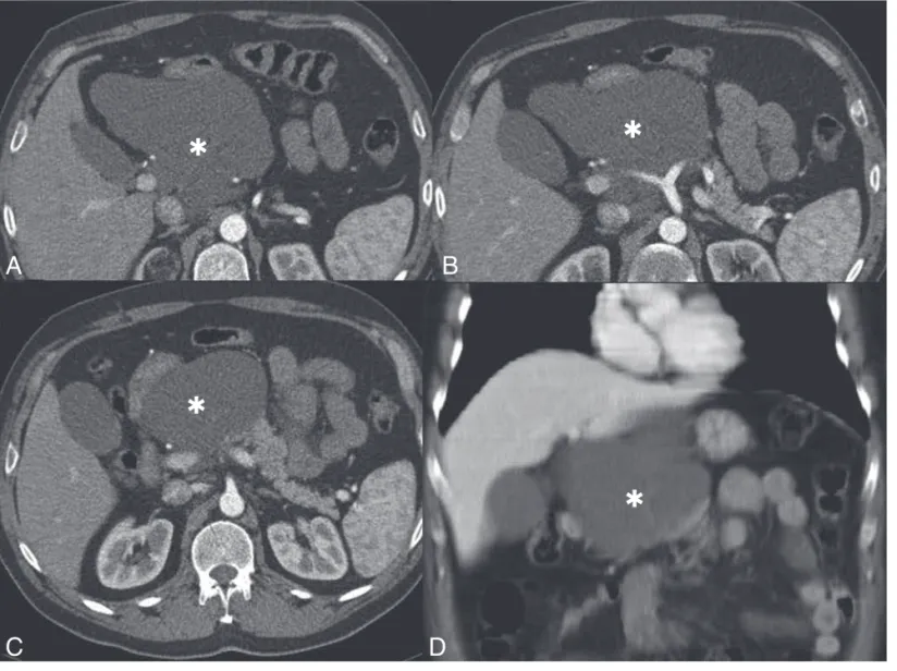
Fig. 1. — Contrast-enhanced MDCT
axi-al (A-C) and coronaxi-al reformatted scan(D): a large hypodense multiloculated cystic mass is demonstrated over the pancreas (asterisk).
A
B
D
C
98 JBR–BTR, 2014, 97 (2)
The patient was operated on with the preoperative diagnosis of pan-creatic cystic neoplasia. At laparato-my, upper peritoneal cavity was found to be almost fully occupied by a multicystic, septated mass contain-ing a clear yellowish fluid, ariscontain-ing from the corpus of pancreas. There was no evidence of invasion into other adherent organs and main vas-cular structures such as portal vein and truncus coeliacus. Distal pancre-atectomy including the cyst and the distal pancreatic tissue was per-formed.
Macroscopically, the surface of the cystic mass was shiny and smooth, and serial sectioning re-vealed multiple cysts ranging 1 to 3 cm in diameter, filled with yellow-ish fluid. Histologically, the mass was composed of variable-sized cys-tic spaces lined by flattened endo-thelium, consistent with lymphatic vessels. Lymphoid aggregates were detected in stroma and nearly all of them were filled with yellowish pro-teinous material (Fig. 3). The diagno-sis of cystic lymphangioma was con-firmed histologically.
discussion
Cystic lymphangiomas are benign cystic tumors that are the result of congenital lymphatic malformations causing obstruction of the lymphatic flow leading to lymphangiectasia. branches of the truncus coeliacus
were seen through the lesion with-out invasion signs. Thus, the mass had not invaded any adjacent ab-dominal organs such as the liver or kidney. There was no evidence of lymphadenopathy on the MDCT scan neither (Fig. 1 A-D).
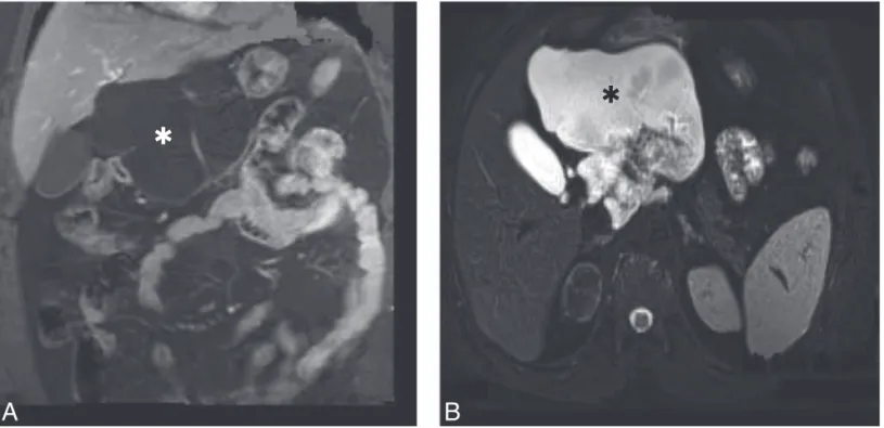
Abdominal MRI scan showed a large, multiloculated cystic lesion with typically T1 hypointense, T2 hy-perintense signals, Thick fibrous septa were seen hypointense on T1 and T2-weighted images. A granular pattern due to high internal density at the posterior component of the cystic masswas also noted (Fig. 2B). form the left lobe of the liver to the
splenorenal angle, lying over the pancreas and aorta. The pancreas could not be identified separately from the mass.
Contrast-enhanced multidetector computed tomography (MDCT) scan revealed 15 x 11 x 10 cm non- enhancing, well-defined lobulated cystic mass with multiple septations located over the pancreas. The attenuation coefficient of the mass was in the +10 to 15 HU range gener-ally. The mass extended from the tail of the pancreas to left liver lobe. Sol-id component of the lesion was not detected in the mass. The arterial
Fig. 2. — Lobulated pure cystic lesion without any solid component is seen on coronal plane contrast-enhanced, fat saturated SE
T1 image (A). B: Lympangioma shows heterogeneous-hyperintense signal on fat- saturated TSE T2 image. Granular pattern due to high internal density at the posterior component of the cystic mass on fat- saturated TSE T2 image is noted.
✱
✱
✱
Fig. 3. — Histology shows pancreatic tissue (star), lymphatic
vessels (thick arrow), and aggregates of lymphoid cells (thin arrow).
A
B
GIANT PANCREATIC CYSTIC LYMPANGIOMA— MACIN et al 99 and MR imaging may be helpful for a diagnostic approach to complex pancreatic cystic masses. Although rare, intraabdominal lymphangio-mas should be included in the differ-ential diagnosis of abdominal cystic lesions. The exact diagnosis still mainly relies on pathological exami-nation.
references
1. Koenig T.R., Loyer E.M., Whitman G.J., et al.: Cystic lymphangioma of the pancreas. Am J Roentgenol, 2001, 177:1090.
2. Colovic R.B., Grubor N.M., Micev M.T., et al.: Cystic lymphangioma of the pancreas. World J Gastroenterol, 2008, 14: 6873-6875.
3. Lyngdoh T.S., Konsam R., Th B., Marak B.: Giant cystic lymphangioma of pancreas. ANZ J Surg, 2008, 78:673-674.
4. Allen J.G., Riall T.S., Cameron J.L., et al.: Abdominal lymphangiomas in adults. J Gastrointest Surg, 2006, 10: 746-751.
5. Chung J.C., Kim H.C., Chu C.W., et al.: Huge cystic lymphangioma of the pancreas. Can J Surg, 2009, 52: 303-305.
6. Igarashi A., Maruo Y., Ito T., et al.: Huge cystic lymphangioma of the pancreas: report of a case. Surg Today, 2001, 31: 743-746.
7. Ghatak S., Ray S., Sanyal S., et al.: An unusual cause of acute abdomen in adults: Giant cystic lymphangioma of the pabcreatic head. A clinical case and literature review. J Pancreas, 2011, 12: 266-270.
8. Hanelin L.G., Schimmel D.H.: Lymph-angioma of the pancreas exhibiting an unusual pattern of calcification.
Radiology, 1977, 122: 636.
9. Cutillo D.P., Swayne L.C., Cucco J., Dougan H.: CT and MR imaging in cystic abdominal lymphangiomato-sis. J Comput Assist Tomogr, 1989, 13: 534-536.
10. Shinozaki K., Yoshimitsu K., Honda H., et al.: Abdominal cystic tumors con-taining small amount of fat in the septa: report of two cases. Abdom
Imaging, 2001, 26: 315-318.
MDCT is considered superior to US, and provides additional informa-tion about the characteristics of the lympangioma. Cystic lympangiomas typically appear as well encapsulat-ed, water-density, multiloculated masses. The septa can usually be seen and rare calcifications may be present. Otherwise, we could not see any calcification in this case.
MR imaging can demonstrate uni-or multi-locular cysts with septa in abdomen, an assessment of the rela-tionship of lesions to neighboring organs with multiplanar imaging. The capability of MR imaging is to provide images in multiple planes without loss of resolution that may demonstrate additional pathologies like as internal hemorrhage, rupture, or other else exactly (9).
T2-weighted images showed mul-tiloculated hyperintense areas which corresponded to dilated lymphatic spaces. The septa of the lesion ap-peared as hypointense bands which corresponded to fibrous tissue.
Histopathological examination plays an essential role in diagnosis. Cystic lymphangiomas are multi-loc-ulated soft cystic masses composed of a combination of variable size, dilated lymphatic channels divided by thin septa. Flat endothelial cells line are seen in the cyst walls as well as focal aggregates of lymphoid tissue (10).
A complete surgical excision is curative for lymphangioma. Incom-plete excision is the only reason for recurrent disease (3). Depending on the tumor location and size, com-plete excision may require pancreat-ic resection, such as a Whipple pro-cedure or distal pancreatectomy. In our case, we needed to perform dis-tal pancreatectomy for the removal of the lesion.
In conclusion, it is often difficult to establish a precise diagnosis of a pancreatic cystic masses. However, combined evaluation with MDCT These malformations result in
block-age of the lymphatic flow; thus, cys-tic dilatation of lymphacys-tic channels occurs (1, 2). Another hypothesis is an inflammation leading to obstruc-tion along the lymphatic channels (1-3). Lymphangioma is mostly detect-ed in the pdetect-ediatric age group patients. They are mostly localized in the neck (75%) and the axilla (20%) with cystic and cavernous spaces (1). Abdomi-nal lymphangiomas account for less than 1% of all lymphangiomas and are more frequent in female adults (4). These masses generally present with vague abdominal symp-toms such as abdominal distension, pain, nausea, vomiting, and palpable mass if it is big enough. However, months or years may pass from the onset of symptoms until the diagno-sis especially in adults. Occasionally, acute abdomen can occur due to the complications such as intestinal ob-struction, rupture or internal hemor-rhage. Although a lymphangioma is a benign lesion, it can be locally inva-sive and can recur after incomplete excision of the cysts (5). Pancreatic pseudocysts, mucinous and serous cystadenomas, and congenital cysts should be considered for the differ-ential diagnosis of gross abdominal cystic lesions.
Most reports in the literature describe the body and tail of the pan-creas as the most common regions involved by lymphangioma (6). Re-cent studies showed that tumor size may vary between 3 and 30 cm in diameter (7).
Lymphangiomas typically appear as complex cystic masses on ultra-sonography (US) due to internal sep-ta and calcifications. They also may have internal echoes if they are sec-ondarily infected. Rarely, calcifica-tions could be demonstrated by US with acoustic shadow sign (8). There-fore, US can sometimes differentiate various cystic lesions, but ancillary studies are needed.