Ankara Üniv Vet Fak Derg, 50,103-106,2003
EvaIuation of kidney abnormalities in cows using transrectal
ultrasonography and urinalysis*
Ramazan DURGUTı, Ramazan GÖNENCI
2,Sefa ÇELİK
3,Ramazan BAL\ M. Enes ALTUG
2 Departments of lInternal Medicine, 2Surgery, 'Biochemistry, 4Physiology, Faculty of Veterinary Medicine, Mustafa KemalUniversity, Antakya/Hatay
Summary: For this study, 100 Holstein crossbreed cows aged between 4 to 8 years, admitted to Antakya Slaughterhouse, were used. After clinical examination, urine samples were collected by catheterization from urinary bladder for urine analyses using reagent dipsticks and microscopy. In ultrasonography; abnormal appearances were observed in 18 cows, including smail kidney with echogenicity in two, a hydronephrosis in one, renal ca1culi İn two, alteration of the corticomedullary architecture in one, dilated renal pelvis and irregularly shaped kidneys with echogenic cortex in four, patchy hyperechoic cortex and poor demarcalion between cortex and medulla in five, and renomegaly in three. Urinalyses revealed the presence of leukocyte, erythrocyte, phosphate and/or siliceous crystals in the 18 cows. Therefore, it is concluded that both transrectal ultrasonography and urine analysis appears to be adequate for diagnosis of renal diseases.
Key words: Cow, kidney, ultrasonography, urine analysis
İneklerde böbrek bozukluklarının
transrektal ultrasonografl ve idrar analizi ile değerlendirilmesi
Özet: Bu çalışmada Antakya mezbahanesine getirilen, 4-8 yaşlı, 100 Holştayn melezi inek kullanıldı. Klinik muayeneden sonra idrar kesesinden kateterle toplanan idrar örnekleri dipstik test çubukları ve mikroskobik yöntemle analiz edildi. Ultrasonografide; 2 inekte ekojenik küçük böbrek, birinde makrokist, ikisinde böbrek taşı, birinde kortikomeduller yapıda değişiklik, dördünde renal pelviste genişleme ve düzensiz yapı ile birlikte kortekste ekojenite, beşinde korteks ve medulla arasında hafif demarkasyon ve hiperekojenik korteks parçası ve üçünde renomegali olmak üzere toplam 18 hayvanda anormal görüntüler saptandı. Bu 18 İneğin idrar analizlerinde lökosit, eritrosit ve silisyum-fosfat kristalleri görüldü. Sonuçta transrektal ultrasonografi ve idrar analizlerinin birlikte böbrek hastalıklarının tanısında yeterli olabileceği kanısına varıldı.
Anahtar kelimeler: Böbrek, idrar analizi, inek, ultrasonografi
Introduction
In smal1 animals, ultrasound can be used to assist in the differentiation between acute and chronic renal diseases, and the renal masses caused by hematomas, cysts, abscesses or neoplasia (1,3). In large animals, application of transrectal ultrasonography has recently been used for diagnosis of some caudal abdominal abnormalities (6,7). However, in practice transrectal ultrasonography is used for diagnosis of large animal pregnancy mostly and also urolithiasis (2,4,5). The information of the kidneys and ureters obtained by ultrasonography can help the best treatment decision, or to confirm appropriateness of curative therapy (3,5,8,11). The aim of the study was to investigate the kidney lesions in cows admitted to Antakya Slaughterhouse in Hatay province, using ultrasound scanner equipped with a 6.0 MHz and 8.0 MHz transrectal transducers, and urine analyses.
Materials and Methods
For this study, 100 Holstein crossbreed cows, aged between 4 to 8 years, admitted to Antakya Slaughter-house, were screened in Summer 2001. They were sub-jected to clinical, ultrasonographical and biochemical examinations.
Af ter clinical examination, urine samples were obtained by catheterization from urinary bladder for testing and analysed within 15 minutes after the col1ection using reagent dipsticks. The urine samples were centrifuged at 1500 rpm for 5 minutes. Af ter deyanting, the supematant sediments were resuspended in physiological sa1İne. Urine cytology was examined 10 field s using x40 lens to identify east, cel1s, and crystals. Kidney function was evaluated by testing urine concentration with a veterinary refractometer.
Kidneys and ureters were examined ultrasonograp-hically. Ultrasonographic measurements were performed
104 Ramazan Dutgut - Ramazan Gönenci - Sefa Çelik - Ramazan Bal - M. Enes Altuğ
i
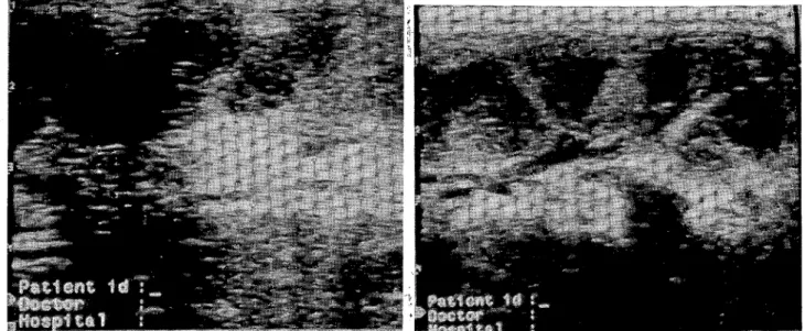
Figure
ı.
An ultrasonographic \iew show s a marked increase in echogenicity of the renal cortex and the enhanced definition of corticomedullary junction withih the medulla.i
ii
i
iFigure 2. An ultrasonographic appearancds of a kidney. Note that the kidney is smail echogenic with los~ of definition of the corticomedullary junction.
definition of the corticomedullary junction (Figure 2). In the left kidney of another animal, ultriasonograPhY revealed a hydronephrosis, about 7 cm in ,diameter and renomegaly (Figure 3). In fouranimals, 6n increased echogenicity of the renal cortex and medlulla, dilated renal pelvis and irregularly shaped kidneys Jere observed (Figure 4). In the left kidneys of five aniJals, a patchy hyperechoic cortex and poor demarcation bktween cortex and medulla were observed (Figure 5). In ihree animals,
renomegaly, increased cortical an~ medullar
echogenicity, ill-defined are as of hypereclhogenicity in the cortexis, and lack of visualizaltion of the corticomedulary junctions were observ~d. The renal pelvises were dilated and mild hyperechoic debrises were present (Figure 6). In the right kidney of one animaL, a dilated anechoic pelvis, an alteration of the medullary and cortical architecture, and an enlarged anechoic ureter and an anechoic stmcture (4 cm in diameter) were detected. Ultrasonographic examination results of 18 cows with renal abnormalities were shown in Table
ı.
No ultrasonographic abnormalities were dete ted in the restof the animals.
i
Of the 100, in 18 cows, urinalysis showed the
i
presence of leukocyte, erythrocyte, phosphate or siliceous crystals, and the affected cows excreted fr;equently diluted urine with densities ranging between 1005 and 1015. Analysis of urine with dipsticks of thJ 18 cows with kidney problems revealed existence o~ blood in ll, protein in 8 and glucose in 3 cows. ResJılts of urinalyses of the 18 cows were shown in Table
ı.
i
Mostly the left kidneys anp ureters were identified as structurally involved in ultrasonographic examination. Right kidneys of the cowswere ton difficult to examine thoroughly, especially in huge and/or tall animals. In all the cows, the transducer of 6.01 MHz resulted in greater
depth of tissue penetration, acid lesser image detail in kidneys and ureters, whereas
i
transducer of 8.0 MHz resulted in lesser depth of tissue penetration and a greateri
image detai1. Ultrasonographicı renal abnormalities were observed in 18 cows. In twoı of the 18 animals with
lesion, the marked increase inı echogenicity of the renal cortex with some degrees of afoustic shadowing and the en hane ed definitian of corticdmedullary junction within the medulla were detected (lFigure 1). In an other two animals. the left kidneysl were typically small (approximately 4x4.5 cm) an1d echogenic. with lass of
i
using a scanner 100 LC Yet ulttasound machine (Pie
i
Medical Equipment B.Y., Philipsweg 6227 AI
Maastricht, The Netherlands) withla 6.0/8.0 MHz LA DF
i
.
bYet TRD (401670, 401 8 11) transrectal ultrasonıc pro e. i
i
Results i
Clinical observation includ~d weight loss, anemia, lifting their back dorsally or hybersensitivity in kidney palpation in four animals. These
ianimals also frequently
attempted to urinate, and their tails seemed to be pumping or twitching during thb examination. Physicalexamination and rectal p~lpation showed no
abnormalities of urethra, bladde~ neck, and local lymph i
Ankara Üniv Vet Fak Derg, SO, 2003 105
Figure 3. An ultrasonographie view of hydronephrosis in a kıdııey.
Figure 5. A patehy hyperechoic cortex and poor demareation between eortex and meduııa in ultrasonography.
Figure 4. An u1trasonographie view of an increased eehogenieity of renal cortex and meduııa with renal pelvis. Note the irregularly shaped kidney.
Figure 6. A view of a dilated renal pelvis in ultrasonography. Note debrises accumulated in the renal pelvis, with a mild hyperechoic appearance.
Table I. Evaluation of ultrasonographic examİııation and minalysis in 18 cows with renal lesions Number of cases 2 2 1 4 5 3 VItrasonographicfindings
Echogenic renal cortex, Enhanced definition of cörticorriedLiııary junction Echogenic smaıı kidney, los of corticomeduııary junction Renomagaly due to macrocysts Eehogenie meduııa and cortex, dilated renal pel vis, irregular shape of kidneys Patch hyperechoie eortex, poor
demareation between cortex and medtı1la Renomegaly, eortieomedullary
echogenicity, hyperechogenie ill-defined areas in eortex, lack of
visualization in the corticomeduııary junction Dilated aneehoie pelvis, eortieomeduııary altetation, enlarged aneehoic meters
Vrinalysis
Amorph urate, calcium oxalate crystals, phosphate and silieeous erystals, epithelial ceııs, 8-10 erythrocytes Abundant Vrate and mic acid erystals, 10-
ı
4 leukocytes,7 -8 erythroeytes casts
Squamous epithelial eeııs, triple phosphate crystals 9- 10 granular and i0- i2 leukoeyte casts, 4-5 leukocytes
10- 14 erythrocyte, 7-8 epithelial casts,
Abundant renal epithelial eeııs, 20-2S erythroeytes
106 Ramazan Du~gut - Ramazan Gönenci - Sefa Çelik - Ramazan Bal - M. Enes Altuğ
Discussion and Cohclusion
Ultrasonography is partibularly helpful in determining the possible etioıdgy of urinary tract disorders, such as cysts and stonelformation (1-3,12). In the catlle industry, this technologyı is not used commonly because of the east of the machine and the perception that rectal palpation is sufficiently ıeliable. Nevertheless, competent rectal palpator can sim~ly not be accurate as a competent ultrasonographeI.
i
The extents and characteristics of lesions can be evaluated without invasion. Whereas, ability for recial palpation to identify the pathologies is limited. In thid study, ultasonography provided diagnostic information, and helped to determine the severity of renal diseases. As iwe did in this research, less costly portabIe ultrasound equipment will help to.. . i
examll1e urınary tract ın cows.
Depth of tissue penetratiort of sound waves, and image resolutian is dependent up~m and inversely related to the frequency of the transduceI. The study presented
i •
here, a 6.0 MHz transducer resulted ın greater depth of tissue penetration and lesser imlge detai!, whereas, 8.0
i
MHz transducer resulted in lesser depth of tissue penetration and a greater image qetail. Thus, we believed that an ultrasaund scanner equipped with a 6.0 MHz transducer appear to be more sa~isfactory for examining
kidneys and ureters.
i
Results of urinalysis and Isensitivity of the urine using by dipstick test stripe is helpful as far as combining
i
with the other clinical evaluation. Casts, crystals, mucous
i
erythrocyte can be used as indexes for damage to the kidney tubules (9-12), and the bresence of renal tubular ceııs and/or leukocytes may iddicate infectian (10,11).
For this reasons, in every suspected cattle urinary tract
i
abnormalities or diseases, urinaİysis should be done as a general screening to check for
i
diagnosing early kidneydisease.
i
The disturbance of calciı.jm and phosphorus ratio and change in urine volume are important risk factors
i
(5,11) for stone formation, which might account for a higher incidence of stone forma~ion during summer in the, present study. The pres en ce ofi microhematuria detected by dipstick in the study presepted here may imply the presence of kidney disease.
i
In conclusion, convincingevidence presented in this study suggests that for
i
diagnosis, non-invasive examination method s, namely, lultrasoIlography and urinecytology appear to be sufficienl. Therefore, examination of kidneys by ultrasonography and urinal~'sis has the potential to provide new perspectives ian clinical diagnosis of mild to moderate renal diseases in cowS.
References
I. Chandler KJ, O'Brien K, Huxley JN, 'Fhompson H, i
Fitzpatrick JL (2000): Hydronephrosis and renal failure in two Friesian cows. Yet Rec, 146,646-648./
2. Divers TJ, Reef VB, Roby KA (1989): Nephrolithiasis re-sulting in intermittent ureteral obstruction
Jı
a cow.eor-nell Yet, 79,143-149.
i
3. Hayashi H, Biller DS, Rings DM, Miyabayashi T (1994):
Ultrasonographic diagnosis of pyeıonePhritllis in a cow. J
Am Yet Med Assoc, 205, 736-738.
4. Kennedy S, Rice DA (1987): Renallesioııs bn caıtle fed so-dium hydroxide-treated barley. Yet Pathol, 2~, 265-271.
5. King W, Kimme-Smith C, Winter J (198)>): Renal stone
shadowing: an investigation ot' contributi,1g factorso
Ra-diology, 154,191-196. J
6. Miller CW, Wingfield WE (1981): Appl'cations of ult-rasound to veterinary diagnostics in a vetelrinary teaching hospital. Biomed Sci Instrum, 17,85-90.
7. Miller CW, Wingfield WE, Boon JA (l9g2): Applications
of ultrasound to veterinary diagnostics in
lı
veterinary te-aching hospital. ISA Trans, 21, 101-106.8. Naoi M, Kokue E, Takahashi Y, Kido Y (1985):
w-paroscopic-assisted serial biopsy of the bovine kidney. Am
J Yet Res, 46,699-702.
i
9. Ohba Y, Kitagawa H, Okura Y, Kitoh K, Sasaki Y
(2001): Clinical features of renal tubular &ysplasia, a new
hereditary disease in Japanese Black ca ttik. Yet Rec, 149,
115-118.
!
ıo.
Sato R (1991): Comparative studies on the validity ofrenalfunction tests in the experimenta/ly-indu 'ed bovine glo-merulonephritis. J Yet Med Sci, 53,307-3115.
i
ı.
Sato R, Sano Y, Sato J, Naito Y (1999):)~-acetyl-beta-D-glucosaminidase activity in urine of cow.~ with renal pa-renchymallesions. Am J Yet Res, 60,4
ıo-k
i3.12. Tyler JW, Smith BP, Irvine J (1991 ):/ Hydronephrosis
and pyelonephritis associated with an anomalous vas
de-. i
ferens ın a bu/l. J Am Yet Med Assoc, 198, 871-872.
i
Geliş tarihi: 9.9.2002/ Kabul tarihi: 30.](J.2002
Correspondence address:
Yrd. Doç. Dr. Ramazan Durgut Mustafa Kemal Üniversitesi
Veteriner Fakültesi
Iç Hastalıklar Anabilim Dalı