nostic distinction between BrP and true congenital BrS fo-cuses on a few key features. First, patients with BrP have a reversible underlying condition, such as adrenal insufficiency, hypokalemia, or myocardial ischemia, that elicits or induces the Brugada ECG pattern. Once this underlying condition resolves, there is prompt normalization of the ECG. Second, patients with BrP have a low clinical pretest probability of true congenital BrS as opposed to a high clinical pretest probability in patients with BrS, who have a history of VF, polymorphic VT, nonvagal syncope, and a family history of sudden cardiac. Third, patients with BrP have a negative provocative challenge with a sodium channel blocker, whereas those with true congenital BrS have a positive provocative challenge. Therefore, ECG alone cannot be used as a diagnostic tool. Implantation of an implantable cardioverter defibrillation in an asymptomatic patient without a spontaneous type 1 Brugada electrocardiographic pattern has no benefit.
Semih Kalkan1, Ahmet Güner1, Macit Kalçık2, Çetin Geçmen1, Mehmet Özkan1,3
1Department of Cardiology, Koşuyolu Kartal Training and Research Hospital; İstanbul-Turkey
2Department of Cardiology, Faculty of Medicine, Hitit University; Çorum-Turkey
3School of Health Sciences, Ardahan University; Ardahan-Turkey
Address for Correspondence: Dr. Ahmet Güner, Koşuyolu Kartal Eğitim ve
Araştırma Hastanesi, Kardiyoloji Bölümü, İstanbul-Türkiye Phone: +90 505 653 33 35 Fax: +90 216 500 15 00 E-mail: [email protected]
©Copyright 2018 by Turkish Society of Cardiology - Available online at www.anatoljcardiol.com
DOI:10.14744/AnatolJCardiol.2018.43789
E-page Original Images
Type I Brugada pattern associated with
diabetic ketoacidosis in a patient with
type II diabetes mellitus
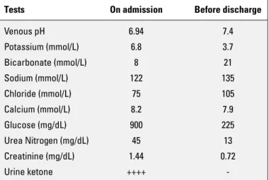
A 42-year old male with known insulin-dependent diabetes mellitus and otherwise unremarkable medical history present-ed with nausea, vomiting, and atypical chest pain for 2 days. His initial vital signs were remarkable for sinus tachycardia (101 bpm) and tachypnea (20 breaths per minute). He did not have a personal or family history of arrhythmia-related symptoms, ventricular tachycardia or fibrillation (VT/VF), or premature sud-den cardiac death (SCD). Electrocardiography (ECG) revealed ST elevations (V1–V2) with associated T wave inversions (Fig. 1a). A baseline ECG performed 6 months ago showed no ab-normalities. Serial troponin T measurements were normal. His initial laboratories were notable for hyperglycemia, hyponatre-mia, hyperkalehyponatre-mia, ketonuria, and metabolic acidosis (Table 1). Upon further questioning, it was found that he had no history of referable cardiac symptoms. There was no family history for SCD, recurrent syncope, or arrhythmia. He was diagnosed with and treated for diabetic ketoacidosis. His clinical status stabi-lized with normalization of electrolytes and pH (Table 1). After the medical treatment, ECG (Fig. 1b) showed improvement, and he had a negative provocative testing with a sodium channel blocker (ajmaline). He had an uneventful discharged and, at 10 months after treatment, he is doing well.
Brugada syndrome (BrS) is a relatively rare genetic chan-nelopathy; however, Brugada pattern is an ECG phenomenon, which is now known as Brugada phenocopy (BrP). The
diag-E-1
Figure 1. (a) Electrocardiography depicts coved-type ST-segment eleva-tion in leads V1-V2 with associated T wave inversions during diabetic ketoacidosis. (b) Electrocardiography indicates resolution of the prior findings before the discharge
a
b
Table 1. Patient’s laboratory results on admission versus before discharge
Tests On admission Before discharge
Venous pH 6.94 7.4 Potassium (mmol/L) 6.8 3.7 Bicarbonate (mmol/L) 8 21 Sodium (mmol/L) 122 135 Chloride (mmol/L) 75 105 Calcium (mmol/L) 8.2 7.9 Glucose (mg/dL) 900 225 Urea Nitrogen (mg/dL) 45 13 Creatinine (mg/dL) 1.44 0.72 Urine ketone ++++