Ankara Üniversitesi Tıp Fakültesi Mecmuası 2007, 60(4)
DAHİLİ BİLİMLER / MEDICAL SCIENCES Olgu Sunumu / Case Report
Iliac Arteriovenous Fistula Secondary To Iliac Aneurysm Rupture:
Multidetector Computed Tomographic Angiography
Findinds
İliyak Anevrizma Rüptürüne Sekonder Gelişen İliyak Arteriyovenöz Fistül: Multidedektör Bilgisayarlı Tomografi k Anjiyografi Bulguları
Aysel Türkvatan, Tülay Ölçer, Turhan Cumhur
Türkiye Yüksek İhtisas Hastanesi, Radyodiagnostik Anabilim Dalı
176
Received: 28.09.2007 • Accepted: 01.03.2007 Corresponding author
Aysel Türkvatan
Türkiye Yüksek İhtisas Hastanesi Radyodiagnostik Anabilim Dalı Sıhhıye, Ankara
Phone : + 90 (312) 306 16 71 E-mail address : [email protected]
Iliac artery aneurysms mostly occur in association with abdominal aor-tic aneurysms, either as an exten-sion of the aortic aneurysm to the common iliac artery or as a sepa-rate aneurysm of the iliac artery (1). In addition, one third of the patients have contralateral iliac aneurysms (2). Spontaneous rup-ture of iliac arterial aneurysm into the iliac venous system is less com-mon (3). Iliac arteriovenuos fistula is a life-threatening condition that requires immediate diagnosis and treatment.
We report a case of iliac arteriove-nous fistula complicating iliac artery aneurysm which was diag-nosed preoperatively using mul-tidetector row computed tomog-raphic (MDCT) angiography. This report underlines the significant role of MDCT angiography as the first imaging method in thoracoab-dominal vascular emergencies.
Case Report
A 70-year-old man presented to the emergency room complaining of acute onset of lower abdomi-nal pain and shortness of breath. Physical examination revealed a pulsatile mass in the abdomen accompanied by a thrill and bru-it, suggestive of an aneurysm and right lower extremity edema. Ele-ctrocardiography showed sinus tachycardia at 103 beats/min, and normal P-waves. His blood pressu-re was 90/35 mmHg. Thoracoab-dominal MDCT was immediately performed with suspicion of aortic aneurysm rupture.
A thoracoabdominal MDCT angiog-raphy was performed using a 16-row multislice CT (Light-speed Ul-tra, GE medical System, WI, USA). Patient was examined while supine and all images were acquired du-ring a single breathhold of 12 sec
Iliac arteriovenous fi stula occurring as a complication of spontaneous rupture of an atherosclero-tic iliac aneurysm is extremely rare. Early diagnosis is important as it allows immediate interventi-on. Herein, we present the imaging fi ndings of iliac arteriovenous fi stula complicating iliac artery aneurysm which was diagnosed immediately using multidetector row CT angiography.
Key Words: iliac artery, arteriovenous fi stula, multidetector computed tomography
Aterosklerotik iliyak arter anevrizmasının spontan rüptürüne sekonder olarak gelişen iliyak arte-riyovenöz fi stül oldukça nadir görülen erken tanı ve tedavi gerektiren bir durumdur. Biz burada multidedektör bilgisayarlı tomofrafi anjiyografi ile kısa zamanda tanı alan iliyak arteriyovenöz fi s-tül olgusunun görüns-tüleme bulgularını sunduk.
Journal of Ankara University Faculty of Medicine 2007, 60(4)
177 Aysel Türkvatan, Tülay Ölçer, Turhan Cumhur
duration, extending from base of the neck to the groin. Imaging pa-rameters were as follows: 120 kV,
400 mA, 16x1.25 mm collimation, speed 27.5 mm/rot, rotation time 0.5 sec, pitch value of 1.375:1. Af-ter deAf-termining the contrast agent transit time using the smart prep bolus technique, we acquired image data during an intravenous injection of 120 ml of iodinated contrast agent iopromid (Ultravist 370, Schering, Berlin, Germany) at a rate of 4 ml/sec. For 3-dimen-sional image reconstruction, the raw CT data were processed on a separate workstation (Advanced Workstation 4.2, GE Medical Sys-tem, WI) with multiplanar refor-matting (MPR), maximum inten-sity projection (MIP) and volume rendering (VR).
MDCT angiography demonstrated a 5 cm diameter infrarenal aortic aneurysm immediately followed by right common iliac artery aneurysm in 6.6 cm diameter and left common iliac artery aneurysm in 3 cm diameter (Figure 1). The right common iliac artery aneurysm had directly fistulized into right common iliac vein
th-rough a well identifiable fistulous communication at the length of 3 cm and diameter of 2.2 cm from the posterolateral aspect of the aneurysm wall (Figure 2). Early enhancement of the enlarged infe-rior vena cava and both common iliac veins and retrograde flow to the renal and iliac veins were de-tected on CT. Additionally, right pleural effusion, right atrial and ventricular enlargement associa-ted with cardiac failure and dela-yed opacification of the renal cor-tex probably associated with renal failure were detected (Figure 3). The patient underwent emergency
operation. Aortailiac aneurysm was repaired with an aortoiliac bi-furcation prosthesis and iliac arte-riovenous fistula was closed with a patch. His postoperative course was uncomplicated.
Discussion
Trauma seems to be the main
cau-Figure 1: Coronal MPR image shows a 5 cm
diameter aortic aneurysm immediately fol-lowed by 6.6 cm diameter right common iliac artery aneurysm. The slightly enlarged inferior vena cava appears simultaneously isodense with the aorta and both common iliac arteries due to left-to-right shunt. (IVC: inferior vena cava, A: aorta, RCIA: right common iliac ar-tery)
Figure 2A: Coronal oblique MIP image shows
the right common iliac artery aneurysm dire-ctly fistulizes into the right common iliac vein through a well-identifiable 2.2 cm diameter fistulous communication.
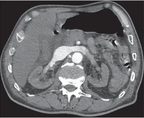
Figure 2B: Axial image shows the fistulous
communication between right common iliac artery and vein. In addition, the left common iliac artery aneurysm is also seen.
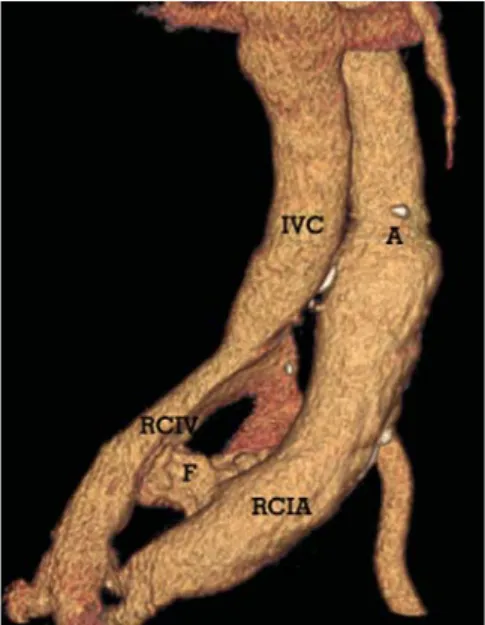
Figure 2C: Volume rendering image shows
iliac arteriovenous fistula. IVC: inferior vena cava, A: aorta, RCIA: right common iliac ar-tery, RCIV: right common iliac vein, F: fistula)
Ankara Üniversitesi Tıp Fakültesi Mecmuası 2007, 60(4)
178 Iliac Arteriovenous Fistula Secondary To Iliac Aneurysm Rupture: Multidetector Computed Tomographic Angiography Findinds
se of arteriovenous fistula betwe-en iliac artery and vein (1). It re-sults from gun shot wounds, stab wounds, seat belt trauma and iat-rogenic injuries in 84% of patients (4). Iatrogenic injury to major ret-roperitoneal vessels producing an arteriovenous fistula is also a well-described complication of lumbar disc surgery (5). Only in remaining 16% of patients is it secondary to spontaneous rupture of an iliac aneurysm (6). In contrast, 80% of all aortacaval fistulas result from aneurysmal rupture (7). Sponta-neous perforation has been desc-ribed secondary to atherosclerotic or infective (tuberculosis, syphilis, mycotic) aneurysm formation (8). Rare causes include Marfan’s synd-rome, Takayasu’s arteritis, Ehlers-Danlos disease. Rarely, neoplasms may also cause a fistula by erosi-on of adjacent arterial and venous structure (3).
The classical clinical triad for aorta-iliac aneurysm with fistula consists
of severe low back or abdominal pain, pulsatile abdominal mass, and machinery-like bruit. This tri-ad is complete in only 50-80% of patients (7). Other clinical signs are various and represent con-sequences of the physiologic de-rangements of the fistula and the left-to-right shunt (9). Fulminant onset high-output cardiac failure that does’nt respond to medical treatment may result from decre-ased peripheral resistance that in-duces a sharp secondary increased cardiac output (10). A large volu-me left-to-right shunt causes inc-reased venous return leading to venous hypertension and then to passive venous congestion, hepa-tomegaly, ascites, or severe indu-ced portal hypertension resulting in upper gastrointestinal bleeding. Lower extremity edema, pulsatile venous dilatation and scrotal ede-ma, priapism, rectal bleeding and vesical bleeding may be seen (9). Renal failure is not uncommon and is most likely caused by the
decreased mean arterial pressure and increased venous pressure in the renal vessels which reduces the glomerular filtration (3). Pul-monary embolization is rare and is thought to be due to the embolic material or atheroma from within the aneurysm (paradoxical embo-lism) or thrombus and clot from the major veins compressed by the aneurysm (1).
Preoperative diagnosis of spontane-ous iliac arteriovenspontane-ous fistula is difficult because the classical triad of abdominal pain, pulsatile lower abdominal mass and abdominal bruit may be absent in up to 50% of patients and may be masked by less specific additional symptoms such as dyspnea, cardiac failure, pain and edema of the legs, or va-rious hemorrhagic complications (7,9). In our patient there were mixed symptoms and signs of high output cardiac failure, renal failure and peripheral venous hi-pertension.
The diagnosis of iliac arteriovenous fistula often made on the basis of imaging, such as Doppler ultra-sound, helical CT, MDCT, MR an-giography and conventional angi-ography.
Ultrasonography has also been used the diagnose of aortailiac arterio-venous fistula. It readily depicts aneurysms and sometimes dete-cts dilatation of the inferior vena cava. Doppler interrogation can show arterialized wave forms in iliac vein and inferior vena cava. Color imaging can demonstrate turbulent, mixed arterial and ve-nous flow at the fistula (11). Un-fortunately, the acoustic window of ultrasonography is frequently limited by superimposed bowel gas.
Traditionally, the aneurysm is diag-nosed by angiographic
demons-Figure 3: Axial view at the level of the renal veins during the arterial phase showing delayed
Journal of Ankara University Faculty of Medicine 2007, 60(4)
179 Aysel Türkvatan, Tülay Ölçer, Turhan Cumhur
REFERENCES
1. Vallina EA, Perez MA, Pascual MF, et al. Ili-ac arteriovenous fistula secondary to iliIli-ac aneurysm rupture associated with pulmo-nary embolism and anuria. Ann Vasc Surg 2000;14:170-3
2. Morrow C, Lewinstein C, Ben-Menachem Y. Spontaneous iliac arteriovenous fistula. J Vasc Surg 1987;6:524-7
3. Gregoric ID, Jacobs MJ, Reul GJ, et al. Spontaneous common iliac arteriovenous fistula manifested by acute renal failure: a case report. J Vasc Surg 1991;14:92-7 4. Zajtchuk R, Yacoub M, Kittle CF.
Spon-taneous arteriovenous fistula between the right common iliac vessels. Surgery 1971;69:194-200
5. Franzini M, Altana P, Annessi V, et al. Iatro-genic vascular injuries following lumbar disc surgery: case report and review of the literature. J Cardiovasc Surg 1987;28:727-30
6. Weimann S, Flora G. Primary arteriove-nous fistula between the common iliac vessels secondary to aneurysmal disease. Surgery 1987;102: 91-5
7. Fenster MS, Dent JM, Tribble C et al. Aor-tacaval fistula complicating abdominal aortic aneurysm: case report and literatu-re literatu-review. Cathet Cardiovasc Diag 1996; 38: 75-9
8. Alarcon M, Quiroga S, Sebastia MC,et al. Arteriovenous fistula secondary to iliac mycotic aneurysm: helical CT findings. Abdom Imaging 2000;25:55-8
9. McAuley CE, Peitzman AB, de Vries EJ, et al. The syndrome of spontaneous iliac ar-teriovenous fistula: a distcint clinical and pathophysiologic entity. Surgery 1986; 99:373-7
10. Turse JC, Dunlap DB. Spontaneous arteri-ovenous fistula between the left common iliac artery and iliac vein. Am J Med Sci 1989;297:190-2
11. Subber SW, Russ PD, Whitehill TA, et al. Diagnosis and localization of spontaneous aortacaval fistula by computed tomograp-hy with angiographic confirmation:case report. Can Assoc Radiol J 1998;49:35-8 12. Frauenfelder T, Wildermuth S, Marincek
B, et al. Nontraumatic emergent abdo-minal vascular conditions: advantages of multi-detector row CT and three-di-mensional imaging. Radiographics 2004; 24:481-96
13. Coulier B, Tilquin O, Etienne PY. Multide-tector row CT diagnosis of aortacaval fis-tula complicating aortic aneurysm:a case report. Emerg Radiol 2004;11:100-3 14. Wang EA, Lee MH, Wang MC, et al.
Iat-rogenic left iliac-caval fistula: imaging and endovascular treatment. AJR 2004; 183:1032-4
15. Schott EE, Fitzgerald SW, McCarthy WJ, et al. Aortacaval fistula: diagnosis with MR angiography.AJR 1997;169:59-61
tration. This technique provides anatomical details of the aneurysm and clearly outlines the aortic branches and their relationship to the aneurysm. But the early chan-ges in the arterial wall and the pe-riaortic soft tissue are not detected on angiography (8).
MDCT angiography has become an important technique in the evalu-ation of the vascular system and has already been proven to yield high accuracy in the assesment of the abdominal aorta and its major branches. In combination with 3D image reconstruction techniques, it may be used to clarify complex vascular anatomies. MDCT angi-ography is the modality of choice for assesment of emergent abdo-minal vascular conditions beca-use it enables the acquisition of high spatial resolution volumetric image data during a single breat-hhold. Unlike catheter angiograp-hy, MDCT angiography not only
depicts the vessels but also allows direct visualization of associated perivascular abnormalities and as-sessment of perfusion in adjacent organs (12,13).
While MDCT angiography is an excellent modalitiy for identifying aortailiac arteriovenous fistulas, MR angiography offers an impor-tant alternative, particularly in pa-tients with contraindications to io-dinated contrast material (14,15). However, diagnosis of fistula might be impossible due to flow artifacts (15).
CT findings of iliac arteriovenous fistula include early enhancement of the venous system, equivalent and simultaneous to that of the aorta and iliac artery, due to rapid contrast flow from the arterial to venous system, loss of the normal anatomic space between the iliac artery and vein and rarely direct visualization of the abnormal
com-munication between the artery and vein. Other possible related findings include distention of the inferior vena cava, iliac veins and renal veins, and retrograde flow to the renal and iliac veins (8,11,13). Delayed opacification of the renal cortex, probably due to the asso-ciation of reduced renal perfusion owing to decreased diastolic arte-rial pressure associated with incre-ased venous pressure in the renal veins may be seen (13).
In conclusion, MDCT angiography, a fast, safe and noninvasive imaging technique, should be preferred as the first imaging modality for diag-nosis of acute thoracoabdominal vascular conditions, since it enab-les the acquisition of high spatial resolution volumetric image data during a single breathhold and not only depicts the vessels but also allows direct visualization of associated perivascular abnorma-lities.