Assessment of sleep pattern and quality before and after
liver transplantation using different methods
Genco Gençdal1 , Gamze Türker2 , Işıl Yazıcı Gençdal3 , Burçak Ekinci4 , Şencan Acar5 , Ayhan Dinçkan6 , Murat Akyıldız7 1Department of Gastroenterology, Üsküdar University School of Medicine, İstanbul, Turkey
2Department of Sleep Disease, and Chest Disease, Yeni Yuzyil University, GOP Hospital, Organ Transplantation Center, İstanbul, Turkey 3Department of Neurology, Bakirkoy Mazhar Osman Mental Health and Neurological Diseases Training and Research Hospital, İstanbul, Turkey 4Department of Neurology, Yeni Yuzyil University GOP Hospital, Organ Transplantation Center, İstanbul, Turkey
5Department of Gastroenterology, Sakarya University School of Medicine, İstanbul, Turkey
6Department of General Surgery, İstinye University School of Medicine, Organ Transplantation Center, İstanbul, Turkey 7Department of Gastroenterology, Koç University School of Medicine, İstanbul, Turkey
ABSTRACT
Background/Aims: Sleep disorders (SDs) are frequently seen in patients with liver cirrhosis. Polysomnography (PSG), actigraphy, and electroencephalogram (EEG) are the common objective methods to diagnose SDs. The most commonly used subjective methods are the Pittsburgh sleep quality index (PSQI) and Epworth sleepiness scale (ESS). We aimed to evaluate the effect of liver transplantation (LTx) on SDs using a combination of objective (PSG and EEG) and subjective (PSQI and ESS) methods.
Materials and Methods: A total of 18 patients with cirrhosis on an LTx waiting list were included in this study. Patient clinical status and biochemical parameters were evaluated. All patients completed the validated Turkish forms of the PSQI and ESS before and 9 months after LTx. All patients underwent EEG and PSG before and 9 months after LTx.
Results: In total, 18 patients with liver cirrhosis (men: 12; 66.7%, mean age: 53.22±10.43 years) were included in this study. Pretrans-plant mean PSQI and ESS scores were 8.4±3.11 and 7.28±3.89, respectively; 9-month posttransPretrans-plant mean PSQI and ESS scores were 4.5±2.8 and 4.72±2.91 (p<0.01), respectively. Before transplantation, metabolic encephalopathy was detected in 6 patients by EEG, whereas metabolic encephalopathy was detected in only 1 patient posttransplant. Posttransplantation PSG sleep duration (all stages) increased relative to pretransplant PSG values. Sleep latency and rapid eye movement latency were found to be reduced compared to the pretransplant values.
Conclusion: This pilot study compared SDs in patients with pre- and post-LTx by combining the subjective and objective methods. Sig-nificant SD improvements were found at the 9th month.
Keywords: Sleep disorders, liver cirrhosis, EEG, polysomnography, PSQI, ESS
INTRODUCTION
Sleep disorders (SDs) are more common in patients with cirrhosis than healthy adults. SDs adversely affect the daily lives and quality of life (QOL) of patients with cirrho-sis. Disease-related disorders, pain, poor sleep hygiene, sleep-warding drug treatments, pruritus, and fatigue are generally the most frequent factors causing sleep-iness in patients with cirrhosis. These factors may be pathophysiologically related to the existing chronic liver disease or impaired liver function. In some studies, how-ever, SDs have been detected in compensated patients with cirrhosis without tense ascites and diuretic therapy. Major complaints in patients with cirrhosis with SD are prolonged time to fall asleep (short sleep latency [SL]), shortened sleep duration, excessive daytime sleepiness
(EDS), poor sleep quality, and frequent nocturnal awak-ening. Insomnia in patients with cirrhosis is reported to be 26%-42% in various studies. The most frequent finding by polysomnography (PSG) studies is decreased rapid eye movement (REM) duration (1-7).
It is thought that SD is the first clinical manifestation of hepatic encephalopathy (HE). HE is classified as overt and covert. Covert HE corresponds to grade I according to the West Haven Criteria. Covert HE affects 80% of the com-pensated and decomcom-pensated patients with cirrhosis. On average, 30%-45% of the patients on the liver transplant list undergo overt HE at least once while waiting for liv-er transplantation (LTx). Cognitive deficits caused by HE before LTx are often cured after LTx. SD is common in HE Cite this article as: Gençdal G, Türker G, Yazıcı Gençdal I, et al. Assessment of sleep pattern and quality before and after liver trans-plantation using different methods. Turk J Gastroenterol 2020; 31(8): 581-7.
This study was presented at the 11th National Hepatology Congress, 17-21 May 2017, Antalya, Turkey. Corresponding Author: Genco Gençdal; [email protected]
Received: October 30, 2018 Accepted: July 25, 2019
© Copyright 2020 by The Turkish Society of Gastroenterology • Available online at www.turkjgastroenterol.org DOI: 10.5152/tjg.2020.18809
population before and after LTx. Recent studies indicate that posttransplant patients have improved general QOL. Neurological complications are more common in liver transplant than in other solid-organ transplants. Neu-rological complications are fewer after living donor LTx than cadaveric LTx. The most important factors affect-ing the cognitive deficits after LTx are pretransplantation HE status, advanced anoxic/ischemic brain injury during surgery, osmotic myelinolysis, immunosuppressive tox-icity, and comorbid diseases. These are prevalent in this patient group before and after transplantation. However, studies investigating SD in liver transplant recipients are limited (8-14).
In this prospective study, we aimed to compare pre and posttransplant sleep quality and SDs in patients with liver transplant using a combination of objective and subjec-tive methods, such as the Pittsburgh sleep quality index (PSQI), PSG, and electroencephalogram (EEG).
MATERIALS AND METHODS Patient population
A total of 18 patients with liver cirrhosis aged 18–70 years who underwent LTx between January 2017 and June 2017 were included in this prospective study. Patients with severe renal insufficiency, taking antidepressant/an-tipsychotic medications as well as with alcohol-induced liver cirrhosis who had consumed alcohol within the prior 6 months, cerebrovascular disease, severe chronic ob-structive pulmonary disease and severe congestive heart disease, severe diabetes mellitus, neurological deficits other than HE (such as Parkinson’s disease and demen-tia), and patients lacking the physical and mental capac-ity to answer the questionnaires were excluded. Before LTx, all the patients completed PSQI and ESS forms and underwent EEG and PSG. PSQI and ESS forms were com-pleted, and EEG and PSG were performed on each pa-tient during the 9th month after LTx. Pre- and post-LTx results were compared.
Pittsburgh sleep quality index questionnaire
This questionnaire is useful for evaluating sleep quality and sleep disturbances over the preceding month and to detect “good” and “poor” Sleepers. PSQI contains 19 items, which cover 7 components, including subjective sleep quality, SL, sleep duration, sleep efficiency, sleep disturbance, use of sleep medications, and daytime dys-function. Each question is scored from 0 to 3, where a score of 3 represents the negative extreme. These com-ponent scores are then summarized to provide the to-tal PSQI score (range: 0-21); scores of >5 identify “poor sleepers.” Completing the questionnaire takes approxi-mately 10 min, and scoring takes 5 min. We used a val-idated Turkish version of the PSQI in this study (15, 16). The PSQI questionnaire was completed by patients in the presence of a doctor. All patients filled these forms pre-transplant and postpre-transplant in the 9th month.
Epworth’s sleepiness scale
ESS was used to detect the subjective levels of EDS. ESS asks the patients to rank their likelihood of doz-ing in 8 different situations, with “0” indicatdoz-ing “would never doze” and “3” indicating “high chance of dozing.” Higher scores (0-24) indicate increasing levels of EDS, and a score of >10 resembles clinically significant EDS. We used a validated Turkish version of the ESS in this study (17, 18).
The ESS questionnaire was completed by the patients in the presence of a doctor. All the patients filled these forms pretransplant and posttransplant in the 9th month.
Electroencephalography
For each patient, a spontaneous eye-closed waking EEG was recorded with digital EEG equipment (Nihon Kohden Corporation, Nishiochiai, Shinjuku-ku, Tokyo, Japan); 21 compatible silver/silver chloride electrodes were affixed to the scalp according to the International 10-20 system. All the electrodes were grounded and referenced at the frontopolar and ear points, respectively. The recording was done for 20-30 min, and the raw data were filtered between a low frequency, 0.5 Hz, and a high frequency, 70 Hz. Impedance was lower than 5 kΩ. Each patient underwent EEG before and 9 months after LTx. Each imaging was done under the control of 2 electroneuro-physiology specialists who were blindfolded to the clinical findings and to each other. The results of the EEGs were reported by the same 2 specialists, and the readings were also assessed according to the Parsons–Smith classifica-tion (19).
MAIN POINTS
• We aimed to evaluate the effect of liver transplantation (LTx) on SDs using a combination of objective (PSG and EEG) and subjective (PSQI and ESS) methods in this pro-spective study.
• This is the first study which compared SDs by subjective and objective methods in pre and posttransplant patients. • Posttransplant results revealed significant improvements in SDs. Future studies with larger populations are needed to enlighten this era.
Polysomnography
PSG was performed using the Alice 5 Polysomnogra-phy System (Respironics İnc., Murrysville, PA, USA) be-fore and 9 months after LTx. PSG including 6-channel EEG (C3-A, C4-2, O1-A2, 02-A2, F3M3, and F4M1), 2 channel EOG (E1M2 and E2M1), submental electromy-ography (EMG), tibialis anterior EMG, nasal airflow, tho-racic and abdominal respiratory movement monitoring, electrocardiography, oxygen saturation monitoring, and nightly polygraphic sleep monitoring with synchronous video recording was performed on all the patients. Hy-popnea assessment was performed according to the American academy of sleep medicine (AASM) task force scoring guidelines (20). The taskforce reached consen-sus on a definition of hypopnea in adults using a 30% drop in the nasal pressure expulsion for 10 s or greater associated with ≥3% desaturation.
According to the AASM criteria, <5 Apnea-hypopnea index (AHI) events/hour is normal; for 5–15 AHI events/ hour, the sleep respiratory disorder is classified as mild; 16-30 AHI events/hour is moderate; and >30 AHI events/ hour is severe (20).
All the patients were informed about the experiment protocol, and verbal and written approvals were obtained. The study protocol was approved by our ethics commit-tee. The study was conducted in accordance with the Helsinki Declaration and the European guidelines for good clinical practice.
RESULTS
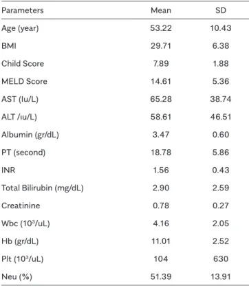
Demographic characteristics of the 18 patients with liv-er cirrhosis (men: 12, 66.7%; age: 53.22±10.43 years) are presented in Table 1 and Figure 1.
According to the Child-Pugh classification, 6 (33.3%) of the 18 patients were classified as group A, 10 (55.6%) were classified as B, and 2 (11.1%) as C.
PSQI versus ESS
According to the pretransplant PSQI scores, 15 (83.4%) of the 18 patients showed a decrease in sleep quality, and 3 (16.6%) of the 18 patients had EDS according to the pretransplant ESS scores.
According to the posttransplant 9-month PSQI scores, 5 (17.8%) of the 18 patients showed a poor sleep quality, and 1 (5.4 %) of the 18 patients had EDS according to the posttransplant ESS scores.
The pretransplant PSQI and ESS scores of the patients were 8.4±3.11 and 7.28±3.89, respectively. The post-transplant PSQI and ESS scores of the patients were 4.5±2.8 and 4.72±2.91, respectively. The differences be-tween pre and posttransplant values were statistically significant (p<0.01) (Table 2).
Table 1. Demographics of the patients.
Parameters Mean SD Age (year) 53.22 10.43 BMI 29.71 6.38 Child Score 7.89 1.88 MELD Score 14.61 5.36 AST (Iu/L) 65.28 38.74 ALT /ıu/L) 58.61 46.51 Albumin (gr/dL) 3.47 0.60 PT (second) 18.78 5.86 INR 1.56 0.43 Total Bilirubin (mg/dL) 2.90 2.59 Creatinine 0.78 0.27 Wbc (103/uL) 4.16 2.05 Hb (gr/dL) 11.01 2.52 Plt (103/uL) 104 630 Neu (%) 51.39 13.91
SD: Standard Deviation; BMI: Body mass index; MELD: Model For End-Stage Liver Disease; AST: Aspartate Aminotransferase; ALT: Alanine Aminotransfer-ase; PT: Prothrombin Time; INR: International Normalized Ratio; Wbc: White Blood Cell; Hb: Hemoglobin; Plt: Platelets; Neu: Neutrophils
Electroencephalogram
In total, 10 of the 18 patients had a normal well-struc-tured EEG with a symmetrical posterior basic rhythm >8 Hz and <13 Hz before and after LTx.
The EEG of 4 of the 18 patients had a predominant theta activity with random delta waves, roughly corresponding to Grade C of the Parsons–Smith classification, and after LTx, all the EEG tracings were normal.
Furthermore, 3 of the 18 patients had a low-frequency alfa rhythm (8 Hz) disturbed by random waves in the the-ta range over both hemispheres, corresponding to Grade B of the Parsons–Smith classification, and after LTx, 2 EEG readings were assessed as normal.
The EEG of 1 of the 18 patients had diffuse asynchronous theta and delta waves over both the hemispheres with triphasic waves, corresponding to Grade D-E of Parsons– Smith’s classification, and after LTx, a low-frequency al-pha rhythm with rare waves in the theta range over both the hemispheres was observed and s assessed as Grade B of the Parsons–Smith classification.
Polysomnography
The duration of all the posttransplant PSG sleep stages increased relative to the pretransplant PSG values. SL and REM latency were reduced relative to the pretrans-plant values. The decrease in SL was statistically signifi-cant (p<0.05). The increase in N1 sleep duration was sta-tistically significant (p<0.05). The comparison of patient PSG parameters between pretransplant and 9-month posttransplant is presented in Table 3.
DISCUSSION
SDs are common in patients with liver cirrhosis and may occur owing to a wide variety of factors, such as pain,
rest-lessness, fatigue, liver-disease-associated itching, poor sleep hygiene, medication use, and impaired liver func-tion. Several mechanisms, such as hyperammonemia, HE, melatonin abnormalities, and circadian rhythm disor-ders, have been demonstrated in the pathophysiology of SDs in patients with cirrhosis. LTx is the most effective treatment for liver cirrhosis. SDs are expected to improve (as are other cirrhosis-related complications) after LTx. However, a small number of studies have found that SDs are still present in patients after LTx. Factors affecting posttransplantation SDs include pretransplantation HE type, advanced anoxic/ischemic brain injury during sur-gery, osmotic myelinolysis, immunosuppressive toxicity, comorbid diseases, and numerous posttransplant medi-cations (1, 5, 7, 13, 14).
PSQI is the gold standard subjective method for sleep quality assessment. PSQI can easily distinguish poor sleepers from good sleepers. Samanta et al. detected 60 (60%) poor sleepers using PSQI in 100 patients with liv-er cirrhosis (21). Xiao et al. detected 214 (62.46%) poor sleepers using PSQI in 341 patients with liver cirrhosis (22). In our study, 15 (83.4%) of the 18 patients showed a poor sleep quality according to the pretransplant PSQI scores. The 9-month posttransplant PSQI scores identi-fied 5 (17.8%) of the 18 patients with a decrease in sleep quality. Pretransplant and 9-month posttransplantation PSQI scores of the patients were 8.4±3.11 and 4.5±2.8, respectively. The difference between pre and posttrans-plant values was statistically significant (p<0.01). EDS is one of the most well-known parameters of sleep-wake abnormalities in patients with liver cirrhosis and is best evaluated by ESS. Samanta et al. investigated 100 patients with liver cirrhosis and detected EDS in 38 (38%) patients by using ESS (21). Montegnese et al. investigat-ed 87 patients with liver cirrhosis and detectinvestigat-ed EDS in 21 (24.1%) patients (6). According to the pretransplant ESS scores in our study, 3 (16.6%) of the 18 patients had EDS. The 9-month posttransplantation ESS scores showed that 1 (5.4 %) of the 18 patients had EDS. Pretransplant ESS and 9-month posttransplantation ESS scores of the patients were 7.28±3.89 and 4.72±2.91, respectively. The difference between pre and posttransplantation values was statistically significant (p<0.01).
Subjective methods, such as PSQI and ESS, are very use-ful for screening, but they do not provide sufficient in-formation about sleep structure and staging. The gold standard for SDs is PSG. Adults often need a total sleep time (TST) of about 7-9 hours. Sleep has 4 stages: NREM Table 2. PSQI and ESS results.
Pretransplantation n (%) Postransplantation n (%) p Poor Sleepers (PSQI>5) 15 (83.4%) 5 (17.8%) p<0.05 Excessive Day-time Sleepiness (ESS>10) 3 (16.6%) 1 (5.4%) p<0.05 PSQI Score (Mean) 8.4±3.11 4.5±2.8 p<0.05 ESS Score (Mean) 7.28±3.89 4.72±2.91 p<0.05
(nonrapid eye movement sleep; N1, N2, and N3) and REM sleep. A complete sleep cycle takes an average of 90–110 min. Usually there are 4 to 5 cycles per sleep time. A sleep cycle begins with NREM (N1, N2, N3) and con-tinues with REM. TST is composed of 70%–80% NREM and 20%–25% REM sleep (20). Findings of recent sleep studies suggest that the informative value of sleep refer-ence data in healthy individuals is limited because of the high interindividual and intraindividual variation in sleep
variables. Hertenstein et al. performed a PSG study with 206 healthy adults aged 19–73 years. According to this study, the reference levels of sleep stages were TST (min), 393.5±50.1 (women), 391.7±52.0 (men); N1 sleep (%), 8.9±4.6 (women), 9.2±4.9 (men); N2 sleep (%), 52.3±8 (women), 52.1±9.7 (men); REM sleep (%) 17.4±4.9 (wom-en), 18.7±4.9 (men); N3 sleep (%), 8.6±7.2 (wom(wom-en), 6.6±6.7 (men) (23). PSG studies in cirrhosis are very few. Teodoro et al. investigated SDs in patients with cirrhosis; Table 3. Evaluation of Polysomnography parameters before and after liver transplantation.
Parameters
Pre-Transplantation Post- Transplantation
p
Mean SD Mean SD
TRT (min) 362.78 37.80 374.50 21.57 0.142
TST (min) 242.40 53.08 273.33 52.12 0.133
Sleep Lat. (min) 40.56 29.77 22.14 24.06 0.049996
REM Lat. 145.08 89.30 113.53 66.75 0.184 SE 67.31 15.86 72.99 13.21 0.528 Arousals/h 27.84 22.47 16.23 16.12 0.170 Total Arousals 113.22 93.87 74.83 72.34 0.267 AHI 15.54 12.96 22.36 16.67 0.055 PLMI 20.87 40.20 22.47 44.09 0.774 Mean SpO2 96.06 2.92 96.11 1.49 0.811 Nadir SpO2 86.78 8.74 85.94 11.20 0.887 ODI 6.62 8.69 9.93 13.55 0.896 NREM (min) 207.94 44.58 236.94 47.85 0.058 REM (min) 28.81 17.03 36.56 22.75 0.513 REM (%) 11.40 5.57 13.31 7.66 0.528 N1 (min) 9.44 5.12 20.17 11.11 0.003 N1 (%) 4.29 2.94 8.14 6.00 0.028 N2 (min) 180.92 48.68 197.03 42.31 0.396 N2 (%) 74.48 10.45 72.19 8.64 0.557 N3 (min) 23.92 21.44 21.19 15.19 0.831 N3 (%) 10.18 9.24 7.28 4.40 0.349
Deep Sleep (min) 23.72 21.66 21.19 15.19 0.906
ST: Standard deviation; TRT: Total recording time; min: Minute; TST: Total sleep time; Sleep Lat: Latency to sleep onset; REM Lat: REM sleep latency; SE: Sleep efficiency; Arousals/h: Number of arousals per hour of sleep; AHI: Apnea-hypopnea index; REM: Rapid eye movement; NREM: Non-Rapid eye movement; PLMI: Periodic Limb movement index; Mean SpO2: Mean oxy-hemoglobin saturation; Nadir SpO2: Minimal oxy-hemoglobin saturation; ODI: Oxygen desatu-ration index N1:NREM Stage 1; N2: NREM Stage 2; N3:NREM Stages 3 and 4;
they reported that the sleep duration decreased, sleep efficiency decreased, SL increased, and REM latency in-creased in such patients (4). In our study, pretransplant and 9-month posttransplant PSG results revealed that posttransplant TST and duration of all the sleep stages (REM and NREM [N1, N2, and N3]) increased relative to pretransplantation. SL and REM latency were found to be reduced. However, only the increase in S1 sleep and the decrease in SL were found to be statistically significant. Periodic limb movements per hour and AHI values were similar between the 2 groups.
EEG reflects postsynaptic activity of the pyramidal cells of the brain cortex that is extremely sensitive to the in-fluence of toxic, metabolic, and pharmacological factors. In patients with HE, the basic rhythmic activity of the EEG progressively slows down, and the reactivity to eye open-ing reduces and finally disappears. The other component is “triphasic waves,” which are not specific to this metabolic encephalopathy, and can be seen in moderate/severe HE. In this study, after LTx with the reversibility of bioelectric brain dysfunction, visual EEG performances significantly improved as in studies with spectral EEG (24, 25).
There are some limitations to this study. The major lim-itation is the small population size. PSG is an expensive and specialized method that is not practiced everywhere. Therefore, the number of patients included in this study was limited. Another limitation is that single-night PSG recordings were performed in this study. As PSG may be a first-night effect, some authors suggest two-night hos-pitalization for PSG. Adding a second-night PSG recording may reduce the adjustment process experienced by the patient on the first night. Another limitation was that spec-tral analysis of the EEG could not be performed. Specspec-tral EEG assessment has been found to be more reliable than qualitative visual EEG reading in the diagnosis of overt and minimal HE, especially in the accurate detection of mild HE. LTx improves the liver function, and cirrhosis complica-tions are expected to disappear after transplantation. A small number of SD studies and data are available in this patient group. Ours is the first study to compare SDs by subjective and objective methods in pre and posttrans-plant patients. Posttransposttrans-plant results revealed significant improvements in SDs at 9 months. Further studies with larger populations and longer follow-ups are required. Ethics Committee Approval: Ethics committee approval was
re-ceived for the study from the Ethics Committee of Yeni Yüzyıl Uni-versity (21.07.2017/035).
Informed Consent: Written informed consent was obtained from all
participants.
Peer-review: Externally peer-reviewed.
Author Contributions: Concept – G.G., G.T., I.Y.G., M.A.; Design -
G.G., G.T., I.Y.G., M.G.; Supervision – G.G., G.T., A.D., M.A.; Resource – G.G., S.A., M.A., A.D.; Materials - G.G., G.T., B.E., I.Y.G., S.A., A.D., M.A.; Data Collection and/or Processing – G.G., G.T., I.Y.G., B.E., S.A.; Anal-ysis and/or Interpretation – G.G., G.T., I.Y.G., B.E.; Literature Search – G.G., G.T., I.Y.G.; Writing – G.G., G.T., I.Y.G.; Critical Review - G.G., I.Y.G., A.D., M.A.
Conflict of Interest: The authors have no conflict of interest to declare. Financial Disclosure: The authors declared that this study has
re-ceived no financial support. REFERENCES
1.Córdoba J, Cabrera J, Lataif L, Penev P, Zee P, Blei AT. High prevalence of sleep disturbance in cirrhosis. Hepatology 1998; 27: 339-45. [CrossRef] 2. Hamdan AJ, Al Enezi A, Anwar A, et al. Prevalence of insomnia and sleep patterns among liver cirrhosis patients. J Circadian Rhythms 2014; 12: 2. [CrossRef]
3. De Cruz S, Espiritu JRD, Zeidler M, Wang TS. Sleep disorders in chronic liver disease. Semin Respir Crit Care Med 2012; 33: 26-35. [CrossRef]
4. Teodoro VV, Júnior MAB, Lucchesi LM, et al. Polysomnographic sleep aspects in liver cirrhosis: a case control study. World J Gastro-enterol 2013; 19: 3433-8. [CrossRef]
5. Mostacci B, Ferlisi M, Baldi Antognini A, et al. Sleep disturbance and daytime sleepiness in patients with cirrhosis: a case control study. Neurol Sci 2008; 29: 237-40. [CrossRef]
6. Montagnese S, Middleton B, Skene DJ, Morgan MY. Night-time sleep disturbance does not correlate with neuropsychiatric impairment in patients with cirrhosis. Liver Int 2009; 29: 1372-82. [CrossRef] 7. Montagnese S, De Pittà C, De Rui M, et al. Sleep-wake abnormalities in patients with cirrhosis. Hepatology 2014; 59: 705-12. [CrossRef] 8. Vilstrup V, Amodio P, Bajajetal J. Hepaticencephalopathy in chron-ic liver disease:2014 Practchron-ice Guideline by the Amerchron-ican Association for the Study Of Liver Diseases and the European Association for the Study of the Liver. Hepatology 2014; 60: 715-35. [CrossRef] 9. Groeneweg M, Quero JC, De Bruijn I, et al. Subclinical hepatic en-cephalopathy impairs Daily functioning. Hepatology 1998; 28: 45-9. [CrossRef]
10. Stewart CA, Malinchoc M, Kim WR, Kamath PS. Hepatic enceph-alopathy as a predictor of survival in patients withend-stageliverdis-ease. Liver Transpl 2007; 13: 1366-71. [CrossRef]
11. Ichikawa T, Naota T, Miyaaki H, Miuma S, Isomoto H,Takeshima F, et al. Effect of an oral branched chain amino acid-enriched snack in cirrhotic patients with sleep disturbance.[CrossRef]Hepatol Res 2010; 40: 971e8.
12. Saner F, Gu Y, Minouchehr S, et al. Neurological complications after cadaveric and living donor liver transplantation. J Neurol 2006; 253: 612e7. [CrossRef]
13. Senzolo M, Ferronato C, Burra P. Neurologic complications after solid organ transplantation. Transpl Int 2009; 22: 269e78. [CrossRef] 14. Amodio P, Biancardi A, Montagnese S, et al. Neurological compli-cations after orthotopic liver transplantation. Dig Liver Dis 2007; 39: 740e7. [CrossRef]
15. Buysse DJ, Reynolds CF 3rd, Monk TH, Berman SR, Kupfer DJ. The Pittsburgh Sleep Quality Index: a new instrument for psychiatric practice and research. Psychiatry Res 1989; 28: 193-213. [CrossRef] 16. Ağargün MY, Kara H, Anlar O, et al. Pittsburgh Uyku Kalitesi İndek-si’nin Geçerliği ve Güvenirliği. Turk Psikiyatri Derg 1996; 7: 107-11. 17. Johns MW. A new method for measuring daytime sleepiness: the Epworth sleepiness scale. Sleep 1991; 14: 540-5. [CrossRef] 18. Izci B, Ardic S, Firat H, et al. Reliability and validity studies of the Turkish version of the Epworth Sleepiness Scale. Sleep Breath 2008; 12: 161-8. [CrossRef]
19. Parsons-Smith BG, Summerskill WJH, Dawson AM, Sherlock S. The electroencephalogram in liverd isease. Lancet 1957; 2: 867-71. [CrossRef]
20. Berry RB, Budhiraja R, Gottlieb DJ, et al. Rules for scoring respi-ratory events in sleep: update of the 2007 AASM Manual for the Scoring of Sleep and Associated Events. Deliberations of the Sleep Apnea Definitions Task Force of the American Academy of Sleep Medicine. J Clin Sleep Med 2012; 8: 597-619. [CrossRef]
21. Samanta J, Dhiman RK, Khatri A, Thumburu KK, Grover S, Duseja A, Chawla Y. Correlation between degree and quality of sleep distur-bance and the level of neuropsychiatric impairment in patients with liver cirrhosis. Metab Brain Dis 2013; 28: 249-59. [CrossRef] 22. Xiao G, Ye Q, Han T, Yan J, Sun L, Wang F. Study of the sleep qual-ity and psychological state of patients with hepatitis B liver cirrhosis. Hepatol Res 2018; 48: E275-E282. [CrossRef]
23. Hertenstein E, Gabryelska A, Spiegelhalder K, el al. Reference Data for Polysomnography-Measured and Subjective Sleep in Healthy Adults. J Clin Sleep Med 2018; 14: 523-32. [CrossRef] 24. Ciancio A, Marchet A, Saracco G, et al. Spectral electroenceph-alogram analysis in hepatic encephalopathy and liver transplanta-tion. Liver Transpl 2002; 8: 630-5. [CrossRef]
25. Campagna F, Montagnese S, Schiff S, Biancardi A, Mapelli D, Angeli P et al. Cognitive impairment and electroencephalographic alterations before and after liver transplantation: what is reversible? Liver Transpl 2014; 20: 977-86. [CrossRef]