Overlooked Infection in a Dog with Degenerative Discospondylitis
and Hip Joint Osteoarthritis: Ruminant Originated Brucellosis
(Dejeneratif Diskospondilitis ve Koksafemoral Osteoartritisli bir
Köpekte Gözden Kaçan Enfeksiyon: Ruminant Kökenli Brusellozis)
A. Alkan KUŞÇU
1M. Özay BEDİZCİ
1Sırrı AVKİ
2
Hülya TÜRÜTOĞLU
31 2 3
Petcity Vet. Clinic, Cevat Paşa Mahallesi, Nazım Demircioğlu Sokak, TR-17000 Çanakkale - TURKEY
Mehmet Akif Ersoy University, Faculty of Veterinary Medicine, Department of Surgery, TR-15030 Burdur - TURKEY Mehmet Akif Ersoy University, Faculty of Veterinary Medicine, Department of Microbiology, TR-15030 Burdur - TURKEY
Dear Editor,
Canine degenerative osteoarthritis and discospondylitis are serious disorders of locomotor system with many etiologic factors contributing to pathogenesis[1,2]. Canine
brucellosis caused by Brucella canis is one of these factors and enlargement of lymph nodes, uveitis, osteomyelitis, polyarthritis, glomerulonephritis, pyogranulomatous dermatitis, epididymitis, orchitis, scrotal dermatitis (some- times scrotal necrosis) are other clinical signs of this infection [3-6]. This letter is written to underline the
possibility of canine brucellosis caused by other Brucella species, and to share an interesting case of dog brucellosis originated from ruminants with colleagues.
A 4½ years old male Labrador dog was brought to the clinic with complaints of reluctance to walk; inability to jump into a car; growling when his back was touched and lethargy. As it was learned from the patient’s story, the complaints were become apparent during past 6 months, but since two years he does not want to sit after walking due to swelling and pain in the testicles, and therefore he had been castrated. On physical examination, body temperature, pulse and respiratory rates were within normal values and pain was recorded on palpation of the left hip joint and lumbar region. When raised on hind legs, it was noted that he could not tolerate this position more than 30 seconds. Laboratory examination revealed that WBC count (19.57 x10⁹/L), % values of basophils and
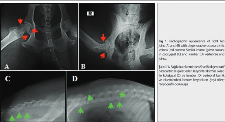
neutrophils were increased and % value of lymphocytes was decreased. Serum CRP level was quite high (119 mg/L). On radiographic examination, signs of degenerative osteo-arthritis were observed in right hip joint (Fig. 1A and B)
and lumbar-coccygeal vertebrae (Fig. 1C and D). A Brucella
infection had aroused suspicion due to the orchitis and castration story. His serum sample gave negative result
for Brucella canis with an immunochromatographic test kit (Brucella IC®, Biopronix, Agrolabo SpA, Italy), but it was positive with Rose Bengal plate test (SeroLam®, Seromed, Istanbul) which detects IgG and IgM formed against smooth
Brucella species. According to the test results, dog was
thought to be infected with one of these bacteria species:
Brucella melitensis (sheep and goats), Brucella abortus (cattle)
or Brucella suis (swine). While this development has been shared with the owner, it was learned that the dog had run around in neighboring goat farm since his puppyhood. Isolation of specific bacteria was not able because synovial fluid sampling of the relevant joints was not possible. Based on the serological tests results and owner’s last information, the dog’s disease was considered as ruminant originated brucellosis, and concerned pathologies (orchitis, diskospondylitis and degenerative right hip osteoarthritis) were thought to be related to this overlooked and chronic infection. Dog was treated for 2 months with doxycycline (2 times a day 300 mg, oral route, Monodoks® 100 mg capsules, Deva, Istanbul) and streptomycin sulfate (1 g IM and in 1, 3, 5 and 8th weeks for 7 days, Streptomycin® vial, IE
ULAGAY, Istanbul). After 1 month of therapy, blood serum CRP level was found in normal levels (9 mg/L). By future phone calls with the owner, it was learned that the dog can swim, get in the car, play ball and has no more back pain.
The presented case teaches us 3 important lessons: 1- Dogs with epididymitis, orchitis, scrotal dermatitis, uveitis, osteoarthritis or discospondylitis may be candidates of canine brucellosis. 2- During serological examinations, we must not only refer with Brucella canis specific tests. 3- Serological tests for B. melitensis, B. abortus or B. suis infections must absolutely be in diagnostic protocols particularly for dogs whose close contact with farm animals or have the possibility of eating farm animals’ placenta or drinking their milk.
İletişim (Correspondence)
+90 248 2132102
[email protected]Journal Home-Page: http://vetdergi.kafkas.edu.tr
online SubmiSSion: http://vetdergikafkas.org
LETTER to EDITOR
Kafkas Univ Vet Fak Derg
20 (6): 983-984, 2014
DOI: 10.9775/kvfd.2014.12111
984
Overlooked Infection in a Dog ...
REFERENCES
1. Lipowitz AJ, Newton CD: Degenetative joint disease and traumatic arthritis. In, Newton CD and Nunamaker DM (Ed): Textbook of Small Animal Orthopaedics. 1029-1042, JB Lippincott Comp, Philadelphia, 1985.
2. Betts CW: Osteomyelitis of the vertebral body and the intervertebral disk: Discospondylitis. In, Newton CD and Nunamaker DM (Ed): Textbook of Small Animal Orthopaedics. 725-730, JB Lippincott Comp, Philadelphia, 1985.
3. Anderson GI, Binnington AG: Discospondylitis and orchitis associated with high Brucella titre in a dog. Can Vet J, 24, 249-252, 1983.
4. Xavier MN, Paixão TA, den Hartigh AB, Tsolis RM, Santos RL: Pathogenesis of Brucella spp. Open Vet Sci J, 4, 109-118, 2010.
5. Keid LB, Soares RM, Morais ZM, Richtzenhain LJ, Vasconcellos SA:
Brucella spp. isolation from dogs from commercial breeding kennels in
Sao Paulo state, Brazil. Braz J Microbiol, 35, 161-166, 2004.
6. Almeida AC, Santorelli A, Bruzadelli RMZ, Oliveira MMNF: Sero-epidemiology of canine brucellosis caused by B. canis and B. abortus in Alfenas, MG, Brazil. Arq Bras Med Vet Zootec, 56 (2): 275-276, 2004.
Fig 1. Radiographic appearance of right hip joint (A) and (B) with degenerative osteoarthritic lesions (red arrows). Similar lesions (green arrows) in coccygeal (C) and lumbar (D) vertebrae and joints.
Şekil 1. Sağ kalça ekleminde (A) ve (B) dejeneratif osteoartritis’e işaret eden lezyonlar (kırmızı oklar) ile koksigeal (C) ve lumbar (D) vertebral kemik ve eklemlerdeki benzer lezyonların (yeşil oklar) radyografik görünüşü.