New indicator of cellular ischemia in coronary slow-flow
phenomenon: Cell-free DNA
Koroner yavaş akım fenomeninde hücresel iskemi için yeni gösterge:
Serbest DNA
1Department of Cardiology, Yeni Yüzyıl University Faculty of Medicine, İstanbul, Turkey; 2Department of Medical Genetics, İstinye University Faculty of Medicine, İstanbul, Turkey; 3Department of Medical Biology, Erzincan University Faculty of Medicine, Erzincan, Turkey
Mustafa Yolcu, M.D.,1 Ali Dogan, M.D.,1 Nuri Kurtoglu, M.D.,1
Veysel Sabri Hancer, M.D.,2 Mehmet Gürbüzel, M.D.3
Objective: Coronary slow-flow phenomenon (CSFP) is de-fined as the delayed arrival of coronary blood flow to the distal vascular bed in at least 1 major epicardial coronary artery. Cell-free DNA (cfDNA) is a type of DNA that circu-lates freely in the blood once released from nucleated cells. The aim of this study was to determine if the level of cfDNA, which is an indicator of ischemia at the cellular level, was increased in CSFP.
Methods: The study included 46 patients in total: 23 pa-tients with CSFP and 23 with a normal coronary angiogram (NCA). The level of cfDNA, and clinical, biochemical, and angiographic features of the groups were compared.
Results: The mean age was 53.8±10.3 years for the CSFP patient group and 56.6±9.4 years for the NCA patient group. There was no statistically significant difference between the groups in terms of basal clinical characteristics or labora-tory data. The plasma cfDNA level was 5.04±2.37 ng/µL in the CSFP patients and 2.28±1.09 ng/µL in the NCA group (p<0.001).
Conclusion: Several invasive and noninvasive studies con-ducted on patients with CSFP have revealed myocardial ischemia. The results of this study demonstrated that the level of cfDNA was significantly increased in patients with CSFP as a result of ischemia at the cellular level caused by microvascular disruption.
Amaç: Koroner yavaş akım fenomeni (KYAF) en az bir ma-jör epikardiyal koroner arterde kan akımının distal damar yatağına geç ulaşması olarak tanımlanır. Serbest hücre-sel DNA (shDNA), hücre nükleuslarından serbestleşen ve dolaşımda serbest olarak tespit edilebilen DNA tipidir. Bu çalışmada, KYAF’de hücresel düzeyde iskeminin göster-gesi olan shDNA düzeylerinin artıp artmadığını göstermeyi amaçladık.
Yöntemler: Bu çalışmaya, 23 KYAF’lı ve 23 anjiyografik ola-rak normal koroner arterlere (NKA) sahip toplam 46 hasta alındı. Grupların shDNA düzeyleri, klinik, biyokimyasal ve anjiyografik özellikleri karşılaştırıldı.
Bulgular: Ortalama yaş KYAF grubunda 53.8±10.3 ve NKA grubunda 56.6±9.4 idi. Laboratuvar bulguları ve bazal kli-nik karekteristikleri yönünden gruplar arasında istatistik-sel anlamlı fark yoktu. Plazma shDNA düzeyleri KYAF’da 5.04±2.37 ng/µL, NKA’da 2.28±1.09 ng/µL olarak tespit edil-di (p<0.001).
Sonuç: Farklı invaziv ve noninvaziv çalışmalar KYAF’da miyokardiyal iskeminin varlığını göstermiştir. Çalışmamızda KYAF’lı hastalarda mikrovasküler bozukluğa bağlı olarak, hücresel düzeyde iskemi ve hasarın göstergesi olan shDNA düzeyinin anlamlı şekilde arttığını gösterdik.
Received:November 18, 2019 Accepted:March 16, 2020
Correspondence: Dr. Mustafa Yolcu. Yeni Yüzyıl Üniversitesi Tıp Fakültesi, Kardiyoloji Anabilim Dalı, İstanbul, Turkey
Tel: +90 212 - 615 38 38 e-mail: [email protected]
© 2020 Turkish Society of Cardiology
ABSTRACT ÖZET
C
oronary slow-flow phenomenon (CSFP) is de-fined as the delayed arrival of coronary blood flow to the distal vascular bed in at least 1 major epicardialartery without any stenosis in the epicardial coronary arteries.[1] Routine coronary angiographies have re-vealed a CSFP incidence of 1% to 7%.[2–4] Tambe et
al.[5] described CSFP in 1972 in 6 patients who presented with chest pain and they suggested that this phenomenon could develop due to ab-normalities in coro-nary microcircula-tion. Several studies have reported an association between CSFP and sudden cardiac death, life-threatening arrhythmias, myocar-dial ischemia, recurrent acute coronary syndromes, and congestive heart failure.[6–8] Though a number of studies performed in biopsies of the left and right ventricles of the patients with CSFP have shown that CSFP can cause luminal narrowing, changes in nor-mal morphology of the nucleus, thickening of the capillary endothelium, and small vessel disease, such as pyknosis, the possible role of dynamic components affecting the microvascular structure remains to be demonstrated in order to support these findings.[9,10]
Cell-free DNA (cfDNA) is a type of DNA that circulates freely in the blood after being released from nucleated cells.[11,12] The origin of cfDNA is not clear; however, it has been proposed that cfDNA is released into circulation as a consequence of cell ly-sis, necrotic death, apoptoly-sis, and active release.[13] In 1948, Mandel et al.[14] reported that some nucleic acids circulated in the blood. In several studies conducted shortly afterwards, it was suggested that cfDNA may occur as a result of metastasis. The quantitative mea-surement of cfDNA isolated from serum and plasma samples of patients with diseases such as rheumatoid arthritis, systemic lupus erythematosus, leukemia, chronic glomerulonephritis, and cancer has been ob-served to be generally higher than in normal individ-uals.[15–19] A review of the literature demonstrates that, with the development of technical measurements, the quantitative value of cfDNA has been investigated in many diseases, as well as whether genetic analyses such as next-generation sequencing can be performed using cfDNA.[20,21]
CSFP is an angiographic observation defined by the delayed passage of contrast media in the
epicar-dial vessels. Various studies have shown an increased risk of sudden cardiac death, life-threatening arrhyth-mias, and congestive heart failure in CSFP. These risks are increased even without the presence of coro-nary ischemia-induced stenosis, suggesting different etiopathogenetic conditions in these patients. The level of cfDNA is an important parameter that indicates cel-lular ischemia and may be a valuable sign demonstrat-ing silent myocardial ischemia in these patients. The aim of the present study was to demonstrate whether the level of cfDNA was increased in CSFP.
METHODS Study population
This prospective study included 8350 patients who underwent coronary angiography between August 2017 and May 2019. CSFP was detected in 23 con-secutive patients. An additional 23 subjects with a normal coronary angiogram (NCA) were randomly selected for comparison. In total, the study population consisted of 46 patients. The research was designed as cross-sectional, observational study. Patients with CSFP had slow flow in all 3 of their coronary arter-ies; patients who had slow flow in 1 or 2 coronary arteries were not included in the study. The indication for coronary angiography was typical angina and a positive exercise test or a perfusion defect observed with myocardial perfusion scintigraphy (MPS). The patients’ clinical characteristics, including age, sex, smoking status, and dyslipidemia, hypertension (HT), and diabetes mellitus (DM) findings were recorded. Hemogram findings; kidney and liver function tests; troponin, C-reactive protein, lipid parameters; and thyroid hormones were evaluated in all of the patients. Potential study participants in both groups were ex-cluded for a previous history of percutaneous coro-nary intervention, moderate-severe valvular disease, left ventricular dysfunction (ejection fraction <50%), left ventricular hypertrophy, myocardial infarction (MI), rhythm other than sinus, chronic systemic ill-ness, chronic obstructive lung disease and/or cor pul-monale, renal failure, congenital heart disease, active infection, or neoplastic disease, with the concomitant presence of slow flow and stenotic lesions. Patients using nitrate medication before or during the coro-nary angiography and those with unstable angina pec-toris or acute coronary syndrome were also excluded from the study if the coronary angiography detected
Abbreviations:
CAD Coronary artery disease CSFP Coronary slow-flow phenomenon cfDNA Cell-free DNA
DM Diabetes mellitus FFS Fractional flow reserve HT Hypertension
LAD Left anterior descending artery LCX Left circumflex artery MI Myocardial infarction MPS Myocardial perfusion scintigraphy NCA Normal coronary angiogram RCA Right coronary artery ROC Receiver operating characteristic TFC TIMI frame count TIMI Thrombolysis in Myocardial Infarction
mmHg or the use of antihypertensive medication. DM was defined as a fasting blood glucose value of ≥126 mg/dL or the use of hypoglycemic agents. Hyperc-holesterolemia was defined as a baseline cholesterol level of >200 mg/dL and/or a low-density lipoprotein cholesterol level of >130 mg/dL or the use of statin medications. Those who had regularly smoked in the previous year were considered current smokers.
This study was approved by the institutional re-view committee and medical ethics committee (date: 30/04/2019, no: 04/11). Written informed consent was obtained from all of the patients.
Coronary angiography
A femoral arterial approach was used for selective left and right coronary angiography and an independent expert interventionalist analyzed the angiographic data. Iohexol was used as the opaque substance in all patients. Left heart catheterization was performed using the Judkins technique. Coronary arteries were displayed at the cranial and caudal angles in the right and left oblique view at 15 frames per second. The patient angiograms were re-evaluated by 2 interven-tional cardiologists to determine the Thrombolysis in Myocardial Infarction (TIMI) frame count (TFC) for each coronary artery. The first frame used was the frame in which the opaque material was delivered to the coronary artery ostium with visualization of the coronary artery, and the last frame was the frame necessary to image the distal landmark of the opaque material. The first posterolateral branch of the right coronary artery (RCA), left circumflex artery (LCX), and distal bifurcation of the left anterior descending artery (LAD) were defined as the distal section. The difference between the first and last frames was eval-uated as the TFC. The frame counts of the LAD were divided by 1.7 to correct for the longer length of this vessel. After the correction, a value of 25 or more for all vessels was accepted as CSFP.[22]
An NCA was defined as the lack of angiographic atherosclerosis during routine coronary angiography. These patients had angiographically normal coronary arteries and no provocative vasoreactivity test was performed.
Cell-free circulating DNA measurement
Angiography was performed after 12 hours of fasting.
diately after diagnosis and centrifugation was initiated. Samples of approximately 10 mL of peripheral blood were centrifuged at 2500xg for 10 minutes. The super-natant was transferred to a new sterile tube which was again centrifuged at 16,000xg for 10 minutes, and this new supernatant was put into a new sterile tube and kept at -80C°. The Plasma/Serum Cell-Free Circulat-ing DNA Purification Micro Kit (Cat. 55500; Norgen Biotek Corp., Thorold, Ontario, Canada) was used for the DNA isolation. The quantity of DNA was mea-sured using a NanoDrop ND1000 spectrophotometer (Thermo Fisher Scientific, Inc., Waltham, MA, USA). Statistical analysis
Statistical analysis was performed using SPSS for Windows, Version 16.0 (SPSS Inc., Chicago, IL, USA). Percentages, means, and SD were calculated. The Kolmogorov-Smirnov test was used to determine the normality of distribution. A t-test was used to compare parametric variables between groups. A chi-square test was used to compare non-parametric val-ues and percentages. Statistical significance was es-tablished as p≤0.05. Receiver operating characteristic (ROC) curves were generated and used to determine the sensitivity and specificity of cfDNA and the cut-off value for predicting coronary slow flow.
Power analysis was conducted for cfDNA for both the CSFP and NCA groups. To obtain a precision of 10% at a type I error level of 5% with 80% power, the necessary sample size was determined to be 24. The study plan was to include at least 46 patients.
RESULTS
Results were obtained from 46 patients who under-went coronary angiography: 23 patients with iso-lated CSFP and 23 with an NCA. The mean age was 53.8±10.3 years in the CSFP patients and 56.6±9.4 years in the NCA patients. There was no statistically significant difference between the groups in terms of HT, hyperlipidemia, DM, family history of CAD, smoking status, age, or gender. The serum glucose, urea, creatinine, triglycerides, low-density lipopro-tein, high-density lipoprolipopro-tein, total cholesterol, thy-roid stimulating hormone, and hematocrit levels were not significantly different between groups (Table 1).
The indication for coronary angiography was a positive treadmill exercise test (ST depression) or
RCA in patients with CSFP. The mean corrected TFC for the LAD was 18.1±2.7, the TFC for the CX was 17.9±2.9 and the TFC for the RCA was 18.0±2.7 in the NCA group (Table 2).
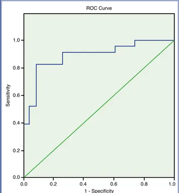
The mean plasma cfDNA level was 5.04±2.37 ng/ µL in the CSFP patients and 2.28±1.09 ng/µL in the NCA group (p<0.001) (Table 2, Fig. 1). According to ROC analysis, the area under the curve for cfDNA was 0.887. The cut-off value of cfDNA to detect coro-nary slow flow was 3.21, with a sensitivity of 82.6 and the presence of ischemia observed with MPS. In the
CSFP group, 13 patients had a positive treadmill ex-ercise test and 10 patients had ischemia findings on MPS. In the NCA group, 11 patients had a positive treadmill exercise test and 12 patients had ischemia findings on MPS. There was no statistically signifi-cant difference between the groups with regard to the treadmill exercise test or the MPS ischemia. All of the patients had slow flow in all 3 coronary arteries: the mean corrected TFC for the LAD was 36.2±8.2, the TFC for the CX was 33.2±6.9 and 32.9±7.5 for the
Table 1. Clinical and biochemical features of the patients
CSFP (n=23) NCA (n=23) p-value Age (years)* 53.8±10.3 56.6±9.4 0.337 Gender (male/female)* 13/10 11/12 0.559 Hypertension, n (%)* 8 (35) 9 (39) 0.763 Diabetes mellitus, n (%)* 7 (30) 6 (26) 0.746 Hyperlipidemia, n (%)* 8 (35) 7 (30) 0.756
Family history of CAD, n (%)* 6 (26) 5 (22) 0.732
Smoking, n (%)* 10 (43) 9 (39) 0.767
Fasting blood glucose (mg/dL)** 109.6±11.7 106.1±10.8 0.528
Urea (mg/dL)** 34.3±10.5 34.1±8.6 0.952 Serum creatinine (mg/dL)** 0.86±0.19 0.86±0.18 0.994 Total cholesterol (mg/dL)** 198±39 190±38 0.506 Triglycerides (mg/dL)** 165±56 148±55 0.304 LDL cholesterol (mg/dL)** 125±33 120±23 0.526 HDL cholesterol (mg/dL)** 40±9 41±8 0.667 TSH (mIU/L)** 1.57±0.96 1.46±0.82 0.671 Hematocrit** 42.6±2.8 42.9±2.8 0.714
*Chi-square test; **t-test. CAD: Coronary artery disease; CSFP: Coronary slow-flow phenomenon; HDL: High-density lipoprotein; LDL: Low-density lipoprotein; NCA: Normal coronary angiogram; TSH: Thy-roid-stimulating hormone.
Table 2. Treadmill exercise test, myocardial perfusion scintigraphy, TFC, and cfDNA measurement
CSFP (n=23) NCA (n=23) p-value
Positive treadmill exercise test, n (%) 13 (57) 11 (48) 0.382
Ischemia on MPS, n (%) 10 (43) 12 (52)
LAD-corrected TFC* 36.2±8.2 18.1±2.7 <0.001
Circumflex artery* 33.2±6.96 17.9±2.9 <0.001
Right coronary artery* 32.9±7.5 18.0±2.7 <0.001
Hs-troponin T* 7.83±3.83 7.96±3.23 0.902
cfDNA (ng/µL)* 5.04±2.37 2.28±1.09 <0.001
*T-test. cfDNA: Cell-free DNA; CSFP: Coronary slow-flow phenomenon; Hs-troponin: High-sensitivity tro-ponin; LAD: Left anterior descending artery; NCA: Normal coronary angiogram; TFC: Thrombolysis in Myocardial Infarction frame count.
a specificity of 91.3% (Fig. 2). DISCUSSION
cfDNA is DNA found in plasma that is released from cells, particularly as a result of ischemic and apop-totic events. In the present study, it was found that the level of cfDNA was significantly increased in cases of CSFP.
CSFP can be detected through delayed dye opaci-fication in the epicardial coronary arteries during an angiography, in the absence of obstructive CAD.[23] The exact mechanism of action in CSFP is not clear. Various disorders, such as diffuse atherosclerosis, in-creased platelet aggregation, inin-creased microvascular inflammation, and endothelial and microvascular dys-functions have been proposed as responsible for the development of CSFP.[24,25]
Mosseri et al.[10] reported that the small coronary arteries of patients with CSFP can reveal proliferation of the myointimal layer, hypertrophy of the media layer, hyperplasia of the fibromuscular layer, swelling of the capillary endothelial cells, and degeneration of endothelial cells. Furthermore, there are lipofuscin deposits and the myocardium is hypertrophic.
Yilmaz et al.[26] analyzed the high incidence of metabolic syndrome in patients with CSFP and the proposition that pathogenesis may be an early phase of atherosclerosis. They suggested that metabolic dis-orders disrupt the vascular structure. Sezgin et al.[27]
reported that coronary resistance may be increased due to endothelial dysfunction in the small coronary arteries, which results in myocardial ischemia in pa-tients with normal coronary arteries presenting with angina. Cin et al.[28] found that patients with CSFP had diffuse intimal thickening and widespread calci-fication along the vessel wall. With respect to these findings, we suggest that CSFP can be considered dif-fuse atherosclerosis involving the epicardial coronary arteries and the microvascular system.
Pekdemir et al.[29] examined epicardial coronary arteries that looked normal in cases of CSFP using the fractional flow reserve (FFR) and intravascular ul-trasound, and it was suggested that a decreased FFR may be related to increased resistance in the epicar-dial coronary arteries due to diffuse atherosclerotic disease. Camsari et al.[30] and Pekdemir et al.[31,32] assessed high levels of endothelin-1 and low nitric oxide concentration in the peripheral blood and the coronary sinus both at rest and after atrial pacing with exercise-induced stress and demonstrated that en-dothelial functions were disrupted. Mangieri et al.[33] detected glycogen content, mitochondrial abnormali-ties, and thickening of vessel walls with luminal size reduction in the endomyocardial biopsies of 20 pa-tients with CSFP. Narimani et al.[23] observed that the
Figure 1. Cell-free DNA (ng/µL) measurement. CSFP: Cor-onary slow-flow phenomenon; NCA: Normal corCor-onary an-giogram. 95% CI DNA 6.00 7.00 5.00 4.00 3.00 2.00 1.00 CSFP Coronary NCA Sensitivity 0.8 0.6 0.4 0.2 0.0 0.0 0.2 0.4 0.6 0.8 1.0 1.0 1 - Specificity
Figure 2. Receiver operating characteristic (ROC) curve (area under the curve: 0.887).
CSFP have demonstrated the presence of myocardial ischemia. In our study, we showed that the level of cfDNA, a marker of cellular ischemia and damage, was significantly increased as a result of ischemia at the cellular level caused by microvascular disruption in patients with CSFP.
Conclusion
Although it was thought to be a benign phenomenon by many clinicians, our study has shown that CSFP actually reveals subclinical findings. A high level of cfDNA in patients with CSFP suggests that impaired myocardial perfusion and the resulting ischemia cause myocardial cell lysis or rapid apoptosis, which leads to earlier programmed cell death. The high level of cfDNA in these patients might be a sign of increased cardiovascular risk. As reported in recent studies, ev-idence of ischemia at the cellular level indicates that this phenomenon cannot be considered a totally be-nign condition.
Limitations
Although the present study provided significant re-sults, it has also some limitations. A larger number of patients could better represent the population. The lack of follow-up is a second limitation. In addition, exclusion of isolated coronary artery slow flow is another limitation of the study. Investigation of the relationship between isolated CSFP and cfDNA is a matter for future study.
Financial disclosure: This research received no specific grant from any funding agency, commercial or not-for-profit sectors.
Ethical statement: This study was approved by the insti-tutional review committee and medical ethics committee (date: 30/04/2019, no: 04/11).
Peer-review: Externally peer-reviewed. Conflict-of-interest: None.
Authorship contributions: Concept: M.Y., M.G.; Design: M.Y., A.D., M.G.; Supervision: M.Y., N.K.; Materials: M.Y., M.G., V.S.H.; Data: M.Y., M.G., V.S.H.; Analysis: M.Y.; Literature search: M.Y., M.G., A.D.; Writing: M.Y., M.G., A.D., N.K.; Critical revision: M.Y., A.D., N.K.
REFERENCES
1. TIMI Study Group. The Thrombolysis in Myocardial In-farction (TIMI) trial. Phase I findings. N Engl J Med 1985;312:932–6. [CrossRef]
lateral s ′ and e ′ waves were lower in patients with CSFP in a tissue Doppler study.
Studies of myocardial perfusion scan and cardiac stress tests have already shown ischemia in 30% to 80% of these patients.[34] Goel et al.[35] observed a pos-itive stress test more commonly in patients with CSFP than NCA patients (71% vs 42%; p<0.01). Cesar et al.[35] have found perfusion abnormalities in 13 of 17 CSFP patients using MPS. Sadr-Ameli et al.[36] noted ischemia in 109 of 217 patients in a long-term follow-up study using noninvasive techniques such as my-ocardial perfusion imaging, stress echocardiography, and exercise stress testing, and reported that CSFP cannot be considered a totally benign condition.
Acute cellular injury or programmed cell death are the main reasons for circulating cfDNA and it can be used as a marker of cellular damage. After apoptosis, it can be detected in the plasma or serum of healthy individuals.[37,38] Recent studies have reported a high level of cfDNA in cases of autoimmune diseases, sepsis, trauma, pregnancy-associated disorders, infec-tion, and a variety of malignancies.[37,38]
Chang et al.[39] found that the average concentration of cfDNA in 55 patients with MI was >10-fold higher than that of 274 normal controls. Jing et al.[40] also found that the cfDNA level was significantly higher in patients with MI compared with controls. Further-more, Destouni et al.[41] reported that the concentration of cfDNA was significantly higher in patients with MI than in a healthy control group using real-time poly-merase chain reaction testing of the β-globin gene during hospitalization. In a study performed using a DNA-based Alu assay, it was demonstrated that the level of cfDNA was significantly higher in patients with acute coronary syndrome when compared with the controls.[42]
CSFP is characterized by delayed passage of contrast substance to the epicardial vessels and the etiopathogenesis has not yet been fully determined. Studies of MI have revealed that the level of cfDNA released from the myocardium after ischemia was statistically significantly high. Previous studies have also shown that CSFP is associated with different dis-eases, such as subclinical atherosclerosis, endothelial dysfunction, inflammation, coronary microvascu-lar dysfunction, and small vessel disease. Invasive and noninvasive studies conducted on patients with
18. Koffler D, Agnello V, Winchester R, Kunkel HG. The occur-rence of single-stranded DNA in the serum of patients with systemic lupus erythematosus and other diseases. J Clin In-vest 1973;52:198–204. [CrossRef]
19. Leon SA, Shapiro B, Sklaroff DM, Yaros MJ. Free DNA in the serum of cancer patients and the effect of therapy. Cancer Res 1977;37:646–50.
20. Ponti G, Maccaferri M, Manfredini M, Kaleci S, Mandrioli M, Pellacani G, et al. The value of fluorimetry (Qubit) and spectrophotometry (NanoDrop) in the quantification of cell-free DNA (cfDNA) in malignant melanoma and prostate can-cer patients. Clin Chim Acta 2018;479:14–19. [CrossRef]
21. Francaviglia I, Magliacane G, Grassini G, Girlando S, Medic-ina D, Lazzari C, et al. Identification and monitoring of so-matic mutations in circulating cell-free DNA by next-genera-tion sequencing in patients with lung adenocarcinoma. Cancer Research 2018;78:5580. [CrossRef]
22. Ong P, Camici PG, Beltrame JF, Crea F, Shimokawa H, Sechtem U, et al. International standardization of diagnostic criteria for microvascular angina. Int J Cardiol 2018;250:16– 20. [CrossRef]
23. Narimani S, Hosseinsabet A, Pourhosseini H. Effect of Coronary Slow Flow on the Longitudinal Left Ventricu-lar Function Assessed by 2-Dimensional Speckle-Tracking Echocardiography. J Ultrasound Med 2016;35:723–9. [CrossRef]
24. Cakmak M, Tanriverdi H, Cakmak N, Evrengul H, Cetemen S, Kuru O. Simvastatin may improve myocardial perfusion abnormality in slow coronary flow. Cardiology 2008;110:39– 44. [CrossRef]
25. Gökçe M, Kaplan S, Tekelioğlu Y, Erdoğan T, Küçükosman-oğlu M. Platelet function disorder in patients with coronary slow flow. Clin Cardiol 2005;28:145–8. [CrossRef]
26. Yilmaz H, Demir I, Uyar Z. Clinical and coronary angio-graphic characteristics of patients with coronary slow flow. Acta Cardiol 2008;63:579–84. [CrossRef]
27. Sezgin AT, Sigirci A, Barutcu I, Topal E, Sezgin N, Ozdemir R, et al. Vascular endothelial function in patients with slow coronary flow. Coron Artery Dis 2003;14:155–61. [CrossRef]
28. Cin VG, Pekdemir H, Camsar A, Ciçek D, Akkus MN, Par-maksız T, et al. Diffuse intimal thickening of coronary arteries in slow coronary flow. Jpn Heart J 2003;44:907–19. [CrossRef]
29. Pekdemir H, Cin VG, Ciçek D, Camsari A, Akkus N, Döven O, et al. Slow coronary flow may be a sign of diffuse ath-erosclerosis. Contribution of FFR and IVUS. Acta Cardiol 2004;59:127–33. [CrossRef]
30. Camsarl A, Pekdemir H, Cicek D, Polat G, Akkus MN, Döven O, et al. Endothelin-1 and nitric oxide concentrations and their response to exercise in patients with slow coronary flow. Circ J 2003;67:1022–8. [CrossRef]
31. Pekdemir H, Cicek D, Camsari A, Akkus MN, Cin VG, Doven O, et al. The relationship between plasma endothelin-1, nitric oxide levels, and heart rate variability in patients with coro-flow: a distinct angiographic subgroup in syndrome X.
Angi-ology 2001;52:507–14. [CrossRef]
3. Beltrame JF, Limaye SB, Horowitz JD. The coronary slow flow phenomenon-a new coronary microvascular disorder. Cardiology 2002;97:197–202. [CrossRef]
4. Saya S, Hennebry TA, Lozano P, Lazzara R, Schechter E. Coronary slow flow phenomenon and risk for sudden cardiac death due to ventricular arrhythmias: a case report and review of literature. Clin Cardiol 2008;31:352–5. [CrossRef]
5. Tambe AA, Demany MA, Zimmerman HA, Mascarenhas E. Angina pectoris and slow flow velocity of dye in coronary arteries-a new angiographic finding. Am Heart J 1972;84:66– 71. [CrossRef]
6. Cutri N, Zeitz C, Kucia AM, Beltrame JF. ST/T wave changes during acute coronary syndrome presentation in patients with the coronary slow flow phenomenon. Int J Cardiol 2011;146:457–8. [CrossRef]
7. Horjeti B, Goda A. Acute ischemia manifestation in a pa-tient with coronary slow flow phenomenon. J Electrocardiol 2012;45:277–9. [CrossRef]
8. Marinescu MA, Löffler AI, Ouellette M, Smith L, Kramer CM, Bourque JM. Coronary microvascular dysfunction, mi-crovascular angina, and treatment strategies. JACC Cardio-vasc Imaging 2015;8:210–20. [CrossRef]
9. Oktay V, Arat Özkan A. Coronary slow flow. Turk Kardiyol Dern Ars 2016;44:193–5. [CrossRef]
10. Mosseri M, Yarom R, Gotsman MS, Hasin Y. Histologic evi-dence for small-vessel coronary artery disease in patients with angina pectoris and patent large coronary arteries. Circulation 1986;74:964–72. [CrossRef]
11. Lo YM, Tein MS, Lau TK, Haines CJ, Leung TN, Poon PM, et al. Quantitative analysis of fetal DNA in maternal plasma and serum: implications for noninvasive prenatal diagnosis. Am J Hum Genet 1998;62:768–75. [CrossRef]
12. Lo YM, Rainer TH, Chan LY, Hjelm NM, Cocks RA. Plasma DNA as a prognostic marker in trauma patients. Clin Chem 2000;46(3):319–23. [CrossRef]
13. Song H, Nan Y, Cheng XW. Circulating cf-DNA: a promising, noninvasive tool for assessment of early cardiometabolic risk. Atherosclerosis 2014;233(1):307–9. [CrossRef]
14. Mandel P, Metais P. Les acides nucléiques du plasma sanguin chez l’homme. [Article in French] C R Seances Soc Biol Fil 1948;142:241–3.
15. Pathak AK, Bhutani M, Kumar S, Mohan A, Guleria R. Cir-culating cell-free DNA in plasma/serum of lung cancer pa-tients as a potential screening and prognostic tool. Clin Chem 2006;52:1833–42.
16. Bendich A, Wilczok T, Borenfreund E. Circulating DNA as a possible factor in oncogenesis. Science 1965;148:374–6. 17. Tan EM, Schur PH, Carr RI, Kunkel HG. Deoxybonucleic acid
(DNA) and antibodies to DNA in the serum of patients with systemic lupus erythematosus. J Clin Invest 1966;45:1732–
specificity for the diagnosis of myocardial infarction. Int J Mol Med 2015;35:72–80. [CrossRef]
38. Jung K, Fleischhacker M, Rabien A. Cell-free DNA in the blood as a solid tumor biomarker-a critical appraisal of the literature. Clin Chim Acta 2010;411:1611–24. [CrossRef]
39. Chang CP, Chia RH, Wu TL, Tsao KC, Sun CF, Wu JT. Ele-vated cell-free serum DNA detected in patients with myocar-dial infarction. Clin Chim Acta 2003;327:95–101. [CrossRef]
40. Jing RR, Wang HM, Cui M, Fang MK, Qiu XJ, Wu XH, et al. A sensitive method to quantify human cell-free circulating DNA in blood: relevance to myocardial infarction screening. Clin Biochem 2011;44:1074–19. [CrossRef]
41. Destouni A, Vrettou C, Antonatos D, Chouliaras G, Traeger-Synodinos J, Patsilinakos S, et al. Cell-free DNA levels in acute myocardial infarction patients during hospitalization. Acta Cardiol 2009;64:51–7. [CrossRef]
42. Cui M, Fan M, Jing R, Wang H, Qin J, Sheng H, et al. Cell-Free circulating DNA: a new biomarker for the acute coro-nary syndrome. Cardiology 2013;124:76–84. [CrossRef]
nary slow flow. Ann Noninvasive Electrocardiol 2004;9:24– 33. [CrossRef]
32. Pekdemir H, Polat G, Cin VG, Camsari A, Cicek D, Akkus MN, et al. Elevated plasma endothelin-1 levels in coronary sinus during rapid right atrial pacing in patients with slow coronary flow. Int J Cardiol 2004;97:35–41. [CrossRef]
33. Mangieri E, Macchiarelli G, Ciavolella M, Barillà F, Avella A, Martinotti A, et al. Slow coronary flow: clinical and histopathological features in patients with otherwise nor-mal epicardial coronary arteries. Cathet Cardiovasc Diagn 1996;37:375–81. [CrossRef]
34. Seyis S. Effect of Coronary Slow Flow on Intrinsicoid Deflec-tion of QRS Complex. Cardiol Res Pract 2018;2018:2451581. 35. Barutcu İ, Sezgin AT, Güllü H, Esen AM. Slow coronary flow
phenomenon associated with exercise-induced myocardial ischemia. Turkish Journal of Thoracic and Cardiovascular Surgery 2005;13:295–7.
36. Sadr-Ameli MA, Saedi S, Saedi T, Madani M, Esmaeili M, Ghardoost B. Coronary slow flow: Benign or ominous?. Ana-tol J Cardiol 2015;15:531–5. [CrossRef]
37. Lou X, Hou Y, Liang D, Peng L, Chen H, Ma S, et al. A novel Alu-based real-time PCR method for the quantitative de-tection of plasma circulating cell-free DNA: sensitivity and
Keywords: Cell-free nucleic acids; coronary slow flow phenomenon; myocardial ischemia
Anahtar sözcükler: Serbest nükleik asit; koroner yavaş akım feno-meni; miyokardiyal iskemi.