11
Case Study
Nasolabial Cyst: Management of a Maxillofacial Soft Tissue Lesion Pınar Erçal1*, Osman Ertuğ Erçal2, Mustafa Ramazanoğlu3, Osman Zeki Gümrü3 1Department of Oral Surgery, Faculty of Dentistry, Altınbaş University, Istanbul, Turkey. 2Private Practice, Istanbul, Turkey.
3Department of Oral Surgery, Faculty of Dentistry, Istanbul University, Istanbul, Turkey.
Submitted: February 8, 2018; Accepted: June 26, 2018
Abstract: Nasolabial cysts are rare non-odontogenic soft tissue cysts that occur in the upper lip and nasal vestibulum. It has been suggested that these cysts develop from the epithelium of nasolacrimal duct. It represents itself as an expansion in the soft tissue causing esthetic problems in the maxillofacial region as well as nasal obstruction in rare cases. In this case study, we present the diagnosis and the surgical treatment of a nasolabial cyst with an intraoral approach.
Key Words: Nasolabial, Non-odontogenic cyst, Intraoral approach
Address of Correspondence: Pınar Ercal - [email protected]
Department of Oral Surgery, Faculty of Dentistry, Altınbas University, Kartaltepe Mahallesi, Incirli Caddesi No: 11-A, 34144 Bakırköy/Istanbul, Turkey
Introduction
Nasolabial cyst is a rare developmental non-odontogenic cyst of jaws that is located in the soft tissue between nasal ala and nasolabial sulcus deriving from non-odontogenic epithelium (Neville et al., 2002). It is also known as Klestadt’s cyst, nasal vestibular cyst, nasal ala cyst, nasal mucoid cyst or nasoalveolar cyst (Toribio and Roehrl, 2011). Two theories have been suggested for its etiology. First theory proposes that the cyst arises from remnant epithelial cells in the mesenchyme after the fusion of the lateral and medial nasal processes. The second theory, which is widely accepted, suggests that the cyst originates from the remaining epithelium of nasolacrimal duct extending between the maxillary prominence and the lateral nasal process (Schuman, 1981; Toribio and Roehrl, 2011). The induction for the development of nasolabial cysts is not yet understood.
Nasolabial cyst is most commonly seen in adults in the fourth and fifth decades of life and a predilection exists for woman with a female to male ratio of 3:1 (Neville et al., 2002). It usually represents itself as a painless, slow-growing lesion localized in the soft tissue. An expansion can be noticed most commonly
12
in fossa canina, upper lip, gingivolabial sulcus, nasal ala and vestibulum (Tezer et al., 2004). As the lesion is located inside the soft tissue, there is no evidence in conventional radiographs in the absence of bone destruction (Aquilino et al., 2008). Diagnosis is usually made by clinical findings and physical examination. Contents of the cysts and resultant bone destruction can further be examined by MR and CT imaging methods (Karadağ et al., 2011).
Description of the Case
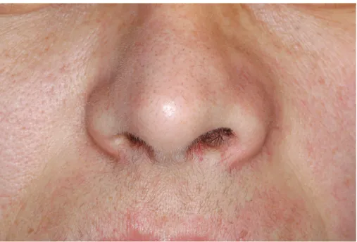
A 43-year-old woman presented with nonpainful lip and cheek swelling complaints (Figure 1). These symptoms were evident for the last 2 years. Extraoral examination revealed a fluctant, partially mobile and nontender lesion extending from left nasolabial sulcus to left nasal ala, partially obstructing the inferior nasal passage (Figure 2). There was no symptom upon intraoral examination. Panaromic radiography showed no pathology. Paranasal sinus CT revealed a cystic mass of 4x5 cm in the soft tissue extending to nasal floor and a concave bone resorption due to the lesion (Figure 3). Clinical and radiographical findings suggested the lesion to be a nasolabial cyst.
Figure 1. Expansion in the left nasolabial sulcus
The lesion was removed under local anesthesia via an intraoral approach. A sulcular incision was made and a full thickness flap was raised to expose the lesion. The cyst was excised from surrounding soft tissues through blunt dissection. After the removal of the cyst, bone resorption due to cyst pressure in nasal floor was confirmed but nasal mucosa was found intact (Figure 4). The operation site was closed by primary intention and the lesion was submitted for histopathological examination.
13 Histopathological evaluation revealed a fibrous cyst wall lined with pseudostratified columnar epithelium
with cilia. Areas of squamous metaplasia in epithelium and minor salivary gland lobules along with hyperemic vascular sections were detected in the fibrous wall. Histopathological findings supported the clinical diagnosis as nasolabial cyst.
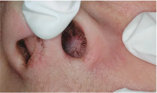
Figure 2. Appearance of the cyst from the nasal cavity.
Figure 3. CT showing the borders of the lesion from aperture piriformis extending to the nasal cavity, located in the soft tissue.
14
Discussion
Nasolabial cyst is the only non-odontogenic cyst located in soft tissue in the oral region. The cyst is mostly unilateral and left-sided but bilateral cases are also reported in the literature (Ural et al., 2007; Patil et al., 2016). Most common symptoms include nasal obstruction, vestibular swelling and pain during palpation. Depending on its effect on the nasal cavity, difficulty in breathing from nose, obstruction and rhinorrhea can be seen (Tiago et al., 2008). Clinical manifestations in this case study are consistent with reported cases in the literature.
Figure 4. Removal of the cyst via sublabial approach.
CT and MR are most commonly used methods for detecting the extent of the lesion (Aquilino et al., 2008). Transcutaneous ultrasonography, a simple office-based diagnostic tool, can also be a first-line imaging tool for the diagnosis of nasolabial cyst (Yeh et al., 2017). Panoramic radiographs, which are frequently used in dentistry, give no evidence of the lesion unless there is deformity in the lateral and anterior edges of the nasal floor. For this reason, asymptomatic nasolabial cysts might not be identified with conventional radiographies in dentistry.
Histopathological examination of nasolabial cysts usually reveals a cuboid basal layer, a double-layered epithelium with a prismatic luminal layer and goblet cells within the epithelium. In addition, multilayered or pseudostratified cuboid and prismatic epithelium can line the cyst (Toribio and Roehrl, 2011). As the histological findings of the nasolabial cyst is similar to that of the nasolacrimal duct lining, the theory supporting the origin of the cyst to be of nasolacrimal duct is more supported. Along with clinical findings, diagnosis of nasolabial cyst can be determined by microscopy easily. Furthermore, immunohistochemical studies of this lesion have been reported recently (Friedrich et al., 2012).
15 The differential diagnosis should be made with dentoalveolar abscess, nasopalatine duct cyst, mucus
extravasation cyst, salivary gland adenoma, epidermoid cyst, and globulomaxillary cyst (Sumer et al., 2010). Dentoalveolar abscess and cysts are related to teeth and are located inside the bone. Nasopalatine duct cyst, also known as incisive canal cyst, is an anterior midline lesion in maxilla and located in the labial or oral region. Glubulomaxillary cysts are located between lateral incisor and canine teeth, localized in the maxillary bone as well. Clinical and radiographic features such as the growth rate of the lesion and its localization inside soft tissue are helpful in distinguishing between nasolabial cyst and other lesions of this area.
Conventional treatment method for nasoalveolar cysts is enucleation of the lesion with a sublabial approach (Tiago et al., 2008). Once the lesion is completely removed, recurrence is not seen. Malignant transformation of this lesion has not been reported in the literature (Toribio and Roehrl, 2011). However, due to the lesion’s proximity and communication with teeth, contamination to odontogenic reasons might lead to odontogenic infection of the cyst (Martini et al., 2016). Other less commonly used approaches for the treatment are transnasal endoscopic marsupialization and sclerosing agent injection into the lesion. Transnasal marsupialization, especially in large cysts, is a faster method than enucleation via sublabial aproach, and patients treated with this method have less postoperative pain (Su et al., 1999; Chao et al., 2009; Lee et al., 2009). However, disadvantages of this method include mucus accumulation and cyst recurrence. Therefore, in this case, we preferred enucleation via sublabial approach considering the size of the lesion and the minimal risk of recurrence. No complications were observed during and after the surgery.
In conclusion, nasolabial cyst is a rare non-odontogenic soft tissue cyst in the maxillofacial region. Patients are treated by oral surgeons, plastic surgeons or ENT specialists according to the appearance of their complaints. Therefore, when a mass in the nasal vestibule is detected, nasolabial cyst should always be kept in mind in terms of differential diagnosis before invasive procedures are performed. Treatment of this lesion has high success rates with minimal recurrence and its long-term prognosis is very good.
Conflict of Interests
Authors declare no conflict of interests
References
Aquilino, R.N., Bazzo, V.J., Faria, R.J., Eid, N.L., Boscolo FN. (2008) Nasolabial cyst:presentation of a clinical case with CT and MR images. Revista Brasileira de Otorrinolaringologia, 74(3), 467-471.
Chao, W.C., Huang, C.C., Chang, P.H., Chen, Y.L., Chen, C.W., Lee, T.J. (2009). Management of nasolabial cysts by transnasal endoscopic marsupialization. Archives of Otolaryngology Head & Neck Surgery, 135(9), 932-935. Friedrich, R.E., Scheuer, H.A., Assaf, A.T, Zustin. J. (2012). Nasolabial cyst: case report with respect to immunohistochemical findings. In Vivo, 26(3), 453-458.
16
Karadağ, D., Beriat, G.K., Akıncı, T., Yinanç, M.A. (2011). Nasolabial cyst: MRI findings: case report. Türkiye Klinikleri Journal of Medical Sciences, 31(3), 731-734.
Lee, J.Y., Baek, B.J., Byun J.Y., Chang, H.S., Lee, B.D., Kim, D.W. (2009). Comparison of conventional excision via a sublabial approach and transnasal marsupialization for the treatment of nasolabial cysts: a prospective randomized study. Clinical and Experimental Otorhinolaryngology, 2(2), 85-89.
Martini, E.C., Coppla, F.M., Campagnoli, E.B., Bortoluzzi, M.C. (2016). Nasolabial cyst associated with odontogenic infection. Case Reports in Dentistry, 2016, 8690593.
Neville, B.W., Damm, D.D., Allen, C.M., Bouquot, J.E. (2002). Oral & Maxillofacial Pathology, 2nd ed. WB Saunders, Philadephia, USA.
Patil, A. R., Singh, A. P., Nandikoor, S., Meganathan, P. (2016). Bilateral nasolabial cysts- case report and review of literature. Indian Journal of Radiology and Imaging, 26(2), 241-244.
Schuman, D.M. (1981). Nasolabial cysts: mechanisms of development. Ear, Nose & Throat Journal, 60(9), 389-394.
Su, C.Y., Chien, C.Y., Hwang, C.F. (1999). A new transnasal approach to endoscopic marsupialization of the nasolabial cyst. The Laryngoscope, 109 (7), 1116-1118.
Sumer, A.P., Celenk, P., Sumer, M., Telcioglu, N.T., Gunhan, O. (2010) Nasolabial cyst: a case report with CT and MRI findings. Oral Surgery, Oral Medicine, Oral Pathology, Oral Radiology, 109(2), 92-94.
Tezer, M.S., Özlügedik, S., Ünal, A., Günhan, Ö., Aydemir, G. (2004) Giant Nasolabial Cyst: Case Report. Kulak Burun Boğaz ve Baş Boyun Cerrahisi Dergisi, 12(1), 7-10.
Tiago, R.S., Maia, M.S., Nascimento, G.M., Correa, J.P., Salgado, D.C. (2008). Nasolabial cyst:diagnostic and therapeutical aspects. Brazilian Journal of Otorhinolaringology, 74(1), 39-43.
Toribio, Y., Roehrl, M.H.A. (2011). The nasolabial cyst: a nonodontogenic oral cyst related to nasolacrimal duct epithelium. Archives of Pathology & Laboratory Medicine, 135(11), 1499–1503.
Ural, A., Huntürk, A.M., Tezer, M., Uslu, S.S. (2007). Bilateral nasoalveolar cyst: case report and therapeutic approach. KBB-Forum, 6(2), 66-68.
Yeh, C.H., Ko, J.Y., Wang, C.P. (2017). Transcutaneous ultrasonography for diagnosis of nasolabial cyst. Journal of Craniofacial Surgery, 28(3), e221-222.