ÖZET
Akciğer kanseri dünyada kansere bağlı ölümlerde başı çekmek-tedir, küçük hücre dışı akciğer kanseri (KHDAK) olguların %85’ini oluşturur. Pozitron emisyon tomografi (PET-BT) 2001 yılında kullanıma girişinden itibaren erken tanı, evreleme ve rekürenslerin erken ve doğru tesbitinde kullanılmaktadır. Bu yayında PET-BT ile ilk değerlendirmeye rağmen, torakotomi ve lenf nodu diseksiyonu ile evresi yükselen ve aynı zamanda PET-BT’de yanlış pozitif tutulum nedeniyle rekürens olarak değerlen-dirilen bir olguyu sunmak istedik. İlginç bir şekilde hastanın sigarayla ilintili olmayan bir testis tümörü de vardı. Bu olguda PET-BT’de yanlış pozitif ve yanlış negatiflik aynı zamanda görül-dü. Ek olarak, hastanın daha önce tesbit edilmemiş seminomu vardı. Akciğer kanseri takip ve tedavisi ile ilgilenen klinisyenler PET-BT’de yanlış pozitif ve yanlış negatif sonuçlar olabileceğini, bu nedenle histopatolojik doğrulamanın gerekli olduğunu akılla-rında bulundurmalıdır. Ayrıca, akciğer kanserine sigarayla ilişkili olmasa bile multipl bağımsız primer tümör eşlik edebilir, ancak bu durum prognoza etki etmemektedir.
(Turk Toraks Derg 2012; 13: 174-7)
Anahtar sözcükler: Akciğer kanseri, pozitron emisyon tomog-rafi, yanlış pozitiflik, yanlış negatiflik
Geliş Tarihi: 23.03.2011 Kabul Tarihi: 28.03.2011 ABSTRACT
Lung cancer is the leading cause of cancer-related deaths world-wide and non-small cell lung cancer (NSCLC) accounts for about 85% of the cases. Since its introduction in 2001, positron emis-sion tomography (PET-CT) has been used in early diagnosis, staging, early and accurate detection of recurrences. In this report, we wish to present a case of lung cancer who was upstaged after thoracotomy and lymph node dissection despite a first evaluation with PET-BT and also, during follow up, the patient was misdiagnosed as a recurrence due to false positive uptake on PET-BT. Interestingly, the patient also had a synchro-nous testicular tumor which was not smoking-related. In this case, the false positive and false negative results of PET-BT was seen in the same patient. Additionally, the patient had a semi-noma accompanying the lung cancer, which was not reported previously. Clinicians who deals with management of lung cancer should keep in mind that PET-BT may cause false positive and false negative results, so histopathological confirmation is necessary, and multiple independent primary tumors may accompany to lung cancer, even non-tobacco related ones, but it does not seem to be effective in the prognosis of the patient. (Turk Toraks Derg 2012; 13: 174-7)
Key words: Lung cancer, positron emission tomography, false positivity, false negativity
Received: 23.03.2011 Accepted: 28.03.2011
False Positivity and False Negativity on Positron Emission
Tomography in a Patient with Lung Cancer
Akciğer Kanserli Bir Olguda Pozitron Emisyon Tomografide Yanlış Pozitiflik ve Yanlış
Negatiflik
Evrim Eylem Akpınar1, Ahmet Hakan Haliloğlu2, Emel Öztürk3, Meral Gülhan1
1Department of Chest Diseases, Faculty of Medicine, Ufuk University, Ankara, Turkey 2Department of Urology, Faculty of Medicine, Ufuk University, Ankara, Turkey 3Department of Nuclear Medicine, Faculty of Medicine, Ufuk University, Ankara, Turkey
CASE REPORT OLGU SUNUMU
174
Address for Correspondence / Yazışma Adresi: Evrim Eylem Akpınar, Department of Chest Diseases, Faculty of Medicine, Ufuk University, Ankara, Turkey Phone: +90 505 243 35 82 E-mail: [email protected]
doi:10.5152/ttd.2012.36 INTRODUCTION
Lung cancer has become the number one killer among cancers worldwide. The 2 main types of lung cancer are small cell lung cancer (SCLC) and non-SCLC (NSCLC); NSCLC accounts for approximately 85% of all cases of lung cancer [1]. Multiple independent primary tumors might be seen together with lung carcinoma, especially smoking related ones [2]. Diagnosis is very important for planning the treatment of lung carcinoma as soon as possible. Since its introduction in 2001, inte-grated positron emission tomography-computed tomog-raphy (PET-CT) has been used in early diagnosis, staging, early and accurate detection of recurrences [3-6]. In this
report, we presented a case of lung cancer who was upstaged after thoracotomy and lymph node dissection despite a first evaluation with PET-CT, during follow up the patient was misdiagnosed as a recurrence due to a false positive uptake on PET-CT. Interestingly, the patient also had a synchronous testicular tumor which is not smoking-related.
CASE REPORT
A sixty-seven years-old male patient was admitted to a clinic with dyspnea and hemoptysis. There was a mass detected on thorax computed tomography. On bron-choscopy, the left upper lobe was completely, and
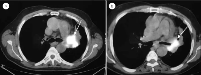
lower lobe almost-completely occluded with endobron-chial lesion. Pathological examination was consistent with epidermoid carcinoma. PET-CT revealed a soft tis-sue mass which was in close relation with the left pul-monary artery, surrounding main bronchus and lober bronchus on the left hilum (Standard uptake value (SUV) max: 13.6) (Figure 1), and a right testicular lesion that was hypometabolic at the centre, had increased 18F -fluorodeoxyglucose (FDG) uptake at the edges (SUVmax: 11.9). The lesion was interpreted as a cyst of the epidydimis on scrotal ultrasonography. Cranial MRI did not show any metastatic lesion. Left extended pneumonectomy and dissection of the lymph nodes performed. Surprisingly, the pathological examination revealed metastasis in the subcarinal lymph node, despite N staging according to PET-CT was 0 and mar-gins of surgery were clear from the tumor. Four cycles of adjuvant vinorelbine-cisplatin treatment were carried out after surgery. Thereafter, the testicular lesion also was resected. Pathological examination revealed a benign cyst and spermatocytic seminoma. After an uro-logical counsultation, it was decided that surgical treat-ment was sufficient. A suspicious thickening was seen on follow up thoracal CT, 17 month after diagnosis. PET-CT showed pathological activity on the medial cru-ris of the left surrenal gland (SUV max: 4.01) (Figure 2). Left surrenalectomy was performed to differentiate a solitary metastasis of lung cancer or primary tumor of the surrenal gland. Another surprise was waiting for us, as the pathology reported a hamartoma.
DISCUSSION
In the management of patients with lung cancer, early diagnosis, staging for treatment planning, and early detection of recurrences are very important issues. PET-CT is a nonivasive and very useful imaging proce-dure for all these aims. However, when using PET in the diagnosis of lung cancer, a false-positive rate of 20-25% is not uncommon [7]. The major causes of false-positive
results in the chest are granulomas, sarcoidosis, histo-plasmosis, tuberculosis, chronic inflammation and Aspergillus infection [8]. Iatrogenic procedures such as placement of chest tubes, percutaneous needle biopsy, mediastinoscopy, talc pleurodesis, radiation pneumoni-tis and oesophagipneumoni-tis might also cause false positive results. Indeterminate adrenal masses on CT are seen in approximately 5-15% of patients with NSLC, but only around 60% represent metastases. PT-CT is effective in discriminating between malignant and benign adrenal masses [4]. A recent large scale study in patients with NSCLC after potentially curative surgery, the diagnostic performance of PET-CT was evaluated, with results as follows: sensitivity 97%, specificity 96% and negative predictive value 99%, but positive predictive value rela-tively low (81%) compared to preceding studies [3]. In this case also, false positivity of the surrenal lesion was misinterpreted as recurrence of lung cancer. Several studies used SUV cut-off 2.5 to differentiate malignant from benign lesions [9,10]. Although this patient’s lesion had a SUV value >2.5, the pathology of the lesion was benign. Solitary extra-pulmonary lesions were
Turk Toraks Derg 2012; 13: 174-7
Eylem Akpınar et al. Positron Emission Tomography in Lung Cancer
175
Figure 1. a, b. Mass was in close-relation with the left pulmonary artery, surrounding main bronchus and lober bronchus on the left hilum (SUVmax: 13.6)
a b
Figure 2. PET-CT showed pathological activity on the medial cruris of the left surrenal gland (SUV max: 4.01)
revealed by PET-CT in 21% of a patient population with newly diagnosed NSCLC; of these,approximately 40% turned out to have a benign or inflammatory lesion and 6% an unrelated malignancy. Thus, confirmation of positive PET findings is recommended [4].
False-negative results are less common and due to small size (<5mm) or well-differentiated malignancies, such as bronchiolo-alveolar carcinomas and carcinoids [10]. Combining FDG-PET and CT is better than CT alone, with a very high negative predictive value (NPV) for stag-ing N2 and N3 disease (95% overall and 99% for indi-vidual nodes) [11]. In this reported case, PET-CT made a mistake in N staging. Although there was no pathologi-cal activity in the mediastinum (Figure 1) a subcarinal lymph node metastasis was detected after surgery. NSCLC patients with stage IIIA (N2) tumors present with substantial heterogenity in the clinical presentation, treatment and prognosis. Therefore, N2 tumors are clas-sified into four subsets in the American College of Chest Physicians (ACCP) guideline [12]. According to this clas-sification, our patient was considered as stage IIIA2 (Nodal-single station metastasis recognized intraopera-tively).The lymph nodes and primary tumor were com-pletely resected, adjuvant platinum-based chemotherapy was carried out postoperatively because he had a good performance status (ECOG 1), as recommended in the ACCP guideline.
The Amerian College of Surgeons defined multiple independent primaries (MIP) as “two or more tumors arising at different sites or at the same site when histo-logic characteristics differ”. When MIP involve a lung cancer, multiplicity may be related to sharing a common etiologic factor such as tobacco smoking or asbestos exposure. In a recent study, Aguilo et al. [2] showed that 70% of the cancers associated with lung cancer were tobacco related. Smoking, lung cancer stage I-II and age above 60 years were found as independent risk factors for developing multiple cancer. Cancer multiplicity was not associated with worse prognosis, and prognosis of the patient is determined by the stage of lung cancer. In a retrospective study including 1105 lung cancer patients, 13 patients had urologic malignancies, 11 were tobacco related, and 12 of them died due to lung cancer, while relapse of urologic malignancies was not seen during follow up of patients. Median survival of NSCLC patients was 18 months [13]. Because our patient was 67 years old and a smoker, there was an increased risk of MIP. However, the second tumor which was syncronously detected in the patient, was a seminoma, which is not tobacco related. Tobacco-related tumors such as rectal carcinoma were reported before the syncronous lung cancer [14]. In a previously reported case, the authors described a lung tumor that metastasized to a testicular seminoma [15]. This was the first reported case of tumor-to-tumor metastases in which seminoma of the testis was the recipient. Our case is different, it is the first
reported case in manner in which the seminoma accom-panied the lung cancer synchronously. The patient is still alive 24 months after the diagnosis of lung cancer, no relapse was detected for both malignancies. The syncro-nous tumor was thought to be ineffective in prognosis of the patient.
In conclusion, although PET-CT is a noninvasive, reli-able imaging technique in the diagnosis, staging, treat-ment planning and follow up of lung cancer, it might make a mistake. In this report, the false positive and false negative results were seen in the same patient. So, histopathological confirmation of PET-CT results is important. MIP, especially tobacco-related ones, may be seen in patients with lung cancer. In this case, the patient had a seminoma accompanying lung cancer, which was not reported previously. Clinicians who deal with the management of lung cancer should be kept in mind that PET-CT may cause false positive and false negative results, and MIP may accompany lung cancer even non-tobacco related ones, but it seems to be inef-fective in the prognosis of the patient.
REFERENCES
1. Molina JR, Yang P, Cassivi SD, et al. Non-small cell lung cancer: Epidemiology, risk factors, treatment and survivor-ship. Mayo Clinic Proceedings 2008;83:584-94. [CrossRef]
2. Aguilo R, Macia F, Porta M, et al. Multiple independent primary cancers do not adversely affect survival of the lung cancer patient. Eur J Cardiothorac Surg 2008;34: 1075-80. [CrossRef]
3. Kanzaki R, Higashiyama M, Maeda J, et al. Clinical value of F18-fluorodeoxyglucose positron emission tomography-computed tomography in patients with non-small cell lung cancer after potentially curative surgery: experience with 241 patients. Interact Cardio Vasc Thorac Surg 2010;10:1009-14. [CrossRef]
4. Fischer BM, Mortensen J. The future in diagnosis and stag-ing of lung cancer: Positron emission tomography. Respiration 2006;73:267-76. [CrossRef]
5. Atasoy Ç, Sönmez D. Imaging in lung cancer. Turkiye Klinikleri J Med Oncol-Special Topics 2008;1:34-46.
6. Şen E, Gönüllü U. Diagnosis and staging in lung cancer. Turkiye Klinikleri J Med Oncol-Special Topics 2008;1:47-57. 7. Stroobants S, Verschakelen J, Vansteenkiste J. Value of
FDG-Pet in the management of non-small cell lung cancer. Eur J Radiol 2003;45:49-59. [CrossRef]
8. Ho Shon I, O’Doherty MJ, Maisey MN. Positron emission tomography in lung cancer. Semin Nucl Med 2002;32: 240-70. [CrossRef]
9. Patz EF Jr, Lowe VJ, Hoffman JM, et al. Persistent or recur-rent bronchogenic carcinoma: detection with PET and 2-[F-18]-2-deoxy-D-glucose. Radiology 1994;91:379-82. 10. Detection of primary and recurrent lung cancer by means of
F-18 fluorodeoxyglucose positron emission tomography (FDG PET). J Thorac Cardiovasc Surg 1995;110:130-9. [CrossRef]
11. Rankin SC. Staging of non-small cell lung cancer (NSCLC): Cancer imaging 2006;6:1-3. [CrossRef]
12. Robinson LA, Ruckdeschel JC, Wagner H Jr, et al. Treatment of non-small cell lung cancer-stage IIIA: ACCP evidence-based clinical practice guidelines (2nd edition). Chest 2007;132(3 Suppl):243-65.
Turk Toraks Derg 2012; 13: 174-7 Eylem Akpınar et al.
Positron Emission Tomography in Lung Cancer
13. Miyazaki K, Satoh H, Kagohashi K, et al. Lung cancer patients with previous or simultaneous urologic cancers. Med Oncol 2008;25:443-6. [CrossRef]
14. Kaya AO, Yıldız R, Öztürk B, et al. Synchronous Squamous Cell Lung Carcinoma and Rectum Adenocarcinoma: Case
Report. International Journal of Hematology and Oncology 2008;18:42-4.
15. Ro JY, Sahin AA, Ayala AG, et al. Lung carcinoma with metastasis to testicular seminoma. Cancer 1990;66:347-53.
[CrossRef] Turk Toraks Derg
2012; 13: 174-7
Eylem Akpınar et al. Positron Emission Tomography in Lung Cancer